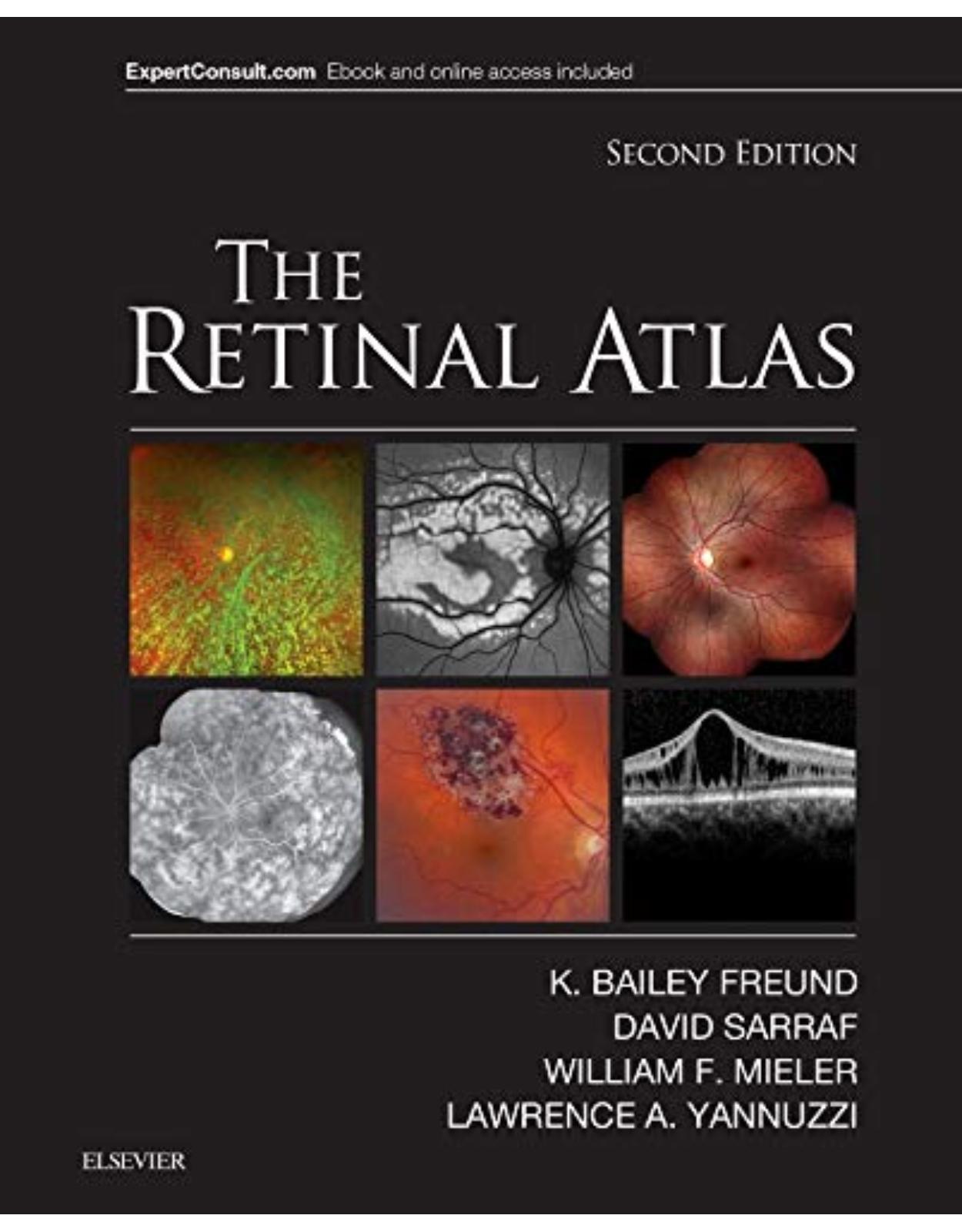
The Retinal Atlas, 2nd Edition
Livrare gratis la comenzi peste 500 RON. Pentru celelalte comenzi livrarea este 20 RON.
Disponibilitate: La comanda in aproximativ 4-6 saptamani
Editura: Elsevier
Limba: Engleza
Nr. pagini: 1200
Coperta: Hardback
Dimensiuni: 216 X 276 mm
An aparitie: 2016
|
With more than 5,000 images, a unique page layout, and comprehensive illustrations of the entire spectrum of vitreous, retina, and macula disorders, The Retinal Atlas, 2nd Edition, is an indispensable reference for retina specialists and comprehensive ophthalmologists as well as residents and fellows in training. For this edition, an expanded author team made up of Drs. K. Bailey Freund, David Sarraf, William F. Mieler, and Lawrence A. Yannuzzi, each an expert in retinal research and imaging, provide definitive up-to-date perspectives in this rapidly advancing field. This award-winning title has been thoroughly updated with new images with multimodal illustrations, new coverage and insight into key topics, and new disorders and classifications, while retaining the innovative page layout that has made it the most useful and most complete atlas of its kind. |
|
|
Features: |
|
|
|
|
New To This Edition: |
|
|
|
|
Table Of Contents: |
|
|
Chapter 1: Normal Chapter 2: Hereditary chorioretinal dystrophies Chapter 3: Pediatric retina Chapter 4: Inflammation Chapter 5: Infection Chapter 6: Retinal vascular disease Chapter 7: Degeneration Chapter 8: Oncology Chapter 9: Macular fibrosis, pucker, cysts, holes, folds, and edema Chapter 10: Non-rhegmatogenous retinal detachment Chapter 11: Peripheral retinal degenerations and rhegmatogenous retinal detachment Chapter 12: Traumatic chorioretinopathy Chapter 13: Complications of ocular surgery Chapter 14: Chorioretinal toxicities Chapter 15: Congenital anomalies of the optic nerve |
|
| An aparitie | 2016 |
| Autor | K. Bailey Freund,David Sarraf, William F. Mieler Lawrence A. Yannuzzi |
| Dimensiuni | 216 X 276 mm |
| Editura | Elsevier |
| Format | Hardback |
| ISBN | 9780323287920 |
| Limba | Engleza |
| Nr pag | 1200 |


Clientii ebookshop.ro nu au adaugat inca opinii pentru acest produs. Fii primul care adauga o parere, folosind formularul de mai jos.