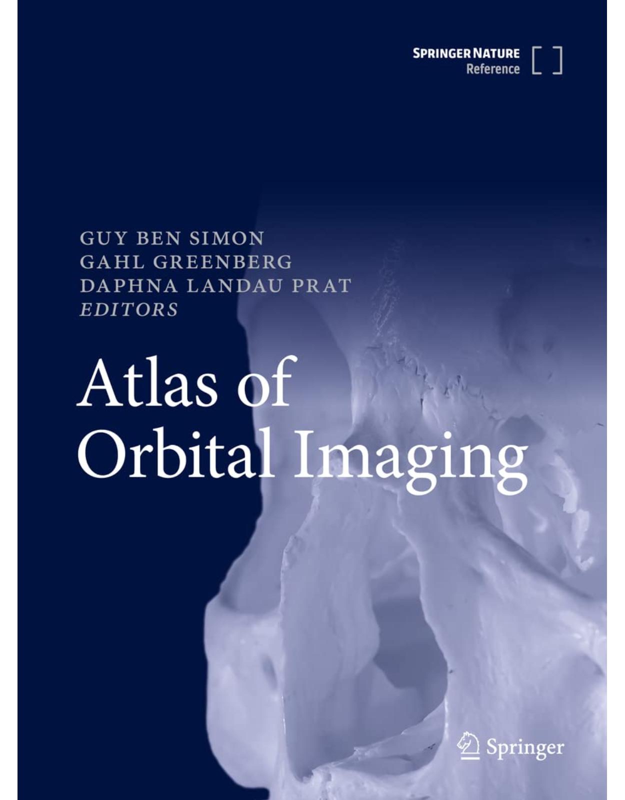
Atlas of Orbital Imaging
Livrare gratis la comenzi peste 500 RON. Pentru celelalte comenzi livrarea este 20 RON.
Disponibilitate: La comanda in aproximativ 4-6 saptamani
Editura: Springer
Limba: Engleza
Nr. pagini: 768
Coperta: Hardcover
Dimensiuni: 20.96 x 4.45 x 27.94 cm
An aparitie: 2022
This book features in-depth descriptions of imaging modalities (MRI, CT, US, PET) for all orbital pathologies, including tumors, vascular anomalies, congenital anomalies, trauma, inflammations and infections. It describes all the imaging features of the pathologies, and includes guidance for differential diagnosis and relevant clinical data.
Atlas of Orbital Imaging serves as a clinical and educational resource for ophthalmologists/orbital surgeon residents, as well as a source of reference for consultants and neuroradiologists at all levels. The illustrations are both highly detailed and depict the orbit in vivid colour, adding to the attractiveness of the chapters. This reference work is a worldwide collaborative effort of all leading orbital surgeons and neuro-radiologists (in Europe, America, Australia and Asia) and provides an indispensable resource for developing skills and knowledge of orbital imaging.
Orbital imaging is an important aspect in the management of oculoplastic and orbital disorders. Correlation of imaging features with clinical presentation, as well as systemic disease, is required. Clinicians cannot rely solely upon the report of a radiologist, who often may not be aware of all symptoms and signs or may not necessarily be experienced in the interpretation of orbital disease. There has been a void in recent textbooks in the teaching of this important art. The authors of this book have painstakingly compiled a comprehensive array of images that provides a systematic and detailed reference source for the state-of-the-art imaging of orbital disease.
It is divided into 96 chapters, covering a wide range of disorders. The photos and images are of an exceptionally high quality. Descriptions are detailed. It is easy to lose oneself, gazing from page to page, becoming increasingly absorbed in the beautiful anatomical detail from chapter 1 onwards. I found this book incredibly appealing.
Part 1 is titled Orbital Anatomy and has 10 sections covering bones, vascular, soft tissue, sinuses, cavernous sinus, Meckel’s cave. What is particularly unique is how four chapters are devoted to the orbital fissures alone. Part 2 comprises nine chapters devoted to orbital imaging modalities: CT, MRI, CTA, CTV, MRA, MRV, intraoperative dynamic imaging, ultrasound of tumours, ultrasound with Doppler, PET CT and lastly, orbital imaging “pearls”. Part 3 comprises eight chapters on congenital malformations and anomalies. There are six chapters on lacrimal gland tumours, three chapters on lacrimal anatomy and disease, seven chapters on primary orbital tumours, 12 chapters on ocular and eyelid tumours that may extend to the orbit, six chapters on osseus and meningeal tumours that affect the orbit, three chapters on optic nerve tumours, six chapters on vascular malformations, nine chapters on orbital inflammatory diseases, nine chapters on orbital infectious diseases, five chapters covering orbital trauma and two chapters covering periocular fillers, now commonplace yet poorly understood by clinicians interpreting orbital or facial imaging. The textbook comprises 742 pages, including index. One really cannot emphasise enough its comprehensive and in-depth coverage of the topic.
Overall, the book was expeditious, succinct and very easy to read, either as a source of reference or for longer reading. It is straightforward, concise, aesthetically pleasing and extremely well presented. Each chapter starts with an abstract, outlining the aims and highlighting key points. Cases are critically organised with high-quality, contemporary imaging technology. Images are of excellent quality and non-ambiguous with precise labelling and use of arrows / asterix to highlight pathology. I was provided an e-copy of the book to review but I am assured that the hard copy version is of an equally high quality of print.
Comparison points and how to differentiate from other diseases are very helpful. For example: “While thyroid eye disease could present with this appearance, the patient had loss of function of the levator with ptosis rather than lid retraction.”
The book fills an important niche by comprehensively describing the appearances of orbital pathologies on orbital imaging. It is an excellent and uniquely positioned book for all ophthalmologists and radiologists, whether experienced or in training, that supersedes any past text on this topic.
Table of Contents:
Front Matter
Pages i-xxvi
Orbital Anatomy
Front Matter
Pages 1-1
Bones of the Orbit
Jack Rootman, Daniel B. Rootman, Bruce Stewart, Stefania B. Diniz, Kelsey A. Roelofs, Liza M. Cohen et al.
Pages 3-23
Vascular Anatomy
Daniel B. Rootman, Bruce Stewart, Jack Rootman
Pages 25-32
Ocular Adnexa, Soft Tissue, and Extraocular Muscles
Jack Rootman, Daniel B. Rootman, Bruce Stewart, Stefania B. Diniz, Kelsey A. Roelofs, Liza M. Cohen et al.
Pages 33-46
Paranasal Sinuses
Jack Rootman, Daniel B. Rootman, Bruce Stewart, Stefania B. Diniz, Kelsey A. Roelofs, Liza M. Cohen et al.
Pages 47-61
Orbital Fissures
Jack Rootman, Daniel B. Rootman, Bruce Stewart, Stefania B. Diniz, Kelsey A. Roelofs, Liza M. Cohen et al.
Pages 63-67
Orbital Fissures: Inferior Orbital Fissure
Jack Rootman, Daniel B. Rootman, Bruce Stewart, Stefania B. Diniz, Kelsey A. Roelofs, Liza M. Cohen et al.
Pages 69-74
Orbital Fissures: Pterygopalatine Fossa
Jack Rootman, Daniel B. Rootman, Bruce Stewart, Stefania B. Diniz, Kelsey A. Roelofs, Liza M. Cohen et al.
Pages 75-78
Orbital Fissures: Infratemporal Fossa
Jack Rootman, Daniel B. Rootman, Bruce Stewart, Stefania B. Diniz, Kelsey A. Roelofs, Liza M. Cohen et al.
Pages 79-84
Cavernous Sinus
Jack Rootman, Daniel B. Rootman, Bruce Stewart, Stefania B. Diniz, Kelsey A. Roelofs, Liza M. Cohen et al.
Pages 85-89
Meckel’s Cave
Jack Rootman, Daniel B. Rootman, Bruce Stewart, Stefania B. Diniz, Kelsey A. Roelofs, Liza M. Cohen et al.
Pages 91-96
Orbital Imaging Modalities
Orbital CT
Denise S. Kim, Remy R. Lobo, Alon Kahana
Pages 99-102
Orbital MRI
Arnaldo Mayer, Gahl Greenberg
Pages 103-111
Orbital CTA/CTV
Denise S. Kim, Remy R. Lobo, Alon Kahana
Pages 113-116
Orbital MRA/MRV
Denise S. Kim, Remy R. Lobo, Alon Kahana
Pages 117-120
Intraoperative Dynamic Imaging
Denise S. Kim, Remy R. Lobo, Neeraj Chaudhary, Alon Kahana
Pages 121-125
Ultrasound of Orbit Tumors and Tumorlike Lesions
Bernadete Ayres, Alon Kahana
Pages 127-147
Orbital Ultrasound with Doppler
Stefania B. Diniz, Robert A. Goldberg
Pages 149-154
Orbital Imaging Modalities
Orbital Positron Emission Tomography/Computed Tomography (PET/CT)
J. Matthew Debnam, Bita Esmaeli
Pages 155-177
Orbital Imaging Pearls
Gahl Greenberg, Daphna Landau Prat, Guy Ben Simon
Pages 179-187
Congenital Malformations/Anomalies
Craniofacial Dystosis
William R. Katowitz
Pages 191-198
Neurofibromatosis
William R. Katowitz
Pages 199-208
Anophthalmia and Microphthalmia
William R. Katowitz
Pages 209-213
Cystic Lesions of the Orbit: Dermoid and Epidermoid Cysts
William R. Katowitz
Pages 215-220
Cystic Lesions of the Orbit: Teratomas
William R. Katowitz
Pages 221-223
Other Cystic Lesions of the Orbit: Encephalocele
William R. Katowitz
Pages 225-232
Congenital Lacrimal Pathologies: Dacryocystocele
William R. Katowitz
Pages 233-238
Pediatric Orbital Vascular Tumors: Infantile Hemangioma
William R. Katowitz
Pages 239-246
Lacrimal Gland Tumors
Lacrimal Gland Prolapse and Dacryops
Oded Sagiv, J. Matthew Debnam, Bita Esmaeli
Pages 249-252
Pleomorphic Adenoma of the Lacrimal Gland
Oded Sagiv, J. Matthew Debnam, Bita Esmaeli
Pages 253-257
Recurrent Pleomorphic Adenoma of the Lacrimal Gland (RLGPA)
Oded Sagiv, J. Matthew Debnam, Bita Esmaeli
Pages 259-262
Lacrimal Gland Carcinoma: Primary
Oded Sagiv, J. Matthew Debnam, Bita Esmaeli
Pages 263-266
Carcinoma Ex-Pleomorphic Adenoma of the Lacrimal Gland
Oded Sagiv, J. Matthew Debnam, Bita Esmaeli
Pages 267-270
Benign (Reactive) Lymphoid Hyperplasia and Lymphoma
Oded Sagiv, J. Matthew Debnam, Bita Esmaeli
Pages 271-275
Lacrimal Pathways
Normal Anatomy of the Lacrimal System
Swati Singh, Mohammad Javed Ali
Pages 279-282
Lacrimal Pathways
Imaging in Lacrimal Drainage Obstruction and Acute Dacryocystitis
Swati Singh, Mohammad Javed Ali
Pages 283-288
Lacrimal Sac Tumors Imaging
Swati Singh, Mohammad Javed Ali
Pages 289-294
Primary Orbital Tumors
Orbital Lymphoma
Jaskirat Aujla, Valerie Juniat, Sandy Patel, Dinesh Selva
Pages 297-305
Orbital Multiple Myeloma
Jaskirat Aujla, Valerie Juniat, Sandy Patel, Dinesh Selva
Pages 307-311
Orbital Soft Tissues Sarcomas/Liposarcoma
Jaskirat Aujla, Valerie Juniat, Sandy Patel, Dinesh Selva
Pages 313-318
Orbital Rhabdomyosarcoma
Ran Ben Cnaan, Dana Niry, Igal Leibovitch
Pages 319-323
Orbital Lipoma
Ran Ben Cnaan, Dana Niry, Igal Leibovitch
Pages 325-329
Solitary Fibrous Tumor of the Orbit
Ran Ben Cnaan, Justin N. Karlin, Dana Niry, Igal Leibovitch, Robert A. Goldberg
Pages 331-338
Granulomatous Orbital Inflammation: Orbital Langerhans Cell Histiocytosis
Alan A. McNab
Pages 339-345
Eye and Ocular Adnexa Tumors with Orbital Extension
Periocular Basal Cell Carcinoma (BCC)
Alon Tiosano, Natalia Michaeli, Iftach Yassur
Pages 349-351
Periocular Squamous Cell Carcinoma (SCC)
Alon Tiosano, Natalia Michaeli, Iftach Yassur
Pages 353-357
Periocular Sebaceous Cell Carcinoma
Alon Tiosano, Natalia Michaeli, Iftach Yassur
Pages 359-363
Ocular Surface Squamous Neoplasia with Orbital Extension
Swathi Kaliki, Ido Didi Fabian
Pages 365-368
Uveal Melanoma with Extraocular Spread
Andrew W. Stacey, Mahmud Mossa-Basha, Ido Didi Fabian
Pages 369-374
Retinoblastoma
Vikas Khetan, Pim de Graaf, Devjyoti Tripathy, Ido Didi Fabian
Pages 375-384
Metastatic Orbital Lesions: Breast Cancer
Oded Sagiv, J. Matthew Debnam, Bita Esmaeli
Pages 385-388
Metastatic Orbital Lesions: Lung Cancer, Prostate Cancer, and Renal Cell Carcinoma (RCC)
Oded Sagiv, J. Matthew Debnam, Bita Esmaeli
Pages 389-394
Metastatic Orbital Lesions: Melanoma
Oded Sagiv, J. Matthew Debnam, Bita Esmaeli
Pages 395-398
Eye and Ocular Adnexa Tumors with Orbital Extension
Orbital Extension of Tumors from the Paranasal Sinuses: Squamous Cell Carcinoma
Oded Sagiv, J. Matthew Debnam, Bita Esmaeli
Pages 399-401
Differential Diagnosis of Malignant Tumors of the Lacrimal Sac
Oded Sagiv, J. Matthew Debnam, Bita Esmaeli
Pages 403-406
Merkel Cell Carcinoma of Eyelid (MCC)
Soltan Khalaila, Rosa Novoa, Benzion Samueli, Erez Tsumi
Pages 407-414
Osseous and Meninges Lesions
Imaging of Orbital Osteoma and Osteosarcoma
Alexandra Manta, Stefania B. Diniz, Robert A. Goldberg
Pages 417-422
Ossifying Fibroma and Chondromyxoid Fibroma of the Orbit
Alexandra Manta, Stefania B. Diniz, Robert A. Goldberg
Pages 423-426
Fibrous Dysplasia
Stefania B. Diniz, Robert A. Goldberg
Pages 427-431
Intraosseous Hemangioma and Cholesterol Granuloma of the Orbit
Liza M. Cohen, Stefania B. Diniz, Robert A. Goldberg
Pages 433-438
Aneurysmal Bone Cyst and Ewing Sarcoma of the Orbit
Stefania B. Diniz, Liza M. Cohen, Robert A. Goldberg
Pages 439-443
Meningioma of the Orbit and Orbital Vicinity
Justin N. Karlin, Robert A. Goldberg
Pages 445-452
Optic Nerve
Optic Nerve Glioma: Pilocytic Astrocytoma
Yoon-Duck Kim
Pages 455-465
Optic Nerve Sheath Meningioma
Yoon-Duck Kim
Pages 467-474
Intrinsic and Extrinsic Etiologies of Optic Nerve Damage
Ofira Zloto, Nina Borissovsky, Judith Luckman, Nitza Goldenberg Cohen
Pages 475-488
Vascular Malformations
Lymphatic Malformations
Kasturi Bhattacharjee, Nirod Medhi, Shyam Sundar Das Mohapatra
Pages 491-497
Venous Malformations (VM) Distensible/Lymphatico-Venous Malformations (LVM)
Kasturi Bhattacharjee, Shyam Sundar Das Mohapatra, Aditi Mehta
Pages 499-505
Orbital Venous Malformations (VM): Nondistensible
Kasturi Bhattacharjee, Nirod Medhi, Shyam Sundar Das Mohapatra
Pages 507-511
Carotid-Cavernous Fistula
Kasturi Bhattacharjee, Nirod Medhi, Shyam Sundar Das Mohapatra
Pages 513-519
Arteriovenous Malformations of the Orbit
Kasturi Bhattacharjee, Aditi Mehta
Pages 521-528
Vascular Malformations
Other Rare Vascular Tumors of the Orbit
Kasturi Bhattacharjee, Vatsalya Venkatraman
Pages 529-535
Orbital Inflammation
Idiopathic Orbital Inflammation (IOI)
Alan A. McNab
Pages 539-544
Orbital Myositis
Alan A. McNab
Pages 545-550
Dacryoadenitis
Alan A. McNab
Pages 551-555
Scleritis
Alan A. McNab
Pages 557-560
Orbital Manifestations of Granulomatosis with Polyangiitis
Alan A. McNab
Pages 561-565
IgG-4 Related Orbital Disease
Alan A. McNab
Pages 567-571
Granulomatous Orbital Inflammation: Orbital Sarcoidosis
Alan A. McNab
Pages 573-576
Xanthogranulomatous Disease of the Orbit
Alan A. McNab
Pages 577-580
Thyroid Eye Disease
Kelsey A. Roelofs, Ezekiel Weis
Pages 581-587
Orbital Infection
Pre-septal Orbital Cellulitis
Kelsey A. Roelofs, Ezekiel Weis
Pages 591-598
Orbital Cellulitis
Kelsey A. Roelofs, Ezekiel Weis
Pages 599-604
Mucocele with Orbital Involvement
Kelsey A. Roelofs, Ezekiel Weis
Pages 605-610
Subperiosteal Orbital Abscess
Kelsey A. Roelofs, Ezekiel Weis
Pages 611-614
Aspergillosis
Kelsey A. Roelofs, Erin D. Wright, Ezekiel Weis
Pages 615-621
Mucormycosis
Kelsey A. Roelofs, Ezekiel Weis
Pages 623-628
Orbital Hydatid Cysts
Milind N. Naik, Dilip K. Mishra
Pages 629-632
Orbital Cysticercosis
Jaee M. Naik, Milind N. Naik
Pages 633-636
Orbital Infection
Granulomatous Orbital Inflammation: Orbital Tuberculosis (TB)
Milind N. Naik, Joveeta Joseph
Pages 637-645
Orbital Trauma and Other Inferred Changes
Orbital Trauma: Orbital Soft Tissue Injuries and Intraorbital Foreign Bodies
Gangadhara Sundar
Pages 649-660
Orbital Trauma: Orbital and Orbitofacial Fractures
Kavya Sundar, Gangadhara Sundar
Pages 661-675
Ocular Trauma and Intrinsic Pathology
Ofira Zloto
Pages 677-681
Nontraumatic Orbital Hemorrhage (NTOH)
Daphna Landau Prat, Gahl Greenberg, Alan A. McNab, Guy Ben Simon
Pages 683-693
Postoperative Changes
Ofira Zloto
Pages 695-703
Periocular Fillers
Ayelet Priel, Don Kikkawa, Gahl Greenberg, Dana Niry, S. Cohen
Pages 705-712
Periocular Fillers–Related Complications: Imaging Features
S. Cohen, Dana Niry, Ayelet Priel
Pages 713-718
Acquired Anophthalmus
Yoav Vardizer, Nina Borissovsky, Daphna Landau Prat
Pages 719-726
Back Matter
| An aparitie | 2022 |
| Autor | Guy Ben Simon, Gahl Greenberg , Daphna Landau Prat |
| Dimensiuni | 20.96 x 4.45 x 27.94 cm |
| Editura | Springer |
| Format | Hardcover |
| ISBN | 9783030624255 |
| Limba | Engleza |
| Nr pag | 768 |


Clientii ebookshop.ro nu au adaugat inca opinii pentru acest produs. Fii primul care adauga o parere, folosind formularul de mai jos.