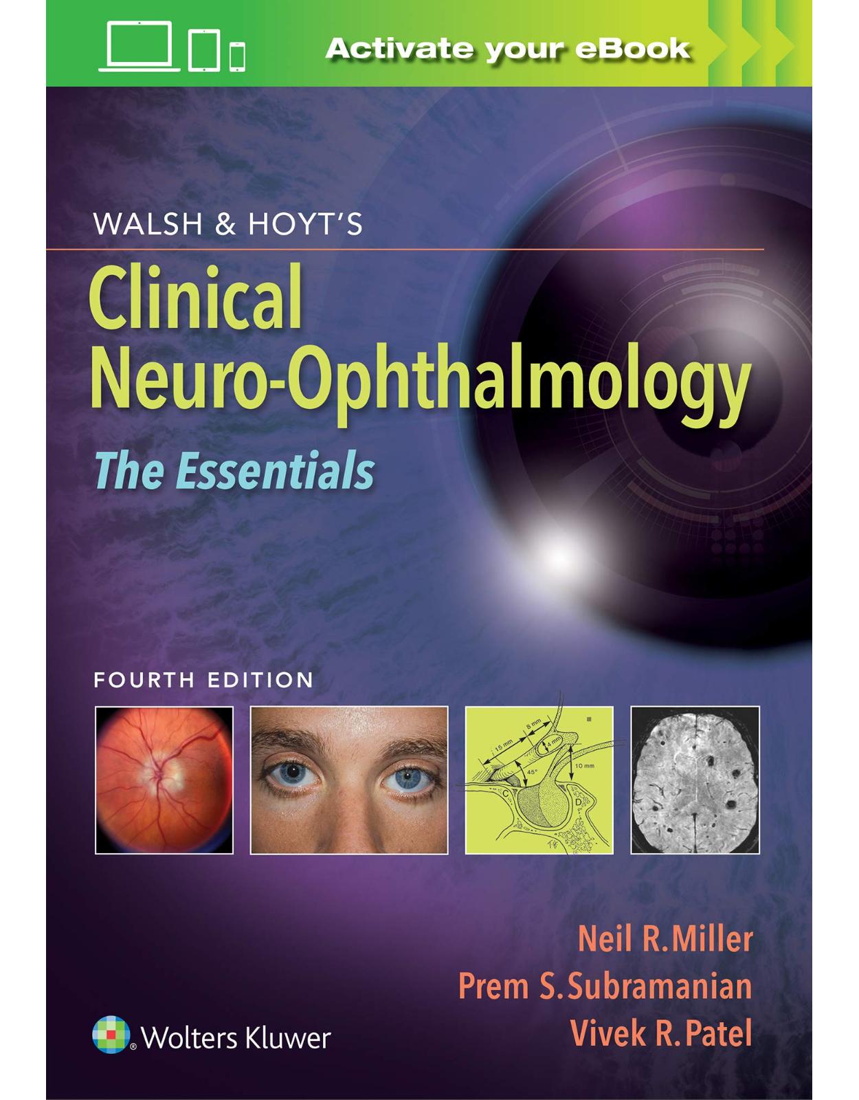
Walsh & Hoyt’s Clinical Neuro-Ophthalmology: The Essentials
Livrare gratis la comenzi peste 500 RON. Pentru celelalte comenzi livrarea este 20 RON.
Disponibilitate: La comanda in aproximativ 4-6 saptamani
Editura: LWW
Limba: Engleza
Nr. pagini: 600
Coperta: Paperback
Dimensiuni: 17.78 x 2.29 x 25.4 cm
An aparitie: 2 Sept. 2020
Description:
Offering authoritative, concise coverage of today’s neuro-ophthalmology, Walsh & Hoyt's Clinical Neuro-Ophthalmology: The Essentials distills the most vital information from the esteemed three-volume parent text, Walsh & Hoyt’s Clinical Neuro-Ophthalmology, into a convenient, single-volume resource. A practical and intuitive organization provides quick access to the essential information you need to effectively evaluate and manage the majority of neuro-ophthalmic conditions. Written by leaders in the field, this must-have fourth edition is a comprehensive, succinct, and clinically relevant reference for your everyday practice.
Table of Contents:
Section I The Afferent Visual System
1 Examination of the Visual Sensory System
History
Clinical Office Examination
Visual Acuity
Contrast Sensitivity
Stereoacuity
Color Vision
Visual Field Examination
Pupillary Examination
Brightness Comparison
Photostress Recovery Test
Cranial Nerves, External Examination, Anterior Segment Examination, and Exophthalmometry
Fundus Examination
Ancillary Testing
Ocular Imaging
Electrophysiologic Testing
2 Anatomy and Physiology of the Retina and Optic Nerve
Anatomy and Physiology of the Retina
Cell Types and Layers of the Retina
Retinal Blood Vessels
Rod and Cone Through Pathways: Functional Considerations
Anatomy and Physiology of the Optic Nerve
Intraocular Optic Nerve (Optic Nerve Head)
Orbital Optic Nerve
Intracanalicular (Intraosseous) Optic Nerve
Intracranial Optic Nerve
Topographic Anatomy of the Optic Nerve
Topographic Diagnosis of Retinal Lesions
Distinguishing Retinal Disease From an Optic Neuropathy by Clinical Examination
Distinguishing Retinal Disease From an Optic Neuropathy by Visual Field
Appearance of the Optic Disc in Retinal Disease
Correlation Between Clinical Findings and Pathology
3 Neuroimaging in Neuro-Ophthalmology
Decision Making Before Ordering Imaging
Computed Tomography
CT Orbits
CT Head
CT Neck
CT Angiography
Magnetic Resonance Imaging
Orbital Disorders
Central Nervous System Lesions
Vascular Disorders
Catheter Angiography and Venography
Other Imaging Modalities (Positron Emission Tomography, Magnetic Resonance Spectroscopy, Functional MRI)
Functional MRI
4 Congenital Anomalies of the Optic Disc
Anomalous Optic Disc Elevation (Pseudopapilledema)
Pseudopapilledema Associated With Optic Disc Drusen
Anomalous Disc Elevation Without Drusen
Optic Nerve Hypoplasia
Clinical Features
Associated Endocrinologic Deficiencies
Associated Central Nervous System Malformations
Systemic and Teratogenic Associations
Segmental Optic Nerve Hypoplasia
Pathogenesis
Anomalous Optic Disc Excavation
Morning Glory Disc Anomaly
Optic Disc Coloboma
Peripapillary Staphyloma
Megalopapilla
Optic Pit
Papillorenal Syndrome
Congenital Tilted Optic Disc Syndrome
Optic Disc Dysplasia
Congenital Optic Disc Pigmentation
Aicardi Syndrome
Doubling of the Optic Disc
Optic Nerve Aplasia
Myelinated (Medullated) Nerve Fibers
5 Topical Diagnosis of Acquired Optic Nerve Disorders
Appearance of the Disc
Optic Disc Swelling
Infiltration of the Optic Disc
Optic Atrophy
Syndromes of Prechiasmal Optic Nerve Dysfunction
Optociliary Shunts and the Syndrome of Chronic Optic Nerve Compression
Distal Optic Nerve Syndrome
Foster Kennedy Syndrome
Bilateral Superior or Inferior (Altitudinal) Hemianopia
Nasal Hemianopia
6 Papilledema
Measurements of Intracranial Pressure
Ophthalmoscopic Appearance of Papilledema
Stages of Papilledema
Other Signs of Papilledema
Chronic Papilledema
Postpapilledema Atrophy
Unilateral or Asymmetric Papilledema
Diagnosis of Papilledema
Course
Symptoms and Signs of Papilledema
Nonvisual Manifestations
Visual Manifestations
Etiology of Papilledema
Intracranial Masses
Disorders of CSF Flow
Syndromes of Elevated Venous Pressure
Trauma
Craniosynostoses
Extracranial Lesions
Pseudotumor Cerebri Syndrome
Diagnosis of PTCS
Etiology of PTCS
Pathophysiology of PTCS Not Associated With Other Factors
Complications
Treatment
Special Circumstances
7 Optic Neuritis
Demyelinating Optic Neuritis
Acute Idiopathic Demyelinating Optic Neuritis
Chronic Demyelinating Optic Neuritis
Asymptomatic (Subclinical) Optic Neuritis
Optic Neuritis in Other Primary Demyelinating Diseases
Myelinoclastic Diffuse Sclerosis (Encephalitis Periaxialis Diffusa, Schilder Disease)
Encephalitis Periaxialis Concentrica (Concentric Sclerosis of Baló)
Causes of Optic Neuritis Other Than Primary Demyelination
Neuromyelitis Optica Spectrum Disorder
Myelin Oligodendrocyte Glycoprotein-Associated Optic Neuritis
Infectious, Parainfectious, and Inflammatory Optic Neuritis
Miscellaneous Causes of Optic Neuritis
Optic Neuritis in Children
Neuroretinitis
Optic Perineuritis
8 Ischemic Optic Neuropathies
Anterior Ischemic Optic Neuropathy (AION)
Arteritic Anterior Ischemic Optic Neuropathy (AAION)
Nonarteritic Anterior Ischemic Optic Neuropathy (NAION)
Posterior Ischemic Optic Neuropathy
Ischemic Optic Neuropathy in Settings of Hemodynamic Compromise
Radiation Optic Neuropathy
Ischemic Optic Disc Edema With Minimal Dysfunction
9 Compressive and Infiltrative Optic Neuropathies
Compressive Optic Neuropathies
Compressive Optic Neuropathies With Optic Disc Swelling (Anterior Compressive Optic Neuropathies)
Compressive Optic Neuropathies Without Optic Disc Swelling (Retrobulbar Compressive Optic Neuropathies)
Visual Recovery Following Decompression
Infiltrative Optic Neuropathies
Tumors
Inflammatory and Infectious Infiltrative Optic Neuropathies
10 Traumatic Optic Neuropathies
Classification
Epidemiology
Clinical Assessment
Pathology
Pathogenesis
Pharmacology
Management
11 Toxic and Deficiency Optic Neuropathies
Etiologic Criteria
Nutritional Optic Neuropathies
Toxic Optic Neuropathies
Clinical Characteristics of Nutritional and Toxic Optic Neuropathies
Differential Diagnosis
Specific Nutritional Optic Neuropathies
Epidemic Nutritional Optic Neuropathy
Isolated Nutritional Optic Neuropathy
Specific Toxic Optic Neuropathies
Primary Toxic Optic Neuropathy: Ethambutol
Demyelinating Optic Neuropathy: Tumor Necrosis Factor-Alpha Inhibitors
Pseudotoxic Optic Neuropathy: Amiodarone
12 Hereditary Optic Neuropathies
Monosymptomatic (Usually) Hereditary Optic Neuropathies
Leber Hereditary Optic Neuropathy
Dominant Optic Atrophy
Other Isolated Hereditary Optic Neuropathies
Hereditary Optic Atrophy With Other Neurologic or Systemic Signs
Progressive Optic Atrophy With Juvenile Diabetes Mellitus, Diabetes Insipidus, and Hearing Loss (Wolfram Syndrome, DIDMOAD)
Complicated Hereditary Infantile Optic Atrophy (Behr Syndrome)
Optic Neuropathy in Hereditary Degenerative or Developmental Diseases
Hereditary Ataxias
Hereditary Polyneuropathies
Hereditary Optic Atrophy Associated With Diffuse White-Matter Disease
13 Topical Diagnosis of Optic Chiasmal and Postchiasmal Visual Pathway Damage
Topical Diagnosis of Optic Chiasmal Damage
Visual Field Defects
Neuro-Ophthalmologic Signs and Symptoms Associated With Optic Chiasmal Damage
Topical Diagnosis of Postchiasmal Visual Pathway Damage
Topical Diagnosis of Optic Tract Damage
Topical Diagnosis of Lateral Geniculate Body Damage
Topical Diagnosis of Optic Radiation Damage
Topical Diagnosis of Occipital Lobe and Striate Cortex Damage
Treatment and Rehabilitation for Homonymous Hemianopia
14 Central Disorders of Visual Function
Segregation of Visual Inputs
Blindsight and Residual Vision
Cerebral Achromatopsia
Prosopagnosia and Related Disturbances of Object Recognition
Acquired Alexia
Disorders of Motion Perception (Cerebral Akinetopsia)
Balint Syndrome and Related Visuospatial Disorders
Positive Visual Phenomena
Visual Perseveration
Visual Hallucinations
Visual Distortions (Dysmetropsia)
Visual Snow Syndrome
Tests of Higher Visual Function
Glossary of Cerebral Visual Deficits
Section II The Pupil
15 Examination of the Pupils and Accommodation
Assessment of Pupillary Size, Shape, and Function
History
Examination
Pharmacologic Testing
Assessment of Accommodation, Convergence, and the Near Response
History
Examination
16 Disorders of Pupillary Function and Accommodation
Disorders of Pupillary Function
Efferent Abnormalities: Anisocoria
Afferent Abnormalities
Light-Near Dissociation
Disturbances During Seizures
Disturbances in Coma
Disturbances in Disorders of the Neuromuscular Junction
Drug Effects
Structural Defects of the Iris
Disorders of Accommodation
Accommodation Insufficiency and Paralysis
Accommodation Spasm and Spasm of the Near Reflex
Disorders of Lacrimation
Generalized Disturbances of Autonomic Function
Section III The Efferent System
17 Examination of Ocular Motility and Alignment
History
Diplopia
Visual Confusion
Blurred Vision
Vestibular Symptoms: Vertigo, Oscillopsia, and Tilt
Examination
Fixation and Gaze-Holding Ability
Range of Eye Movements
Ocular Alignment
Performance of Versions
18 Supranuclear and Internuclear Ocular Motor Disorders
Ocular Motor Syndromes Caused by Lesions of the Medulla
Wallenberg Syndrome (Lateral Medullary Infarction)
Skew Deviation and the Ocular Tilt Reaction
Syndrome of the Anterior Inferior Cerebellar Artery
Syndrome of the Superior Cerebellar Artery
Ocular Motor Syndromes Caused by Lesions of the Cerebellum
Location of Lesions and Their Manifestations
Etiologies
Ocular Motor Syndromes Caused by Lesions of the Pons
Lesions of the Internuclear System: Internuclear Ophthalmoplegia
Lesions of the Abducens Nucleus
Lesions of the Paramedian Pontine Reticular Formation
Combined Unilateral Conjugate Gaze Palsy and Internuclear Ophthalmoplegia (One-and-a-Half Syndrome)
Slow Saccades From Pontine Lesions
Ocular Motor Syndromes Caused by Lesions of the Mesencephalon
Sites and Manifestations of Lesions
Neurologic Disorders That Primarily Affect the Mesencephalon
Ocular Motor Abnormalities and Disease of the Basal Ganglia
Parkinson Disease
Huntington Disease
Ocular Motor Syndromes Caused by Lesions in the Cerebral Hemispheres
Acute Unilateral Lesions
Persistent Deficits Caused by Large Unilateral Lesions
Focal Lesions
Abnormal Eye Movements and Dementia
Ocular Motor Manifestations of Seizures
Eye Movements in Stupor and Coma
Ocular Motor Manifestations of Some Metabolic Disorders
Effects of Drugs on Eye Movements
19 Nuclear and Infranuclear Ocular Motility Disorders
Oculomotor (Third) Nerve Palsies
Congenital
Acquired
Trochlear (Fourth) Nerve Palsies
Congenital
Acquired
Evaluation and Management of Trochlear Nerve Palsy
Abducens (Sixth) Nerve Palsies and Nuclear Horizontal Gaze Paralysis
Congenital
Acquired
Evaluation and Management of Abducens Nerve Palsy
Hyperactivity of the Ocular Motor Nerves
Ocular Neuromyotonia
Superior Oblique Myokymia (Superior Oblique Microtremor)
Synkineses Involving the Ocular Motor and Other Cranial Nerves
20 Disorders of Neuromuscular Transmission
Normal Neuromuscular Transmission
Myasthenia Gravis
Epidemiology
Ocular Versus Generalized MG
Ocular Signs
Nonocular Signs
Diagnostic Testing
Treatment
Congenital Myasthenic Syndromes
Acquired Disorders of Neuromuscular Transmission Other Than MG
Lambert–Eaton Myasthenic Syndrome
Neuromuscular Disorders Caused by Drugs or Toxins
21 Myopathies of Neuro-Ophthalmologic Significance
Developmental Disorders of Extraocular Muscle
Agenesis of the Extraocular Muscles
Anomalies of Extraocular Muscle Origin, Insertion, and Structure
Congenital Adherence and Fibrosis Syndromes
Congenital Myopathies
Muscular Dystrophies
Congenital Muscular Dystrophies
Myotonic Muscular Dystrophies
Oculopharyngeal Muscular Dystrophy
Ion Channel Disorders (MYOTONIA)
Mitochondrial Myopathies
Chronic Progressive External Ophthalmoplegia
CPEO Plus Syndromes
Diagnosis
Encephalomyopathy With Ophthalmoplegia From Vitamin E Deficiency
Thyroid Eye Disease (aka, Dysthyroid Orbitopathy)
The Sagging Eye Syndrome
The Heavy Eye Syndrome
22 Nystagmus and Related Ocular Motility Disorders
General Concepts and Clinical Approach
Normal Mechanisms for Gaze Stability
Types of Abnormal Eye Movements That Disrupt Steady Fixation: Nystagmus and Saccadic Intrusions
Differences Between Physiologic and Pathologic Nystagmus
Methods of Observing, Eliciting, and Recording Nystagmus
Classification of Nystagmus Based on Pathogenesis
Nystagmus Associated With Disease of the Visual System and Its Projections to Brainstem and Cerebellum
Origin and Nature of Nystagmus Associated With Disease of the Visual Pathways
Clinical Features of Nystagmus With Lesions Affecting the Visual Pathways
Acquired Pendular Nystagmus and Its Relationship to Disease of the Visual Pathways
Nystagmus Caused by Vestibular Imbalance
Nystagmus Caused by Peripheral Vestibular Imbalance
Nystagmus Caused by Central Vestibular Imbalance
Periodic Alternating Nystagmus
Seesaw Nystagmus
Nystagmus due to Abnormalities of the Mechanism for Holding Eccentric Gaze
Gaze-Evoked Nystagmus
Convergence-Retraction Nystagmus
Divergence Nystagmus
Centripetal and Rebound Nystagmus
Congenital Forms of Nystagmus
Infantile Idiopathic Nystagmus
Fusional Maldevelopment (Latent) Nystagmus
Spasmus Nutans
Saccadic Intrusions
Square-Wave Jerks
Macrosquare-Wave Jerks (Square-Wave Pulses)
Macrosaccadic Oscillations
Saccadic Pulses, Ocular Flutter, and Opsoclonus
Voluntary Saccadic Oscillations or Voluntary Nystagmus
Oscillations With Disease Affecting Ocular Motoneurons and Extraocular Muscle
Superior Oblique Myokymia (Superior Oblique Microtremor)
Ocular Neuromyotonia
Spontaneous Eye Movements in Unconscious Patients
Treatments for Nystagmus and Saccadic Intrusions
Pharmacologic Treatments
Optical Treatments
Botulinum Toxin Treatment of Nystagmus
Surgical Procedures for Nystagmus
Other Forms of Treatment
Section IV Eyelid
23 Normal and Abnormal Eyelid Function
Examination of Eyelid Function
Anatomy of the Eyelids
Anatomy of the Muscles of Eyelid Opening
Anatomy of the Muscles of Eyelid Closure
Abnormalities of Eyelid Opening
Ptosis
Apraxia of Eyelid Opening
Eyelid Retraction
Abnormalities of Eyelid Closure
Insufficiency of Eyelid Closure
Excessive or Anomalous Eyelid Closure
Section V Nonorganic Disease
24 Neuro-Ophthalmologic Manifestations of Nonorganic Disease
General Considerations
Terminology
Malingering
Münchausen Syndrome
Psychogenic Disturbance
Specific Nonorganic Neuro-Ophthalmologic Disorders
Nonorganic Disease Affecting the Afferent Visual Pathway
Nonorganic Disease Affecting Fixation, Ocular Motility, and Alignment
Nonorganic Disorders of Pupillary Size and Reactivity
Nonorganic Disturbances of Accommodation
Nonorganic Disturbances of Eyelid Function
Nonorganic Disturbances of Ocular and Facial Sensation
Nonorganic Disturbances of Lacrimation
Appendix: List of Videos
Index
| An aparitie | 2 Sept. 2020 |
| Autor | Neil Miller MD, Dr. Prem Subramanian MD, PhD, Dr. Vivek Patel MD |
| Dimensiuni | 17.78 x 2.29 x 25.4 cm |
| Editura | LWW |
| Format | Paperback |
| ISBN | 9781975118914 |
| Limba | Engleza |
| Nr pag | 600 |
| Versiune digitala | DA |


Clientii ebookshop.ro nu au adaugat inca opinii pentru acest produs. Fii primul care adauga o parere, folosind formularul de mai jos.