



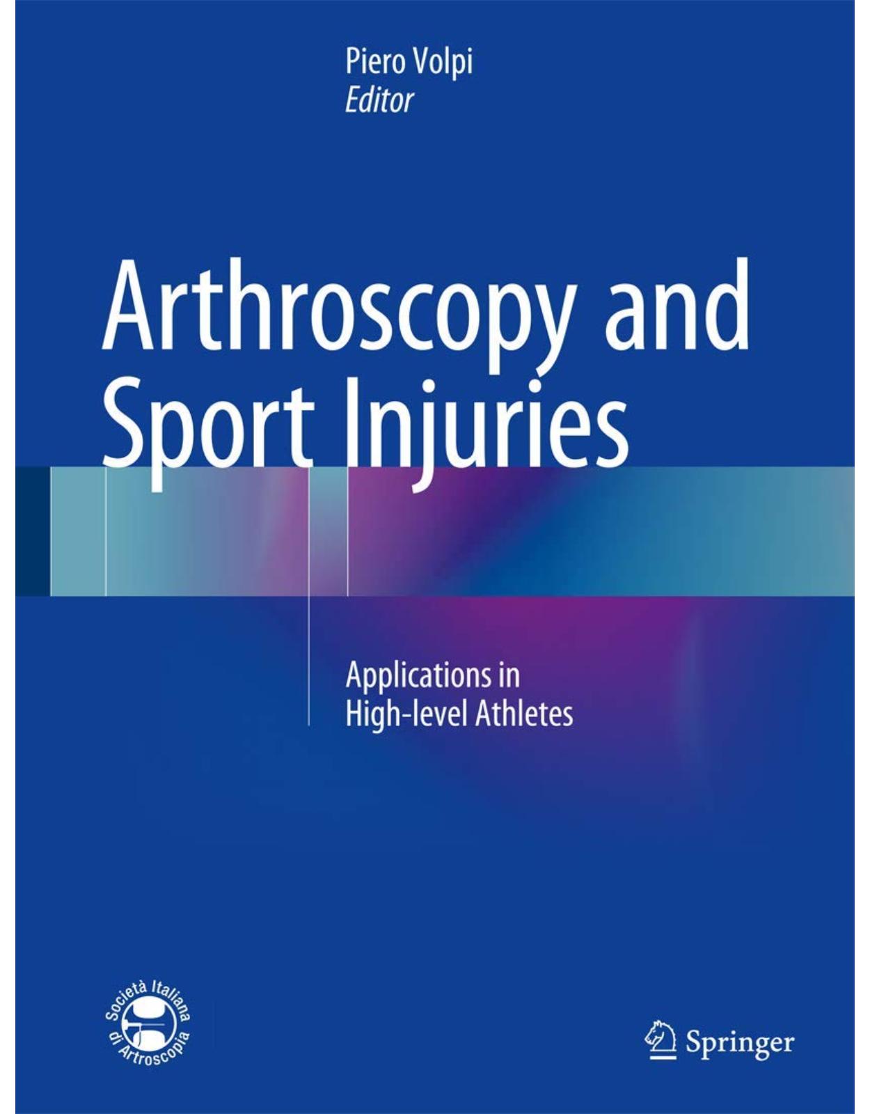
Arthroscopy and Sport Injuries
Livrare gratis la comenzi peste 500 RON. Pentru celelalte comenzi livrarea este 20 RON.
Disponibilitate: La comanda in aproximativ 4-6 saptamani
Autor: Piero Volpi
Editura: Springer
Limba: Engleza
Nr. pagini: 457
Coperta: Hardcover
Dimensiuni: 21.29 x 3.12 x 29.16 cm
An aparitie: 12-12-15
Description:
This book describes the current applications of arthroscopy in a very wide range of sports injuries involving, among other sites, the hip, knee, ankle, shoulder, elbow, and wrist. For each condition, mechanisms of injury are explained and the role of arthroscopy in diagnosis and treatment is described. Relevant information is also provided on the epidemiology and mechanisms of injury in specific sports and on indications for treatment and rehabilitation. The book fully reflects the recent advances that have taken place in arthroscopy, permitting more accurate assessment and more successful management of post-traumatic pathologies. Furthermore, it acknowledges that as a result of the increasing use of new technologies and biomaterials, there is now particular interest in techniques that promote biological healing of articular lesions and permit complete functional recovery. The authors are leading specialists in the field who have aimed to provide practitioners with the clear guidance that they require on the evaluation and treatment of injuries incurred during sporting activity.
Table of Contents:
1: Future in Arthroscopy and Sports Medicine
1.1 Introduction
1.2 Noninvasive Devices for Improved Diagnosis and Prognosis
1.3 Injury Prevention
1.4 Individualized Treatment
1.5 Computer-Assisted Navigation
1.6 Biologics in Sports Medicine
Conclusion
References
2: Injury Risk in the Olympic Games
2.1 Introduction
2.2 Methods
2.2.1 Implementation of Data Collection
2.2.2 Injury and Illness Report Form
2.2.3 Definition of Injury and Illness
2.3 Results
2.3.1 Response Rate and Coverage of the Athletes
2.3.2 Incidence of Injuries
2.3.3 Injury Risk in Different Sports
2.3.4 Injury Location and Type
2.3.5 Injury Mechanism and Circumstance
2.3.6 Injury Severity
2.3.7 Incidence and Distribution of Illnesses (Registered in Vancouver and London Only)
2.3.8 Affected System, Causes, Symptoms, and Severity of Illness
2.4 Discussion
2.4.1 Incidence and Distribution, Type, and Cause of Injuries
2.4.2 Incidence and Distribution, Type, and Cause of Illnesses (Registered in London and Vanco
2.5 Practical Implications and Further Research
Conclusion
References
3: Tennis: Epidemiology and Injury Mechanism
3.1 Introduction
3.2 Epidemiology and Mechanism of Upper Limb Injuries in Tennis Players
3.2.1 The Shoulder
3.2.2 The Elbow
3.3 The Spine
3.3.1 The Cervical Spine
3.3.2 The Lower Back
3.4 Epidemiology and Mechanism of Lower Limb Injuries in Tennis Players
3.4.1 The Ankle
3.4.2 The Knee
3.4.3 The Hip
References
4: Rugby Injuries: Epidemiology and Mechanism
4.1 Introduction
4.2 Definition and Characteristics of Injuries
4.3 Incidence and Severity of Injuries
4.4 Mechanism of Specific Site Injuries
4.4.1 Shoulder
4.4.2 Cervical Spine
4.4.3 Knee
Conclusion
Bibliography
5: Basketball: Epidemiology and Injury Mechanism
5.1 Acute Injuries
5.1.1 Ankle Sprains
5.1.2 Knee Ligament Acute Injuries
5.1.3 Hip and Pelvis Acute Injuries
5.1.4 Upper Extremity Acute Injuries
5.1.5 Wrist Acute Injuries
5.1.6 Back Acute Injuries
5.2 Overuse Injuries
5.2.1 Ankle, Foot, and Lower Leg Overuse Injuries
5.2.2 Knee Overuse Injuries
5.2.3 Hip and Pelvis Overuse Injuries
5.2.4 Back Overuse Injuries
References
6: Football: Epidemiology and Injury Mechanism
6.1 Introduction
6.2 Football Epidemiology
6.3 Methodological Considerations
6.4 Epidemiological Researches in Professional Football
6.5 Injury Incidence in Football
6.6 Risk Factors
References
7: Return to Sport (General Aspects)
7.1 Introduction
7.2 Physical Recovery
7.3 Psychological Recovery
7.4 Discussion
7.5 Summary
References
8: The Groin Pain Syndrome
8.1 Introduction
8.2 Etiology and Clinical Classification
8.3 Injury Mechanisms and Predisposing Factors
8.4 Clinical and Diagnostic Examination
8.4.1 Imaging
8.5 Rehabilitation and Treatment Strategy
8.5.1 Type of Exercise and the Progression of Work Plan
8.5.2 The Intensity, the Frequency, and the Duration of Exercise
8.5.3 Therapeutic Interventions
8.5.4 Surgical Treatment
8.5.5 Inguinal Hernia
8.5.6 Sports Hernia
8.5.7 Adductor Tendinopathy
8.5.8 Osteitis Pubis
8.5.9 Hockey Goalie–Baseball Pitcher Syndrome
8.5.10 Acetabular Labral Tear
8.5.11 Internal Snapping Hip
8.5.12 Osteoid Osteoma
8.5.13 Nerve Entrapment
8.6 Return to Play
Conclusions
References
9: Achilles Tendinopathies
9.1 Etiology
9.2 Injury Mechanism
9.3 Clinical and Diagnostic Examination
9.4 Treatment Strategy
9.4.1 Non-insertional Achilles Tendinopathy
9.4.1.1 Nonoperative Treatment
9.4.1.2 Surgical Treatment
9.4.2 Insertional Achilles Tendinopathy
9.4.2.1 Nonoperative Treatment
9.4.2.2 Operative Treatment
9.5 Rehabilitation and Return to Play
References
10: Achilles Tendon Rupture
10.1 Introduction
10.2 Etiopathogenesis and Injury Mechanism
10.3 Clinical and Diagnostic Examination
10.4 Treatment Strategy
10.5 Percutaneous Achilles Tendon Repair: Surgical Technique
10.6 Rehabilitation and Return to Play
10.7 Discussion
Conclusion
References
11: Patellar and Quadriceps Tendinopathy
11.1 Introduction
11.2 Epidemiology and Predisposing Factors
11.3 Symptomatology and Classification
11.3.1 Jumper’s Knee Classification According to Symptoms
11.4 Radiological Findings
11.5 Anatomo-pathology
11.6 Treatment
11.7 Patellar Tendon Rupture
References
12: Patellar Tendon Rupture
12.1 Introduction
12.2 Etiology
12.3 Clinical and Diagnostic Examination
12.4 Treatment Strategy
12.4.1 Acute Ruptures
12.4.2 Chronic Ruptures
12.5 Outcome
12.6 Rehabilitation and Return to Play
Conclusion
References
13: Hamstring Injuries
13.1 Introduction
13.2 Injury Mechanism
13.3 Diagnosis
13.4 Treatment Strategy
13.5 Rehabilitation and Return to Sport
13.5.1 Stage 1: Protection and Optimal Loading
13.5.2 Stage 2: Aerobic Threshold Workout and Running Recovery
13.5.3 Stage 3: Anaerobic Lactacid and Eccentric Strength Recovery
13.5.4 Stage 4: Return to the Team and Prevention Pro-education
Conclusions
References
14: Return to Sport After Muscle Tendon Lesions
14.1 Introduction
14.1.1 Pathological Processes
14.2 Evaluation
14.3 Managing Healing Process
References
15: Shoulder Imaging Evaluation
15.1 Introduction
15.2 Impingement and Rotator Cuff Tears
15.2.1 Subacromial Impingement Syndrome (SIS)
15.2.1.1 Imaging of Impingement Syndrome: Imaging Modalities
Radiography
Ultrasound
Magnetic Resonance Imaging (MRI)
MR Arthrography
15.2.1.2 Imaging Findings in Impingement Syndrome and Rotator Cuff Tears
Subacromial Bursitis-Bursal Effusion
Tendinosis
Partial-Thickness Tears
Rotator Cuff Tears
Subscapularis Tears and Biceps Tendon Lesions
15.3 Instability
15.3.1 Shoulder Imaging Techniques
15.3.1.1 Radiography
15.3.1.2 CT and CT Arthrography
15.3.1.3 MRI and MR Arthrography
15.3.2 Imaging Findings in Instability
15.3.2.1 Anterior Instability
15.3.2.2 Bony Lesions: Humeral Head Defect
15.3.2.3 Anteroinferior Lesions of the Glenoid Rim
15.3.2.4 Capsulolabral Detachments
15.3.2.5 Posterior Instabilities
Radiographs
Multiplanar Imaging
15.3.2.6 Superior Labral Lesions
Imaging
References
16: Shoulder Management of the First Time Anterior Shoulder Dislocation
16.1 Aetiology
16.2 Diagnosis
16.3 Treatment
Conclusions
References
17: Shoulder Superior Labral Repair
17.1 The Superior Labrum: Anatomy, Role, and Pathology
17.2 Etiology
17.3 Injury Mechanism
17.3.1 Biceps Anchor Anatomy
17.4 Clinical and Diagnostic Examination
17.5 Treatment Strategy
17.6 Rehabilitation and Return to Play
References
18: Shoulder Anterior Instability
18.1 Epidemiology
18.2 Joint
18.3 Injury Mechanism
18.4 Pathoanatomy
18.5 Clinical and Diagnostic Examination
18.6 Treatment Strategy
18.6.1 Nonoperative Management
18.6.2 Operative Management
18.7 Rehabilitation and Return to Play
References
19: Shoulder Instability with Bony Lesions
19.1 Introduction
19.2 Mechanical and Clinical Correlations
19.3 Diagnosis
19.3.1 History
19.3.2 Physical Examination
19.3.3 Imaging
19.4 Instability with Bone Deficiency: Treatment
19.4.1 Hill-Sachs Defects
19.4.2 Glenoid Deficiency
19.5 Postoperative Care and Return to Sports
19.6 Discussion
19.6.1 Indication to the Latarjet as Procedure of Choice According to the Severity Index Scor
References
20: Shoulder Microtraumatic and Atraumatic Instability
20.1 Introduction
20.2 Mechanism of Injury: Overhead Movement
20.3 Pathogenesis
20.4 Role of the Scapulothoracic Joint
20.5 The Proprioceptive System
20.6 Pathological Anatomy
20.7 Clinical Presentation
20.8 Diagnosis
20.9 Imaging Investigations
20.10 Therapy and Return to Competitive Activity
References
21: Posterior Shoulder Instability
21.1 Etiology
21.1.1 Bony Abnormalities of the Glenoid
21.1.2 Bony Abnormalities of the Humeral Head
21.1.3 Soft Tissue Abnormalities
21.2 Mechanism of Injury
21.3 Clinical Examination
21.4 Diagnostic Imaging
21.5 Treatment Strategy
21.6 Operative Technique
21.7 Results of Surgical Management
21.8 Rehabilitation and Return to Play
References
22: Shoulder Rotator Cuff Repair
22.1 Introduction
22.2 Etiology and Injury Mechanism
22.3 Clinical and Diagnostic Examination
22.3.1 Clinical Examination
22.3.2 Radiography
22.3.3 Magnetic Resonance Imaging
22.4 Treatment Strategy
22.4.1 Nonoperative Management
22.4.2 Surgical Treatment
22.5 Rehabilitation and Return to Play
References
23: Treatment of Acute Traumatic Acromioclavicular Dislocations
23.1 Etiology
23.2 Injury Mechanism
23.3 Clinical and Diagnostic Examination
23.4 Treatment Strategy
23.4.1 Treatment Modalities
23.4.1.1 Nonoperative Treatment
23.4.1.2 Surgical Treatment
Open Techniques
Arthroscopic Techniques
23.5 Rehabilitation and Return to Play
References
24: Return to Sport After Shoulder Arthroscopy
24.1 Instability
24.1.1 Anatomy of the Shoulder
24.1.2 Dislocation Mechanism/Incidence
24.1.3 Surgery
24.1.4 Return to Sport After Arthroscopic Surgery for Instability
24.2 Multidirectional Shoulder Instability
24.3 Acute Traumatic Posterior Shoulder Dislocation
24.4 Posterior Instability of the Shoulder
24.4.1 Postoperative Rehabilitation and Return to Sport
24.5 SLAP Lesion
24.6 Rotator Cuff
24.7 Discussion
References
25: Thrower’s Elbow
25.1 Introduction
25.2 Functional Anatomy
25.3 Pathophysiology and Biomechanics
25.4 History and Physical Examination
25.5 Diagnostic Imaging
25.6 Prevention
25.7 Valgus Instability/Ulnar Collateral Ligament Injuries
25.8 Ulnar Neuritis
25.9 Flexor-Pronator Injuries
25.10 Medial Epicondyle Apophyseal Injuries
25.11 Osteochondritis Dissecans of the Capitellum
25.12 Posteromedial Impingement
25.13 Olecranon Stress Fractures
25.14 Summary
Resources
26: Biceps and Triceps Tendon Lesions
26.1 Distal Biceps Lesions
26.1.1 Epidemiology
26.1.2 Etiology
26.1.3 Clinical Presentation
26.1.4 Diagnostic Examinations
26.1.4.1 X-Ray
26.1.4.2 MRI
26.1.4.3 Echography
26.1.5 Treatment
26.1.5.1 Conservative
26.1.5.2 Anatomical or Non-anatomical Repair
26.1.5.3 Partial Ruptures
26.1.5.4 Chronic Ruptures
26.1.5.5 Surgical Technique
Tendon Identification and Evaluation of the Lesion
Tendon Repair at the Radial Tuberosity
Single Anterior Approach Technique
Double Anterior and Posterior Approach Technique
26.1.5.6 Rehabilitation Treatment
Conservative Treatment of the Acute Lesion
Post-surgical Treatment After Tendon Repair
26.1.6 Results
26.2 Triceps Lesions
26.2.1 Mechanism of Lesion
26.2.2 Clinical Picture
26.2.3 Diagnosis
26.2.4 Anatomy
26.2.5 Treatment
26.2.6 Surgical Technique
26.2.7 Results
26.2.8 Rehabilitation
References
27: Medial Epicondylitis
27.1 Introduction
27.2 Etiology and Injury Mechanism
27.3 Clinical and Diagnostic Examination
27.4 Treatment Strategy
27.4.1 Non-operative Treatment
27.4.2 Surgical Treatment
27.5 Rehabilitation and Return to Play
References
28: Lateral Epicondylitis of the Elbow
28.1 Etiology
28.2 Injury Mechanism
28.3 Pathology
28.4 Clinical and Diagnostic Examination
28.5 Differential Diagnosis
28.6 Treatment
28.6.1 Nonsurgical Treatment
28.6.2 Promotion of Healing
28.6.3 Surgical Treatment
28.6.4 Arthroscopy
28.6.5 Open Surgery
28.7 Rehabilitation and Return to Play
References
29: Elbow Instability
29.1 Introduction
29.2 Etiology
29.3 Injury Mechanism
29.3.1 PLRI
29.3.2 Valgus Injuries
29.3.3 Varus Injuries: Posteromedial Dislocation
29.3.4 Complex Injuries
29.4 Clinical Evaluation
29.4.1 Valgus Instability
29.4.2 PLRI
29.5 Diagnostic Imaging
29.6 Treatment Strategy
29.7 Treatment of UCL Lesions
29.8 Treatment of LCL Lesions
Conclusions
References
30: Wrist Ligament Injuries
30.1 Introduction
30.2 Carpal Instabilities
30.2.1 Aetiology
30.2.1.1 Direct Mechanism
30.2.1.2 Indirect Mechanism
30.2.2 Injury Mechanism
30.2.3 Clinical and Diagnostic Examinations
30.2.3.1 Clinical Examination
30.2.3.2 Imaging
Radiography
Magnetic Resonance Imaging (MRI)
Contrast-Enhanced Imaging (MRI Arthrography and CT Arthrography)
Diagnostic Wrist Arthroscopy
30.2.4 Treatment Strategy
30.3 Instability of the Distal Radioulnar Joint
30.3.1 Aetiology
30.3.2 Injury Mechanism
30.3.3 Clinical and Diagnostic Examination
30.3.3.1 Clinical Examination
30.3.3.2 Imaging
Radiography
Magnetic Resonance Imaging (MRI)
Contrast-Enhanced Imaging (MRI Arthrography and CT Arthrography)
30.3.4 Treatment Strategy
30.3.5 Rehabilitation and Return to Play
References
31: Wrist Tendon Lesions
31.1 Introduction
31.2 Etiology and Injury Mechanisms
31.2.1 Types of Injuries in Golf Players
31.2.2 Types of Injuries in Tennis Players
31.3 Pathology
31.3.1 De Quervain’s Tenosynovitis
31.3.2 Intersection Syndrome
31.3.3 Extensor Insertional Tenosynovitis
31.3.4 Extensor Carpi Ulnaris (ECU) Lesions
31.3.5 Flexor Carpi Radialis (FCR) Tenosynovitis
31.3.6 Others Tendons
References
32: Distal Radius Fractures
32.1 Introduction
32.2 Distal Radius Fractures and Sports
32.3 Pertinent Anatomy
32.4 Classification
32.4.1 Frykman’s Classification [8]
32.4.2 Mayo Clinic Classification [9]
32.4.3 Columnar Classification [10]
32.4.4 Fernandez Classification [11]
32.4.5 AO Classification [12]
32.5 Diagnosis
32.6 Treatment
32.6.1 Treatment Strategy
32.6.2 The Role of Conservative Treatment
32.6.3 The Role of Percutaneous Pin Fixation
32.6.4 The Role of External Fixation
32.6.5 The Role of Arthroscopy
32.6.6 The Role of Internal Fixation
32.6.6.1 Dorsal Approach
32.6.6.2 Volar Approach
References
33: Hip Imaging Evaluation
33.1 Plain Radiography
33.2 Computed Tomography
33.3 CT Arthrography
33.4 Ultrasound
33.5 Conventional Magnetic Resonance Imaging
33.6 Magnetic Resonance Arthrography
33.7 Nuclear Scintigraphy
33.8 Hip Arthrography, Injection, Aspiration, and Bursography
33.9 Summary
References
34: Femoroacetabular Impingement (FAI) and Sport
34.1 CAM and PINCER: What Are They?
34.2 CAM and PINCER Physiopathology
34.3 CAM and PINCER Anatomopathology
34.4 Surgical Technique
34.5 Technical Notes
34.6 Rehabilitation
34.7 Outcome and Return to Play
References
35: Hip Tears of the Acetabular Labrum
35.1 Introduction and Etiology
35.2 Clinical and Diagnostic Examination
35.3 Treatment Strategy
35.3.1 Labral Debridement
35.3.2 Surgical Technique for Arthroscopic Repair/Refixation
35.3.3 Primary Labral Repair
35.3.4 Traumatic Posterior Bankart-�Type Labral Tear
35.4 Rehabilitation and Return to Play
References
36: Return to Sport After Hip Arthroscopy
36.1 Premise
36.2 Directions of Rehabilitation
36.3 Objectives to Be Achieved Through the Field Rehabilitation
36.3.1 Recovery of Functional Movement
36.3.2 Recovery Sport-Specific Movements
36.3.3 Recovery of Fitness
36.3.4 Prevention of Reinjury
36.4 Return to Sport and Competition
36.5 Rehabilitation Field
36.5.1 First Phase
36.5.2 Second Phase
36.5.3 Third Phase
36.5.4 Fourth Phase
36.5.5 Fifth Phase
Conclusions
References
37: Medial Meniscus
37.1 Anatomy
37.2 Aetiology and Mechanism of Injury
37.3 Classification of Meniscal Injuries
37.4 Clinical and Diagnostic Examination
37.4.1 Tests
37.4.1.1 McMurray Test [60]
37.4.1.2 Apley Test [61]
37.4.1.3 Finocchietto Sign [62]
37.4.1.4 Childress Test [63]
37.4.2 Imaging Investigations
37.5 Treatment Strategy
37.5.1 Total Meniscectomy
37.5.2 Partial Meniscectomy
37.5.2.1 Complications
37.5.3 Meniscal Repair
37.5.4 Biological Repair
37.6 Rehabilitation and Return to Play
References
38: Lateral Meniscus
38.1 Anatomy
38.1.1 Lateral Meniscal Variants: The Discoid Meniscus
38.2 Injury Epidemiology
38.3 Clinical and Diagnostic Examination
38.3.1 Injury Mechanism in Meniscus Pathology
38.3.2 History
38.3.3 Physical Examination
38.3.4 Imaging
38.4 Therapeutic Approach
Conclusions
References
39: Cartilage Injury Treatment
39.1 Aetiology
39.1.1 Cartilage Layers and Injury Mechanism
39.2 Clinical and Diagnostic Examination
39.2.1 Imaging
39.2.1.1 Plain x-rays
39.2.1.2 CT Arthrography
39.2.1.3 MRI
39.2.1.4 Technetium Scintigraphy
39.3 Operative Interventions
39.3.1 Arthroscopy
39.3.2 Cartilage Defects with Concomitant Joint Injuries
39.4 Treatment Strategy
39.4.1 The Operative Alternatives
39.4.1.1 Microfracture and Other Bone Marrow Stimulation Techniques
39.4.1.2 Osteochondral Grafts
39.4.1.3 ACI
39.4.2 Non-biological Local Repairs
39.4.3 General Comments
39.5 Rehabilitation and Return to Play
39.5.1 The Rehabilitation Process
References
40: Management of Collateral Ligament Injuries (Medial and Lateral) in Competitive Athletes
40.1 Medial Collateral Ligament and Posteromedial Corner
40.1.1 Introduction
40.1.2 Epidemiology
40.1.3 Clinical and Diagnostic Examination
40.1.4 Treatment Strategy
40.1.4.1 Conservative Treatment
40.1.4.2 Surgical Treatment
40.1.4.3 Treatment of Chronic Lesions
40.1.5 Postoperative Management and Rehabilitation
40.2 Lateral Collateral Ligament
40.2.1 Introduction
40.2.2 Clinical and Diagnostic Examination
40.2.3 Treatment of Acute Lesions
40.2.4 Treatment of Chronic Lesions
40.2.5 Postoperative Management and Rehabilitation
References
41: Anterior Cruciate Ligament
41.1 Etiology
41.2 Injury Mechanism
41.3 Clinical and Diagnostic Examination
41.4 Treatment Strategy
41.4.1 Indications
41.4.2 Timing
41.4.3 Graft
41.4.4 Surgical Procedure
41.5 Rehabilitation After ACL Reconstruction
41.6 Return to Play
41.7 Outcome
References
42: Revision Anterior Cruciate Ligament
42.1 Introduction
42.2 Complications and Failures in ACL Reconstruction
42.3 Recurrence of Knee Joint Instability
42.4 Clinical and Instrumental Diagnosis
42.5 Graft Selection
42.6 Treatment Strategy
42.7 Rehabilitation and Return to Sports
References
43: Posterior Cruciate Ligament
43.1 Anatomy
43.2 Biomechanic
43.3 Epidemiology
43.4 Injury Mechanism
43.5 Clinical and Diagnostic Examination
43.6 Conservative Management
43.7 Surgical Indications
43.8 Surgical Timing
43.9 Graft Selection
43.10 Surgical Treatment
43.10.1 Avulsion Fragments
43.10.2 The Transtibial Single-Bundle PCL Technique
43.10.3 The Double-Bundle Technique
43.10.4 The Posterior Inlay Technique
43.10.5 Innovations
43.11 Postoperative Management
43.12 Return to Sport
43.13 Clinical Outcomes
References
44: Posterolateral Instability
44.1 Introduction
44.2 Anatomy and Biomechanics
44.3 Injury Mechanism
44.4 Clinical and Diagnostic Examination
44.5 Treatment Strategies
44.5.1 Surgical Technique
44.5.1.1 The Modified Larson Technique
44.5.1.2 The LaPrade Technique [35]
44.6 Rehabilitation and Return to Sports Activity
References
45: Patellar Instability
45.1 Etiology
45.2 Injury Mechanism
45.3 Clinical and Diagnostic Examination
45.3.1 Clinical Examination
45.3.2 Diagnostic Examination
45.4 Treatment Strategy
45.4.1 Lateral Release
45.4.2 Medial Reefing
45.4.3 Medial Patellofemoral Ligament Reconstruction
45.4.4 Distal Realignment
45.4.5 Trochleoplasty
45.5 Rehabilitation and Return to Play
References
46: Anterior Knee Pain
46.1 Aetiology
46.2 Clinical Presentation
46.3 Clinical Examination
46.4 Diagnostic Examination
46.5 Treatment
46.6 Physical Rehabilitation
46.7 Tape
46.8 Patella Braces
46.9 Foot Orthosis
46.10 Pharmacological Therapy
46.11 Surgical Treatment
Conclusion
References
47: Knee Arthritis in Athletes
47.1 Etiology
47.2 Injury Mechanism
47.2.1 Malalignment
47.2.2 Loss of Meniscal Tissue
47.2.3 Meniscectomy
47.2.4 Meniscal Transplantation
47.2.5 Cartilage Defects
47.2.6 Joint Instability or Laxity
47.3 Treatment Strategy
47.4 Surgical Treatment
Conclusion
References
48: Management of Anterior Cruciate Ligament Lesion in Adolescents
48.1 Risk Factors
48.2 ACL Tear
48.3 Management of ACL Tear
48.3.1 Physeal Sparing Technique (Both Tibia and Femur)
48.3.2 Transphyseal and “All-Inside” Technique
48.4 Conclusions and Guideline for Physician (From the American Academy of Pediatrics) [30]
References
49: Management of the First Patellar Dislocation
49.1 Etiology
49.2 Injury Mechanism
49.3 Clinical and Diagnostic Examination
49.4 Treatment Strategy
49.4.1 Nonoperative Treatment
49.4.2 Surgical Options
49.5 Rehabilitation and Return to Play
References
50: Chronic Ankle Instability
50.1 Anatomy and Biomechanics of the Ankle Ligamentous Complex
50.2 Etiology and Injury Mechanism
50.3 Clinical and Diagnostic Examination
50.3.1 History and Physical Examination
50.3.2 Imaging
50.4 Treatment Strategies
50.4.1 Conservative Treatment
50.4.2 Anatomical Direct Repair
50.4.3 Tenodesis
50.4.4 Arthroscopy
50.4.5 Combined Procedures
50.5 Rehabilitation and Return to Play
References
51: Ankle Impingement (Anterior and Posterior)
51.1 Anterior Ankle Impingement (AAI)
51.1.1 Etiology and Classification
51.1.2 Injury Mechanism
51.1.3 Clinical and Diagnostic Examination
51.1.4 Treatment Strategy
51.2 Posterior Ankle Impingement
51.2.1 Etiology and Classification
51.2.2 Injury Mechanism
51.2.3 Clinical and Diagnostic Examination
51.2.4 Treatment Strategy
51.2.5 Rehabilitation and Return to Play
References
52: Ankle Osteochondral Lesions
52.1 Definition
52.1.1 Etiology
52.1.2 Injury Mechanism and Natural History
52.2 Clinical and Diagnostic Examination
52.3 Treatment Strategy
52.3.1 Acute Lesions
52.3.2 Chronic Lesions
52.3.2.1 Conservative Treatment
52.3.2.2 Retrograde Drilling
52.3.2.3 Microfractures
52.3.2.4 Mosaicplasty
52.3.2.5 Autologous Chondrocyte Implantation
52.3.2.6 Bone Marrow-Derived Cell Transplantation
52.3.2.7 Allograft
52.4 Rehabilitation and Return to Play
52.4.1 Fragment Fixation
52.4.2 Drilling
52.4.3 Microfractures
52.4.4 Mosaicplasty
52.4.5 ACI and BMDCT
Conclusions
References
53: Ankle Arthritis in Athletes
53.1 Background
53.2 Epidemiology
53.3 Physical Examination
53.4 Diagnostics
53.5 Surgical Arthroscopic Procedure
Conclusions
References
54: Return to Sport After Ankle Lesions
54.1 Introduction
54.2 Ankle Instability Rehabilitation After Surgical Treatment
54.3 Degenerative Diseases Rehabilitation After Surgical Treatment
54.4 Tecar and Laser Therapy
54.5 Proprioception
54.6 Discharge and Brace
54.7 Rehabilitation in Water
54.8 NEURAC Rehabilitation
54.9 Computerized Proprioception
54.10 Recovery of Athletic Movement and Return to Sport
References
55: Viscosupplementation in Athletes
55.1 Introduction
55.2 Rationale
55.3 Therapeutic Activities of HA
55.4 HA Preparations
55.5 Clinical Trials
55.5.1 Knee
55.5.2 Hip
55.5.3 Ankle
55.5.4 First Metatarsophalangeal Joint
55.5.5 Shoulder
55.5.6 Elbow
Conclusions
References
56: Use of PRP in Sports Medicine
56.1 Introduction
56.2 Tendinopathy
56.2.1 Elbow Tendinopathy
56.2.2 Achilles Tendinopathy
56.2.3 Patellar Tendinopathy
56.3 Rotator Cuff Tears
56.4 Ligament Injuries
56.4.1 Anterior Cruciate Ligament
56.5 Muscle Injuries
56.6 PRP and Doping
Conclusion
References
57: Use of Scaffolds in Sports Medicine
57.1 Introduction
57.2 Articular Cartilage Scaffolds
57.3 Clinical Evidence in Scaffold Application
Conclusions
References
58: Arthroscopy and Sports Topics in Top Level Athletes: Use of a Brace
58.1 Introduction
58.2 Shoulder
58.3 Elbow
58.4 Wrist
58.5 Knee Braces
58.6 Ankle Braces
Conclusion
References
Bibliography
| An aparitie | 12-12-15 |
| Autor | Piero Volpi |
| Dimensiuni | 21.29 x 3.12 x 29.16 cm |
| Editura | Springer |
| Format | Hardcover |
| ISBN | 9783319148144 |
| Limba | Engleza |
| Nr pag | 457 |


Clientii ebookshop.ro nu au adaugat inca opinii pentru acest produs. Fii primul care adauga o parere, folosind formularul de mai jos.