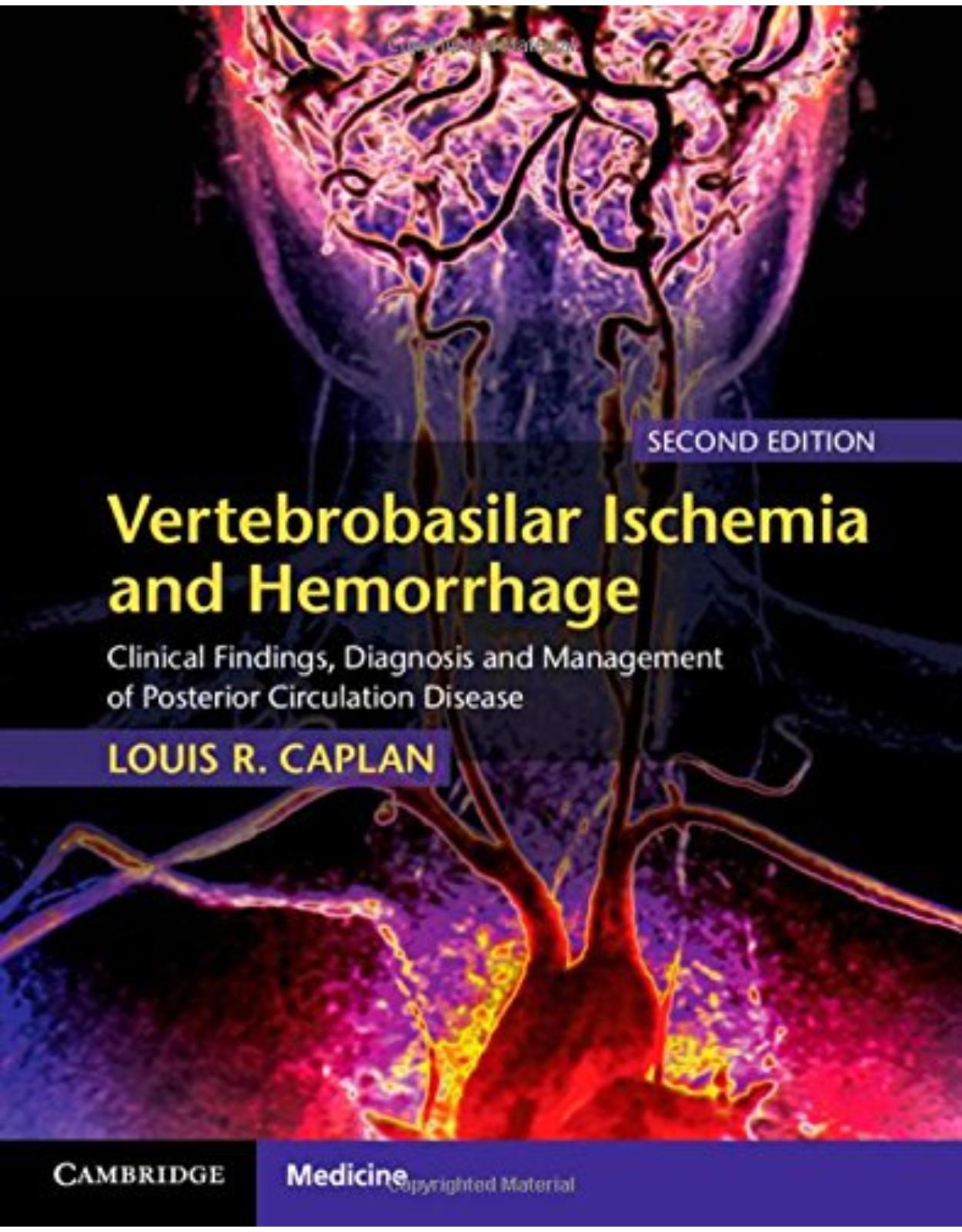
Vertebrobasilar Ischemia and Hemorrhage
Livrare gratis la comenzi peste 500 RON. Pentru celelalte comenzi livrarea este 20 RON.
Disponibilitate: La comanda in aproximativ 4-6 saptamani
Autor: Louis R. Caplan
Editura: Cambridge University Press
Limba: Engleza
Nr. pagini: 608
Coperta: Hardcover
Dimensiuni: 22.23 x 3.81 x 27.94 cm
An aparitie: 2 April 2015
Description:
This comprehensive review of vascular disease in the vertebrobasilar circulation is based on Dr Louis R. Caplan's extensive experience and observation of patients from the New England Medical Center posterior circulation stroke registry. It benefits from an organized, uniform, and coherent analysis of all types of vascular disease involving the posterior circulation, presented by a single author who is one of the world's leading authorities on this topic. This new edition is fully updated throughout, including a review of all the literature published on this topic since the previous edition in 1996. There are major rewrites for the chapters on diagnosis and therapy, inclusion of modern imaging techniques, and extensive illustrations. Essential reading for stroke physicians and neurologists, the book will also be an important source of reference for neurosurgeons, vascular surgeons and endovascular radiologists.
Table of Contents:
Section I General Features of Cerebrovascular Disease in the Posterior Circulation
1 Historical background
Early history
Anatomy and pathology: the sixteenth, seventeenth, and eighteenth centuries
The nineteenth and early twentieth centuries
Development of knowledge about the causes of stroke
Ischemia
Hemorrhage
The modern era (1975–present)
Advances in diagnostic technology
Stroke data banks and registries
Stroke units, stroke specialists, and stroke nurses
Advances in medical and surgical therapy and randomized trials
Acute reperfusion using thrombolytic drugs, thrombus removal with devices, and direct angioplasty and stenting
Interventional techniques to treat potential bleeding cranial lesions: aneurysms and vascular malformations
Posterior circulation strokes
Hemorrhage
Pontine hemorrhage
Cerebellar hemorrhage
Ischemia
2 Basic anatomy and pathology
Brain anatomy
Brainstem and thalamus
Medulla oblongata
Pons
Midbrain
Thalamus
Cerebellum–
Posterior portions of the cerebral hemispheres
Arterial anatomy
Extracranial large arteries
Intracranial large arteries and their branches
Intracranial vertebral artery (ICVA) and branches
Basilar artery
Section
Posterior cerebral arteries (PCAs) and their branches
Arterial supply regions
Venous anatomy
Vascular pathology
Large artery atherosclerosis
Penetrating small artery disease
Arterial dissections
Embolism
Dolichoectasia (dilatative arteriopathy)
Fibromuscular dysplasia (FMD)
Arterial aneurysms and subarachnoid hemorrhage (SAH)
Vascular malformations
Parenchymal hemorrhage (ICH)
Venous occlusions
Systemic hypotension and hypoperfusion
Other less common vasculopathies
Drugs, especially cocaine
MELAS syndrome
Vasculitis
Giant cell (granulomatous, temporal) arteritis
Takayasu disease
Behçet’s disease
Arteritis caused by infectious agents
3 Signs and symptoms and their clinical localization
Visual perception and related deficits
Visual field defects
Visual inattention and neglect
Abnormal visual perceptions and distortions
Complex disorders of visual perception
Lower bank bilateral lesions
Upper bank bilateral lesions
Abnormalities of depth perception
Disorientation for place and space
Other cognitive and behavioral abnormalities including agnosias, alexia, and aphasia
Left hemisphere lesions, including the thalamus
Reading abnormalities
Aphasia
Visual agnosia and visual anomia
Right hemispheral lesions including the thalamus
Abnormal drawing and copying
Visual amnesia and visual hypoemotionality
Nonvisual behavioral and cognitive abnormalities most often associated with bilateral cerebral hemispheral or thalamic lesions
Abulia
Amnesia
Agitation with delirium
Reduced level of consciousness
Anatomy and physiology
Intrinsic brainstem vascular lesions and coma
Extrinsic lesions
Motor abnormalities
Limb abnormalities and gait
Thalamic lesions
Midbrain lesions
Pontine lesions
Medullary lesions
Cerebellum
Movement disorders
Bulbar muscle motor abnormalities
Vestibular and oculomotor abnormalities
The vestibuloocular reflex and its abnormalities
Vestibular and oculomotor abnormalities at various anatomical loci
Lateral medullary and lateral pontine tegmental lesions
Unilateral medial pontine tegmental lesions
Bilateral medial pontine tegmental lesions
Midbrain lesions
Mesencephalic–diencephalic junction and diencephalic lesions
Cerebellar lesions
Sensory abnormalities
Somatosensory
Limbs and trunk
Face and head
Hearing
Taste
Pupillary size and reactivity
Autonomic dysfunctions including abnormalities of cardiac and respiratory control and micturition
Anatomy and physiology
Clinical autonomic and cardiorespiratory abnormalities
4 Diagnosis
Method of clinical diagnosis
The sequential steps in diagnosis
The inductive method: sequential hypothesis generation and testing
Pattern matching
Diagnosis of the and questions should proceed concurrently
Mimicking a computer
The frequency of various findings in the New England Medical Center Posterior Circulation Registry (NEMC-PCR)
Frequency of the location of infarcts within the posterior circulation territories generally and according to stroke mechanisms and arterial lesions
Frequency of symptoms and signs according to posterior circulation territories
Frequency of risk factors according to the presence and location of occlusive vascular lesions
Common patterns of ischemia/infarction
Patterns within the proximal intracranial posterior circulation territory
Patterns within the middle intracranial posterior circulation territory
Patterns within the distal intracranial posterior circulation territory
Diagnosis in the emergency room
Investigations
Brain imaging
Computed tomography (CT)
Magnetic resonance imaging (MRI)
Vascular imaging and functional testing
Ultrasound
Magnetic resonance angiography (MRA)
CT angiography (CTA)
Catheter digital subtraction angiography
Multimodal MRI and CT including perfusion imaging
Blood tests
Evaluation of the heart and aorta
Brainstem reflexes and electrophysiological techniques
Question-driven imaging and laboratory evaluation
5 Treatment
Introduction
Acute ischemic stroke
Maximizing blood flow
Controlling position and activity
Managing blood pressure, blood volume, and cardiac output
Reperfusion strategies
Thrombolysis
Intravenous thrombolysis
Intra-arterial thrombolysis
Bridging thrombolytic treatment
Mechanical devices to remove clot
Stroke prevention
Surgery
Angioplasty and stenting
Surgically bypassing regions of blockage
Prevention of clot formation, propagation, and embolism
Clot formation and pathophysiology
Anticoagulants
Antiplatelet agents
Theoretical utility of the various antithrombotic agents
Trials of antithrombotic agents that include data regarding posterior circulation vascular lesions
Section II Posterior Circulation Ischemia: Specific Vascular Sites and Conditions
6 Extracranial occlusive disease
The development of ideas and information
Subclavian artery occlusive disease
The subclavian steal syndrome
Proximal vertebral artery occlusive disease
Causes, frequency, and epidemiology of arterial lesions at various neck sites
Frequencies and demography
Etiologies
Subclavian artery
Innominate artery
Vertebral arteries
The V segment
Dissections: V and V segments
Neck positioning and cervical manipulation
Cervical vertebral traumatic fractures
Cranio-Cervical junction bone and joint lesions
Cervical spondylitis
Symptoms, signs, and stroke mechanisms
Innominate artery
Subclavian artery
Proximal vertebral artery lesions
NEMC Posterior Circulation Registry (NEMC-PCR) findings: proximal V portion
Dissections of the V and V portions of the vertebral artery in the neck
Diagnostic evaluation
Physical examination of the supplying arteries and the upper limbs
Ultrasound
Vascular imaging: CTA and MRA, and catheter angiography
Treatment
Medical treatment
Atherosclerotic occlusive disease of the proximal vertebral arteries
Cervical vertebral artery dissections
Surgical treatment
Innominate and subclavian arteries
Proximal portion of the vertebral artery
Angioplasty and stenting
Innominate and subclavian arteries
Vertebral artery disease
Summary of present state of therapy
7 Intracranial vertebral arteries and the proximal intracranial territory
Background and development of ideas
Lateral medullary infarction
Hemimedullary and medial medullary infarction
Cerebellar infarction in PICA territory
ICVA disease
Atherosclerosis
Embolism to and from the ICVA
Dissections of the ICVA
Dolichoectasia (dilatative arteriopathy)
Clinical findings in patients with proximal posterior circulation intracranial territory infarcts
Lateral medullary infarcts
Medial medullary infarcts
Cerebellar infarction in PICA distribution
Findings in the New England Medical Center Posterior Circulation Registry
ICVA vascular lesions
Group A. Focal hypoperfusion-related TIAs or infarcts limited to the proximal territory
Group B. Embolism to the ICVA
Group C. ICVA/BA junction disease with middle territory infarction
Group D. Widespread posterior circulation occlusive disease with multifocal hypoperfusion
Group E. Distal territory infarcts due to intra-arterial embolism or hypoperfusion
Outcomes
Proximal intracranial posterior circulation territory infarcts
Conclusions from NEMC-PCR data
Diagnosis and treatment
Diagnosis
Treatment
Antithrombotic treatment
Thrombolysis with or without mechanical devices
Surgical and angioplasty/stenting for stroke prevention
8 Basilar artery
Development of ideas
Early clinico-anatomical necropsy-based reports of basilar artery occlusion
Kubik and Adams’s classical report on the pathology and syndrome of basilar artery occlusion and subsequent series of cases
Transient prodromal symptoms: “vertebrobasilar insufficiency” and its management with anticoagulants
“Top-of-the-basilar” embolism
Improved brain and vascular imaging allowed safer and more rapid clinical diagnosis
Posterior circulation stroke registries
Pathology, pathophysiology, and frequency of vascular lesions
Atherosclerosis
Embolism
Dissection
Aneurysms and dilatative arteriopathy
Other, less common causes
Symptoms and signs
Pontine ischemia and the middle posterior circulation intracranial territory
Motor symptoms and signs
Oculomotor and pupillary signs
Conjugate horizontal gaze and sixth nerve palsies
Internuclear ophthalmoplegia (INO)
One-and-a-half syndrome
Ocular bobbing
Nystagmus
Ptosis
Skew deviation
Pupillary abnormalities
Sensory abnormalities
Coma and reduced level of consciousness
Upper brainstem ischemia as part of the “top-of-the-basilar” syndrome
Oculomotor abnormalities
Pupillary abnormalities
Abnormalities of alertness and behavior
Memory loss and abulia
Other neurological signs
Reports of outcomes in patients with basilar artery disease before the NEMC-PCR and the BASICS studies
Basilar artery lesions in the NEMC Posterior Circulation Registry
Summary conclusions from NEMC posterior circulation data
The Basilar Artery International Cooperative Study (BASICS) registry
Clinical and laboratory diagnosis
Treatment
Reperfusion after acute basilar artery occlusion
Opening the occluded artery
Maximizing blood flow to the ischemic areas
Antithrombotic treatment
Patients with brainstem and cerebellar ischemia who have an acute basilar artery occlusion
Other patients with basilar artery lesions
Anticoagulants and antiplatelet agents
Angioplasty and stenting
9 Posterior cerebral arteries
Background and development of ideas
Occipital lobe anatomy and physiology
Anatomy of the cerebral arterial supply
Clinical studies
Before Foix: late nineteenth and early twentieth century, including Dejerine
Charles Foix
Clinical reports after Foix
Bilateral PCA territory ischemia due to vertebral basilar disease
Clinical, pathological, and neuroradiological studies of the mechanisms of unilateral PCA territory infarcts and their clinical findings
Signs and symptoms of PCA territory infarction correlated with brain and vascular imaging
Pathology and frequency of vascular lesions and stroke mechanisms
Clinical symptoms and signs
Unilateral PCA stenosis and occlusion
Headache
Visual symptoms and findings
Sensory symptoms and findings
Motor dysfunction
Left cerebral hemispheral PCA territory infarction
Right PCA territory cerebral hemispheral infarction
Bilateral PCA territory infarcts
Bilateral inferior calcarine bank temporooccipital ischemia
Bilateral upper calcarine bank parietooccipital lesions
Frequency of various symptoms and signs
PCA and PCA territory vascular lesions in the New England Medical Center Posterior Circulation Registry
Distribution and location of infarctions
Stroke mechanisms
Cardiac-source embolism group ( = 32)
Intra-arterial embolism group ( = 25)
Uncertain embolism source group ( = 8)
Intrinsic PCA occlusive disease ( = 7)
Vasoconstriction ( = 4)
Coagulopathy ( = 3)
Summary and conclusions from the NEMC data
Diagnosis
Treatment
10 Penetrating arteries
Development of ideas about the pathology that causes small deep infarcts
Lacunes
Intracranial branch atheromatous disease
Development of knowledge about the anatomy of posterior circulation branches
Signs, symptoms, and syndromes in penetrating branch artery disease at various brainstem sites
Medulla oblongata
Medial and paramedian medullary infarcts
Lateral medullary infarction
Pons
Anteromedial infarcts
Pure motor hemiplegia (PMH)
Ataxic hemiparesis
Dysarthria–clumsy hand syndrome
“Pure sensory stroke”
Anterolateral infarcts
Lateral tegmental infarcts
Pontine tegmental infarcts of uncertain cause
Midbrain
Midbrain infarcts with predominant oculomotor abnormalities
Midbrain infarcts with prominent motor and/or ataxic abnormalities
Midbrain infarcts with predominant sensory abnormalities
Thalamus
Vascular supply regions
Lateral thalamic infarcts
Pure sensory stroke
Lateral thalamic infarcts (the Dejerine-Roussy syndrome)
Sensorimotor stroke
Polar artery territory infarction
Thalamic–subthalamic artery territory infarcts
Posterior choroidal artery territory infarction
Other thalamic infarcts
The New England Medical Center Posterior Circulation Registry experience
Diagnosis
Treatment of patients with branch artery occlusive disease
11 Cerebellar infarcts
Essential cerebellar brain and vascular anatomy and physiology
Brain anatomy and functions
Vascular anatomy
Development of ideas about cerebellar lesions and infarcts
Cerebellar infarcts: distribution, general clinical signs, outcome, and etiologies
Posterior inferior cerebellar artery (PICA) territory cerebellar infarcts
Anterior inferior cerebellar artery (AICA) territory cerebellar infarcts
Superior cerebellar artery (SCA) territory cerebellar infarcts
Multiple cerebellar artery territory infarcts
Small, nonterritorial cerebellar infarcts
Pseudotumoral space-occupying cerebellar infarcts
Hemorrhagic cerebellar infarcts
Concluding comments
12 Migraine
Background information about migraine
Migraine “auras” and accompaniments
Basilar artery migraine
Vascular and hematological abnormalities
Migrainelike conditions
Reversible cerebral vasoconstriction syndrome
Bartleson syndrome
Strokelike migraine attacks after radiation therapy
Cerebral autosomal dominant arteriopathy with subcortical infarcts and leukoencephaly
Differentiation of migrainous accompaniments from atherostenosis-related brain ischemia
“Migrainous strokes”
Summary and conclusions
13 Venous and dural sinus thrombosis
Anatomy
Development of ideas
Etiologies
Infections
Hormonal factors: pregnancy, postpartum, oral contraceptives
Hematological conditions and coagulopathies
Intracranial tumors
Systemic inflammatory conditions
Dural fistulas
Idiopathic (cause not identified)
Distribution of the venous structures involved
General clinical features
Demography
Mode of onset
Headache
Seizures
Decreased level of consciousness
Focal neurological symptoms/signs and focal brain imaging lesions
Outcomes
Thrombosis of venous structures that drain the structures within the posterior circulation
Lateral sinus thrombosis
Deep vein occlusions
Cortical and cerebellar vein occlusions
Diagnosis
Clinical
D-dimer measurements
Computed tomography
Magnetic resonance imaging
Transcranial Doppler
Treatment
Section III Posterior Circulation Hemorrhage
References
14 Parenchymatous hemorrhage
General considerations
Causes
Hypertension
Trauma
Bleeding diatheses
Cerebral amyloid angiopathy
Other causes
Growth of hematomas
Clinical course and general symptoms and signs
Clinical course
Headache
Decrease in the level of alertness
Vomiting
Other general findings
Historical background
Hemorrhages at various posterior circulation sites
Pontine hemorrhages
Large, middle of the pons, hematomas
Unilateral basal or tegmental small hematomas
Lateral pontine tegmental hematomas
Secondary pontine hematomas
Cerebellar hemorrhages
Thalamic hemorrhages
Ventrolateral (posterolateral) thalamic hematomas
Posteromedial thalamic hematomas
Small anterolateral, posterodorsal, medial, and ventrolateral thalamic hematomas
Midbrain hemorrhages
Medullary hemorrhages
15 Subarachnoid hemorrhage, aneurysms, and vascular malformations
Subarachnoid hemorrhage and intracranial aneurysms
Development of ideas
Distribution of posterior circulation aneurysms and SAH
Clinical findings
Clinical and imaging diagnosis
Treatment
Vascular malformations
Development of ideas
Types and locations of malformations
Cavernous angiomas
Diagnosis
Treatment
Developmental venous anomalies
Treatment
Telangiectasias
Arteriovenous malformations
Diagnosis
Treatment
Dural arteriovenous fistulas
Diagnosis
Prognosis and treatment
Index
| An aparitie | 2 April 2015 |
| Autor | Louis R. Caplan |
| Dimensiuni | 22.23 x 3.81 x 27.94 cm |
| Editura | Cambridge University Press |
| Format | Hardcover |
| ISBN | 9780521763066 |
| Limba | Engleza |
| Nr pag | 608 |











Clientii ebookshop.ro nu au adaugat inca opinii pentru acest produs. Fii primul care adauga o parere, folosind formularul de mai jos.