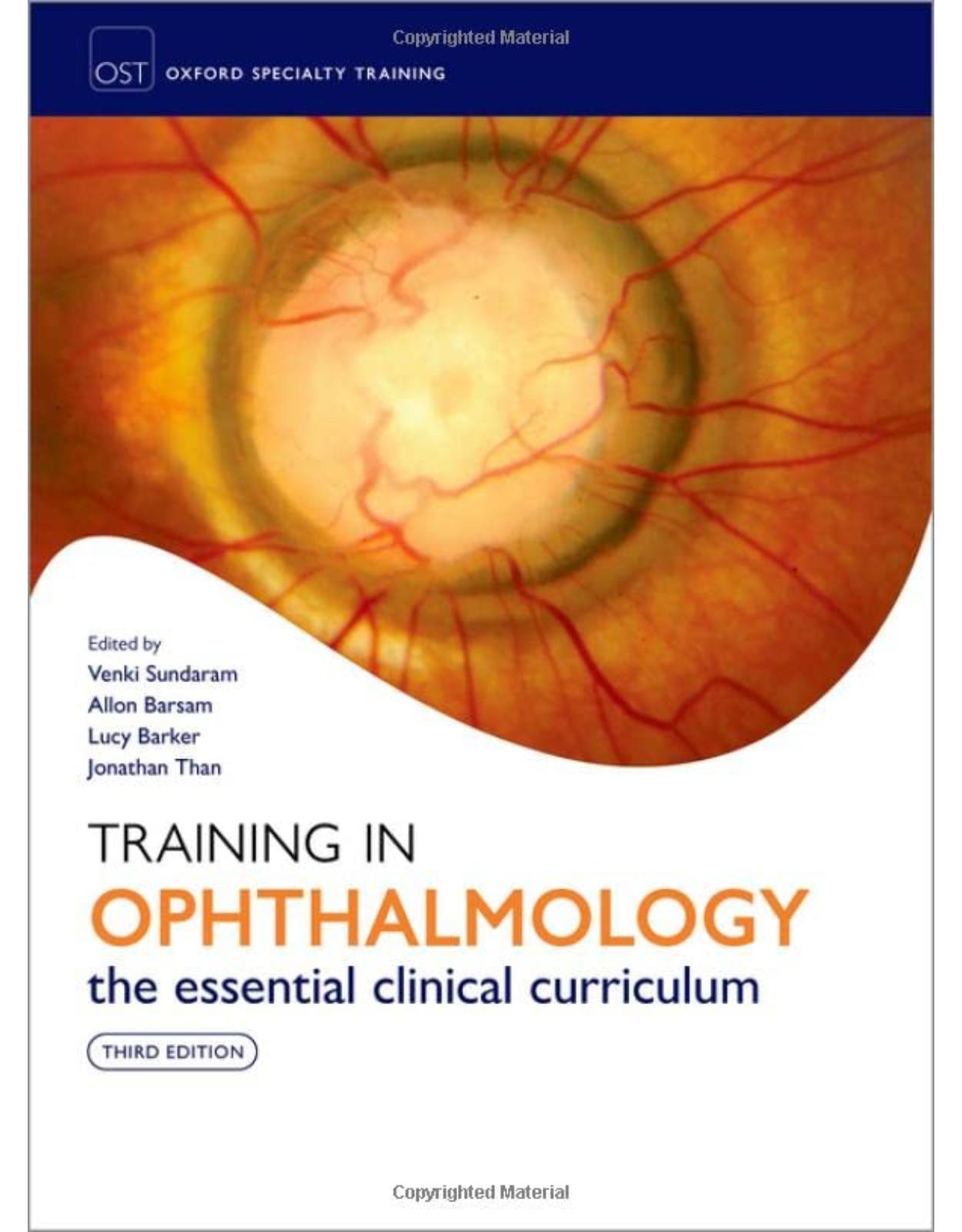
Training in Ophthalmology
Livrare gratis la comenzi peste 500 RON. Pentru celelalte comenzi livrarea este 20 RON.
Disponibilitate: La comanda in aproximativ 4 saptamani
Editura: Cambridge University Press
Limba: Engleza
Nr. pagini: 640
Coperta: Paperback
Dimensiuni:
An aparitie: 29 Jul 2022
Description:
Fully updated for a third edition, Training in Ophthalmology remains the indispensable guide to address the Royal College of Ophthalmologists (RCOphth) syllabus for trainee ophthalmologists, and is essential for all those studying ophthalmology, optometry, and orthoptics. As a theoretical and practical aid, it guides the reader through postgraduate Ophthalmic Specialist Training. Emphasis is placed on the practical assessment and management of key ophthalmic conditions. Clearly laid out and highly illustrated in full colour throughout, each condition is discussed in two to three pages, beginning with general explanations of pathophysiology and clinical evaluation, followed by differential diagnoses and treatment options. This new edition has been fully revised to increase emphasis on instilling an understanding of the rationale of current practice from first principles, with summary tables of seminal studies and distilled guidelines from the RCOphth and NICE. This text will appeal to foundation doctors, specialist trainees in ophthalmology, candidates preparing for the Fellowship of the Royal College of Ophthalmology examination, and consultants and allied practitioners looking for a comprehensive yet accessible guide to the subject.
Table of Contents:
1 Oculoplastics
1.1 Eyelid and nasolacrimal system anatomy
1.2 Lash abnormalities
1.3 Entropion
1.4 Ectropion
1.5 Ptosis
1.6 Benign lid lesions
1.7 Premalignant lesions
1.8 Malignant lid lesions I: clinical assessment
1.9 Malignant lid lesions II: management
1.10 Epiphora
1.11 Acquired nasolacrimal system abnormalities
1.12 Congenital abnormalities
1.13 Miscellaneous
1.14 Practical skills in oculoplastics
2 Cornea and conjunctiva
2.1 Corneal and conjunctival basic science
2.2 History taking for anterior segment disease
2.3 Examination of the anterior segment
2.4 Imaging of the anterior segment
2.5 Blepharitis
2.6 Staphylococcal hypersensitivity disorders
2.7 Dry eye disease
2.8 Conjunctivitis I
2.9 Conjunctivitis II: other infectious causes
2.10 Conjunctivitis III: allergic and miscellaneous
2.11 Conjunctivitis IV: miscellaneous
2.12 Cicatrizing conjunctival disease
2.13 Conjunctival degeneration
2.14 Conjunctival neoplasia I
2.15 Conjunctival neoplasia II
2.16 Corneal degeneration
2.17 Infectious keratitis I: bacterial
2.18 Infectious keratitis II: protozoal/fungal
2.19 Infectious keratitis III: viral; herpes simplex
2.20 Infectious keratitis IV: viral; herpes zoster
2.21 Interstitial keratitis
2.22 Peripheral ulcerative keratitis
2.23 Metabolic and drug-induced keratopathies
2.24 Miscellaneous
2.25 Corneal dystrophies I
2.26 Corneal dystrophies II
2.27 Contact lenses
2.28 Corneal ectasia
2.29 Keratoplasty
2.30 Complications of keratoplasty and graft rejection
2.31 Anterior uveal tumours
2.32 Anterior segment trauma
2.33 Chemical injury
3 Lens
3.1 Lens anatomy and embryology
3.2 Acquired cataract
3.3 Clinical evaluation of acquired cataract
3.4 Treatment for acquired cataract
3.5 Intraoperative complications of cataract surgery
3.6 Infectious postoperative complications of cataract surgery
3.7 Non-infectious postoperative complications of cataract surgery
3.8 Lens dislocation and other abnormalities
3.9 Biometry
3.10 Local anaesthesia
3.11 Operating microscope and phacodynamics
3.12 Intraocular lenses
3.13 Nd:YAG laser capsulotomy
4 Refractive surgery
4.1 Refractive error, aberrations, and presbyopia
4.2 Professional standards in refractive surgery
4.3 Preoperative evaluation for refractive surgery
4.4 Laser refractive surgery
4.5 Other corneal refractive procedures
4.6 Refractive lens surgery and ‘premium’ intraocular lenses
4.7 Phakic intraocular lenses
4.8 Presbyopia correction
5 Vitreoretinal surgery
5.1 Retinal anatomy and physiology
5.2 Posterior segment history taking and examination
5.3 Diagnostic lenses
5.4 Optical coherence tomography
5.5 Ultrasonography
5.6 Retinal photocoagulation
5.7 Vitreous disorders
5.8 Retinal detachment I
5.9 Retinal detachment II
5.10 Peripheral retinal abnormalities
5.11 Macular surgery
5.12 Submacular surgery
5.13 Retinal tumours
5.14 Choroidal tumours I
5.15 Choroidal tumours II
5.16 Vitreoretinopathies
5.17 Posterior segment trauma
6 Medical retina
6.1 Fundus fluorescein angiography
6.2 Abnormal fluorescein angiography
6.3 Indocyanine green angiography
6.4 Fundus autofluorescence imaging
6.5 Electrophysiology
6.6 Diabetic retinopathy I
6.7 Diabetic retinopathy II
6.8 Hypertensive retinopathy
6.9 Retinal vein occlusion I
6.10 Retinal vein occlusion II
6.11 Retinal artery occlusions
6.12 Age-related macular degeneration I
6.13 Age-related macular degeneration II
6.14 Therapies for age-related macular degeneration
6.15 Central serous chorioretinopathy
6.16 Retinal vascular anomalies
6.17 Inherited retinal disorders
6.18 Macular dystrophies
6.19 Choroidal dystrophies
6.20 Miscellaneous retinopathies
7 Uveitis and medical ophthalmology
7.1 Uveal anatomy
7.2 Anterior uveitis
7.3 Intermediate uveitis
7.4 Posterior uveitis
7.5 Specific non-infectious posterior uveitides I
7.6 Specific non-infectious posterior uveitides II
7.7 Viral infectious uveitis
7.8 Parasitic infectious uveitis
7.9 Fungal infectious uveitis
7.10 Bacterial infectious uveitis
7.11 HIV-related disease
7.12 Uveitis: systemic associations I
7.13 Uveitis: systemic associations II
7.14 Scleritis and episcleritis
7.15 Systemic treatment of ocular inflammatory disorders
7.16 Biological agents and periocular treatments of ocular inflammatory disorders
7.17 Intraocular treatments of ocular inflammatory disorders
7.18 Rheumatological disease
7.19 Vasculitis
7.20 Cardiovascular disease and the eye
7.21 Respiratory and skin diseases in ophthalmology
8 Glaucoma
8.1 The optic nerve head
8.2 Aqueous fluid dynamics
8.3 Glaucoma pathogenesis
8.4 Genetics
8.5 Optic nerve head assessment in glaucoma
8.6 Glaucoma imaging devices
8.7 Tonometry and pachymetry
8.8 Gonioscopy
8.9 Perimetry I
8.10 Perimetry II
8.11 Ocular hypertension
8.12 Primary open-angle glaucoma
8.13 Primary angle closure
8.14 Secondary angle closure
8.15 Normal-tension glaucoma
8.16 Steroid-induced glaucoma
8.17 Traumatic glaucoma
8.18 Inflammatory glaucomas
8.19 Pseudoexfoliative glaucoma
8.20 Pigmentary glaucoma
8.21 Neovascular glaucoma
8.22 Malignant glaucoma
8.23 Iridocorneal endothelial syndrome and iridocorneal dysgenesis
8.24 Ocular hypotensive agents I
8.25 Ocular hypotensive agents II
8.26 Laser therapy for glaucoma
8.27 Glaucoma surgery
8.28 Glaucoma clinical trials
9 Paediatric ophthalmology and strabismus
9.1 Anatomy and actions of the extraocular muscles
9.2 Embryology
9.3 Ophthalmic history and examination in children, and adults with strabismus
9.4 Assessment of visual acuity
9.5 Assessment and investigations for a child with poor vision
9.6 Binocular vision and stereopsis
9.7 Assessment of binocular single vision
9.8 Assessment of retinal correspondence
9.9 Assessment of ocular deviation I
9.10 Assessment of ocular deviation II: cover testing
9.11 Assessment of ocular movements
9.12 Hess charts and field of binocular single vision
9.13 Amblyopia
9.14 Concomitant strabismus I: heterophorias
9.15 Concomitant strabismus II: esotropia
9.16 Concomitant strabismus III: exotropia
9.17 Incomitant strabismus I
9.18 Incomitant strabismus II
9.19 Principles of strabismus surgery I
9.20 Principles of strabismus surgery II: complications
9.21 Other procedures in strabismus
9.22 General paediatric development
9.23 Retinopathy of prematurity
9.24 Retinoblastoma
9.25 Congenital cataract
9.26 Paediatric glaucoma
9.27 Paediatric uveitis
9.28 Phacomatoses
9.29 Metabolic and storage diseases
10 Neuro-ophthalmology
10.1 Visual pathway and pupil reflex anatomy
10.2 Cranial nerve anatomy
10.3 Central control of ocular motility
10.4 Neuro-ophthalmic history taking
10.5 Neuro-ophthalmology examination I
10.6 Neuro-ophthalmology examination II
10.7 Neuroimaging
10.8 Chiasmal visual pathway disorders
10.9 Retrochiasmal visual pathway disorders
10.10 Optic nerve disorders I
10.11 Optic nerve disorders II
10.12 Papilloedema and idiopathic intracranial hypertension
10.13 Congenital optic nerve disorders
10.14 Cranial nerve palsies I
10.15 Cranial nerve palsies II
10.16 Nystagmus
10.17 Supranuclear eye movement disorders
10.18 Pupil abnormalities
10.19 Headache
10.20 Non-organic visual loss
10.21 Neuromuscular junction disorders
10.22 Ocular myopathies
11 Orbit
11.1 Orbital anatomy
11.2 Orbital history and examination
11.3 Orbital infections
11.4 Orbital inflammatory disease
11.5 Orbital vascular malformations
11.6 Orbital tumours
11.7 Thyroid eye disease
11.8 Orbital fractures and trauma
11.9 Orbital surgery
12 Microsurgical skills
12.1 Forceps
12.2 Sutures
12.3 Suturing tissues
12.4 Tying a surgeon’s knot with instruments
12.5 Scenario : suturing a leaking corneal wound
12.6 Scenario 2: suturing skin during a lateral tarsal strip operation
12.7 Scenario 3: repairing full-thickness corneal lacerations
12.8 Scenario 4: repairing scleral lacerations
12.9 World Health Organization Surgical Safety Checklist
13 Professional skills and behaviour
13.1 Good medical practice
13.2 Information governance
13.3 Clinical governance and audit
13.4 Clinical leadership and NHS management
13.5 Education and training
13.6 Communication and consent
13.7 Research
13.8 Statistics
13.9 Ophthalmologists in Training Group
13.10 Safeguarding
13.11 Vision and society
Index
| An aparitie | 29 Jul 2022 |
| Autor | Venki Sundaram, Allon Barsam, Lucy Barker, Jonathan Than Matthew D. Gardiner |
| Editura | Cambridge University Press |
| Format | Paperback |
| ISBN | 9780198871590 |
| Limba | Engleza |
| Nr pag | 640 |

Clientii ebookshop.ro nu au adaugat inca opinii pentru acest produs. Fii primul care adauga o parere, folosind formularul de mai jos.