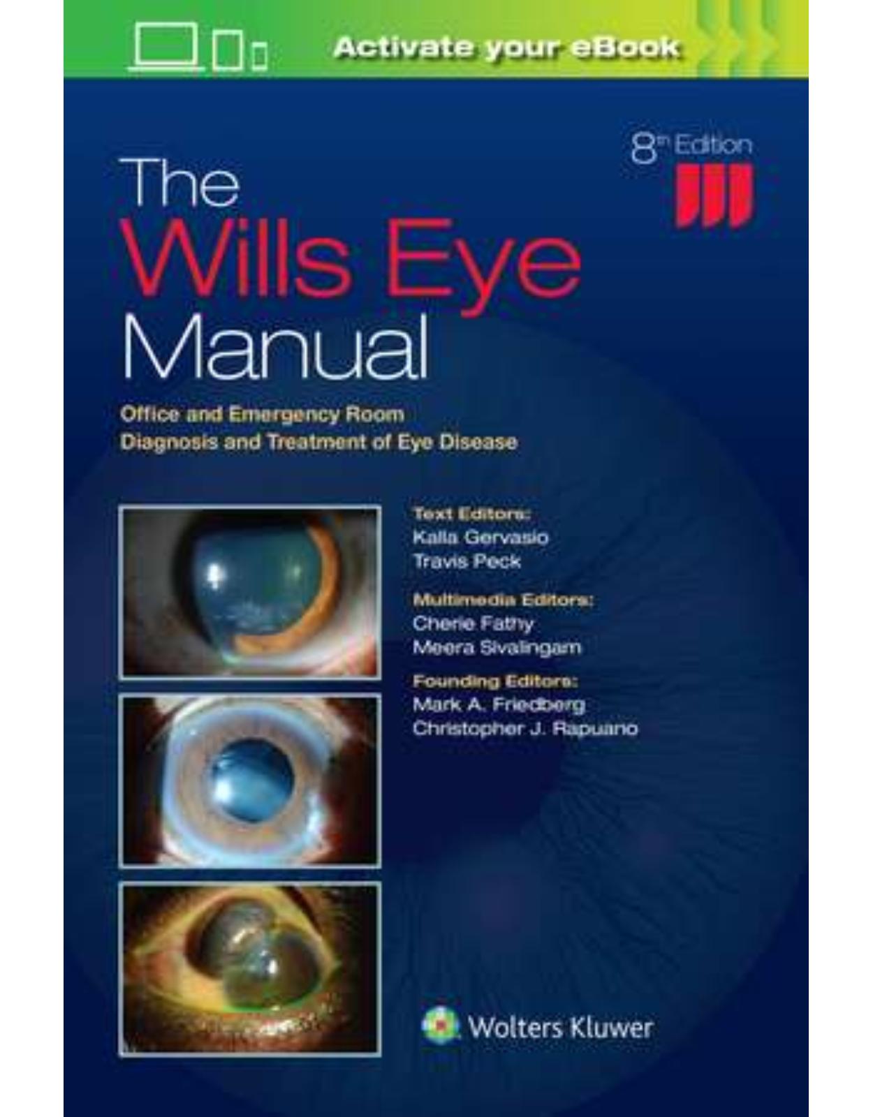
The Wills Eye Manual: Office and Emergency Room Diagnosis and Treatment of Eye Disease
Livrare gratis la comenzi peste 500 RON. Pentru celelalte comenzi livrarea este 20 RON.
Disponibilitate: La comanda in 3-4 saptamani
Editura: LWW
Limba: Engleza
Nr. pagini: 496
Coperta: Paperback
Dimensiuni: 152 x 229 mm
An aparitie: 25 Jun 2021
Description:
A best-selling source of compact, authoritative guidance on the treatment of ocular disorders in a variety of settings, The Wills Eye Manual, 8th Edition, is the comprehensive, high-yield reference of choice for both trainees and seasoned practitioners. It provides highly illustrated information on more than 200 ophthalmic conditions along with proven clinical recommendations from initial diagnosis through extended treatment. The consistent, bulleted outline format makes it ideal for portability and quick reference.
Table of Contents:
Video List
Chapter 1: Differential Diagnosis of Ocular Symptoms
BURNING
CROSSED EYES IN CHILDREN
DECREASED VISION
DISCHARGE
DISTORTION OF VISION
DOUBLE VISION (DIPLOPIA)
DRY EYES
EYELASH LOSS
EYELID CRUSTING
EYELID DROOPING (PTOSIS)
EYELID SWELLING
EYELID TWITCH
INABILITY TO COMPLETELY CLOSE EYELIDS (LAGOPHTHALMOS)
EYES “BULGING” (PROPTOSIS)
EYES “JUMPING” (OSCILLOPSIA)
FLASHES OF LIGHT
FLOATERS
FOREIGN BODY SENSATION
GLARE
HALLUCINATIONS (FORMED IMAGES)
HALOS AROUND LIGHTS
HEADACHE
ITCHY EYES
LIGHT SENSITIVITY (PHOTOPHOBIA)
NIGHT BLINDNESS
PAIN
RED EYE
“SPOTS” IN FRONT OF THE EYES
TEARING
Chapter 2: Differential Diagnosis of Ocular Signs
ANTERIOR CHAMBER/ANTERIOR CHAMBER ANGLE
CORNEA/CONJUNCTIVAL FINDINGS
EYELID ABNORMALITIES
FUNDUS FINDINGS
INTRAOCULAR PRESSURE
IRIS
LENS
NEUROOPHTHALMIC ABNORMALITIES
ORBIT
PEDIATRICS
POSTOPERATIVE COMPLICATIONS
REFRACTIVE PROBLEMS
VISUAL FIELD ABNORMALITIES
VITREOUS
Chapter 3: Trauma
3.1 Chemical Burn
MILD TO MODERATE BURNS
SEVERE BURNS
SUPER GLUE (CYANOACRYLATE) INJURY TO THE EYE
3.2 Corneal Abrasion
3.3 Corneal and Conjunctival Foreign Bodies
3.4 Conjunctival Laceration
3.5 Traumatic Iritis
3.6 Hyphema and Microhyphema
TRAUMATIC HYPHEMA
TRAUMATIC MICROHYPHEMA
NONTRAUMATIC (SPONTANEOUS) AND POSTSURGICAL HYPHEMA OR MICROHYPHEMA
3.7 Iridodialysis/Cyclodialysis
3.8 Eyelid Laceration
NONMARGINAL EYELID LACERATION
MARGINAL EYELID LACERATION
3.9 Orbital Blowout Fracture
3.10 Traumatic Retrobulbar Hemorrhage (Orbital Hemorrhage)
3.11 Traumatic Optic Neuropathy
3.12 Intraorbital Foreign Body
3.13 Corneal Laceration
PARTIAL-THICKNESS LACERATION
FULL-THICKNESS CORNEAL LACERATION
3.14 Ruptured Globe and Penetrating Ocular Injury
3.15 Intraocular Foreign Body
3.16 Firework or Shrapnel-/Bullet-Related Injuries
3.17 Commotio Retinae
3.18 Traumatic Choroidal Rupture
3.19 Chorioretinitis Sclopetaria
3.20 Purtscher Retinopathy
3.21 Shaken Baby Syndrome
Chapter 4: Cornea
4.1 Superficial Punctate Keratopathy
4.2 Recurrent Corneal Erosion
4.3 Dry Eye Syndrome
4.4 Filamentary Keratopathy
4.5 Exposure Keratopathy
4.6 Neurotrophic Keratopathy
4.7 Ultraviolet Keratopathy
4.8 Thygeson Superficial Punctate Keratitis
4.9 Pterygium/Pinguecula
4.10 Band Keratopathy
4.11 Bacterial Keratitis
4.12 Fungal Keratitis
4.13 Acanthamoeba Keratitis
4.14 Crystalline Keratopathy
4.15 Herpes Simplex Virus
4.16 Herpes Zoster Ophthalmicus/Varicella Zoster Virus
VARICELLA ZOSTER VIRUS (CHICKENPOX)
4.17 Interstitial Keratitis
4.18 Staphylococcal Hypersensitivity
4.19 Phlyctenulosis
4.20 Contact Lens–Related Problems
4.21 Contact Lens–Induced Giant Papillary Conjunctivitis
4.22 Peripheral Corneal Thinning/Ulceration
4.23 Delle
4.24 Keratoconus
4.25 Corneal Dystrophies
EPITHELIAL AND SUBEPITHELIAL DYSTROPHIES
CORNEAL STROMAL DYSTROPHIES
DESCEMET MEMBRANE AND ENDOTHELIAL DYSTROPHIES
4.26 Fuchs Endothelial Dystrophy
4.27 Aphakic Bullous Keratopathy/Pseudophakic Bullous Keratopathy
4.28 Corneal Graft Rejection
4.29 Corneal Refractive Surgery Complications
COMPLICATIONS OF SURFACE ABLATION PROCEDURES (PHOTOREFRACTIVE KERATECTOMY, LASER SUBEPITHELIAL KERATECTOMY, AND EPITHELIAL LASER IN SITU KERATOMILEUSIS)
COMPLICATIONS OF LASER IN SITU KERATOMILEUSIS
COMPLICATIONS OF SMALL INCISION LENTICULE EXTRACTION
COMPLICATIONS OF RADIAL KERATOTOMY
Chapter 5: Conjunctiva/Sclera/Iris/External Disease
5.1 Acute Conjunctivitis
VIRAL CONJUNCTIVITIS/EPIDEMIC KERATOCONJUNCTIVITIS
HERPES SIMPLEX VIRUS CONJUNCTIVITIS
ALLERGIC CONJUNCTIVITIS
VERNAL/ATOPIC CONJUNCTIVITIS
BACTERIAL CONJUNCTIVITIS (NONGONOCOCCAL)
GONOCOCCAL CONJUNCTIVITIS
PEDICULOSIS (LICE, CRABS)
5.2 Chronic Conjunctivitis
CHLAMYDIAL INCLUSION CONJUNCTIVITIS
TRACHOMA
MOLLUSCUM CONTAGIOSUM
MICROSPORIDIAL KERATOCONJUNCTIVITIS
TOXIC CONJUNCTIVITIS/MEDICAMENTOSA
5.3 Parinaud Oculoglandular Conjunctivitis
5.4 Superior Limbic Keratoconjunctivitis
5.5 Subconjunctival Hemorrhage
5.6 Episcleritis
5.7 Scleritis
5.8 Blepharitis/Meibomitis
5.9 Ocular Rosacea
5.10 Mucous Membrane Pemphigoid (Ocular Cicatricial Pemphigoid)
5.11 Contact Dermatitis
5.12 Conjunctival Tumors
AMELANOTIC LESIONS
MELANOTIC LESIONS
5.13 Malignant Melanoma of the Iris
Chapter 6: Eyelid
6.1 Ptosis
6.2 Chalazion/Hordeolum
6.3 Ectropion
6.4 Entropion
6.5 Trichiasis
6.6 Floppy Eyelid Syndrome
6.7 Blepharospasm
6.8 Canaliculitis
6.9 Dacryocystitis/Inflammation of the Lacrimal Sac
6.10 Preseptal Cellulitis
6.11 Malignant Tumors of the Eyelid
Chapter 7: Orbit
7.1 Orbital Disease
7.2 Inflammatory Orbital Disease
7.2.1 THYROID EYE DISEASE
SYNONYMS: THYROID-RELATED ORBITOPATHY, GRAVES’ DISEASE
7.2.2 IDIOPATHIC ORBITAL INFLAMMATORY SYNDROME
SYNONYM: INFLAMMATORY ORBITAL PSEUDOTUMOR
7.3 Infectious Orbital Disease
7.3.1 ORBITAL CELLULITIS
7.3.2 SUBPERIOSTEAL ABSCESS
7.3.3 ACUTE DACRYOADENITIS: INFECTION/INFLAMMATION OF THE LACRIMAL GLAND
7.4 Orbital Tumors
7.4.1 ORBITAL TUMORS IN CHILDREN
7.4.2 ORBITAL TUMORS IN ADULTS
7.5 Traumatic Orbital Disease
ORBITAL BLOWOUT FRACTURE
TRAUMATIC RETROBULBAR HEMORRHAGE
7.6 Lacrimal Gland Mass/Chronic Dacryoadenitis
7.7 Miscellaneous Orbital Diseases
Chapter 8: Pediatrics
8.1 Leukocoria
8.2 Retinopathy of Prematurity
8.3 Familial Exudative Vitreoretinopathy
8.4 Esodeviations
8.5 Exodeviations
8.6 Strabismus Syndromes
8.7 Amblyopia
8.8 Pediatric Cataract
8.9 Ophthalmia Neonatorum (Newborn Conjunctivitis)
8.10 Congenital Nasolacrimal Duct Obstruction
8.11 Congenital/Infantile Glaucoma
8.12 Developmental Anterior Segment and Lens Anomalies/Dysgenesis
8.13 Congenital Ptosis
8.14 The Bilaterally Blind Infant
Chapter 9: Glaucoma
9.1 Primary Open-Angle Glaucoma
9.2 Low-Tension Primary Open-Angle Glaucoma (Normal Pressure Glaucoma)
9.3 Ocular Hypertension
9.4 Acute Angle Closure Glaucoma
9.5 Chronic Angle Closure Glaucoma
9.6 Angle Recession Glaucoma
9.7 Inflammatory Open-Angle Glaucoma
9.8 Glaucomatocyclitic Crisis/Posner–Schlossman Syndrome
9.9 Steroid-Response Glaucoma
9.10 Pigment Dispersion Syndrome/Pigmentary Glaucoma
9.11 Pseudoexfoliation Syndrome/Exfoliative Glaucoma
9.12 Lens-Related Glaucoma
9.12.1 PHACOLYTIC GLAUCOMA
9.12.2 LENS PARTICLE GLAUCOMA
9.12.3 PHACOANTIGENIC GLAUCOMA (FORMERLY PHACOANAPHYLAXIS)
9.12.4 PHACOMORPHIC GLAUCOMA
9.12.5 GLAUCOMA CAUSED BY LENS DISLOCATION OR SUBLUXATION
9.13 Plateau Iris
9.14 Neovascular Glaucoma
9.15 Iridocorneal Endothelial Syndrome
9.16 Postoperative Glaucoma
9.16.1 EARLY POSTOPERATIVE GLAUCOMA
9.16.2 POSTOPERATIVE PUPILLARY BLOCK
9.16.3 UVEITIS, GLAUCOMA, HYPHEMA SYNDROME
9.17 Aqueous Misdirection Syndrome/Malignant Glaucoma
9.18 Postoperative Complications of Glaucoma Surgery
BLEB INFECTION (BLEBITIS)
INCREASED POSTOPERATIVE IOP AFTER FILTERING PROCEDURE
LOW POSTOPERATIVE IOP AFTER FILTERING PROCEDURE
COMPLICATIONS OF ANTIMETABOLITES (5-FLUOROURACIL, MITOMYCIN C)
COMPLICATIONS OF CYCLODESTRUCTIVE PROCEDURES
MISCELLANEOUS COMPLICATIONS OF FILTERING PROCEDURES
MISCELLANEOUS COMPLICATIONS OF TUBE-SHUNT PROCEDURES
9.19 Blebitis
Chapter 10: Neuro-ophthalmology
10.1 Anisocoria
10.2 Horner Syndrome
10.3 Argyll Robertson Pupils
10.4 Adie (Tonic) Pupil
10.5 Isolated Third Cranial Nerve Palsy
10.6 Aberrant Regeneration of the Third Cranial Nerve
10.7 Isolated Fourth Cranial Nerve Palsy
10.8 Isolated Sixth Cranial Nerve Palsy
10.9 Isolated Seventh Cranial Nerve Palsy
10.10 Cavernous Sinus and Associated Syndromes (Multiple Ocular Motor Nerve Palsies)
10.11 Myasthenia Gravis
10.12 Chronic Progressive External Ophthalmoplegia
10.13 Internuclear Ophthalmoplegia
10.14 Optic Neuritis
10.15 Papilledema
10.16 Idiopathic Intracranial Hypertension/Pseudotumor Cerebri
10.17 Arteritic Ischemic Optic Neuropathy (Giant Cell Arteritis)
10.18 Nonarteritic Ischemic Optic Neuropathy
10.19 Posterior Ischemic Optic Neuropathy
10.20 Miscellaneous Optic Neuropathies
TOXIC/METABOLIC OPTIC NEUROPATHY
COMPRESSIVE OPTIC NEUROPATHY
LEBER HEREDITARY OPTIC NEUROPATHY
DOMINANT OPTIC ATROPHY
COMPLICATED HEREDITARY OPTIC ATROPHY
RADIATION OPTIC NEUROPATHY
10.21 Nystagmus
CONGENITAL FORMS OF NYSTAGMUS
INFANTILE NYSTAGMUS
LATENT NYSTAGMUS
NYSTAGMUS BLOCKAGE SYNDROME
ACQUIRED FORMS OF NYSTAGMUS
10.22 Transient Visual Loss/Amaurosis Fugax
10.23 Vertebrobasilar Artery Insufficiency
10.24 Cortical Blindness
10.25 Nonphysiologic Visual Loss
10.26 Headache
10.27 Migraine
10.28 Cluster Headache
Chapter 11: Retina
11.1 Posterior Vitreous Detachment
11.2 Retinal Break (Tear)
11.3 Retinal Detachment
RHEGMATOGENOUS RETINAL DETACHMENT
EXUDATIVE/SEROUS RETINAL DETACHMENT
TRACTIONAL RETINAL DETACHMENT
11.4 Retinoschisis
X-LINKED (JUVENILE) RETINOSCHISIS
AGE-RELATED (DEGENERATIVE) RETINOSCHISIS
11.5 Cotton–Wool Spot
11.6 Central Retinal Artery Occlusion
11.7 Branch Retinal Artery Occlusion
11.8 Central Retinal Vein Occlusion
11.9 Branch Retinal Vein Occlusion
11.10 Hypertensive Retinopathy
11.11 Ocular Ischemic Syndrome/Carotid Occlusive Disease
11.12 Diabetic Retinopathy
11.13 Vitreous Hemorrhage
11.14 Cystoid Macular Edema
11.15 Central Serous Chorioretinopathy
11.16 Nonexudative (Dry) Age-Related Macular Degeneration
11.17 Neovascular or Exudative (Wet) Age-Related Macular Degeneration
11.18 Idiopathic Polypoidal Choroidal Vasculopathy
11.19 Retinal Arterial Macroaneurysm
11.20 Sickle Cell Retinopathy (Including Sickle Cell Disease, Anemia, and Trait)
11.21 Valsalva Retinopathy
11.22 Pathologic/Degenerative Myopia
11.23 Angioid Streaks
11.24 Ocular Histoplasmosis
11.25 Vitreomacular Adhesion/Vitreomacular Traction/Macular Hole
11.26 Epiretinal Membrane (Macular Pucker, Surface-Wrinkling Retinopathy, Cellophane Maculopathy)
11.27 Choroidal Effusion/Detachment
11.28 Retinitis Pigmentosa and Inherited Chorioretinal Dystrophies
RETINITIS PIGMENTOSA
SYSTEMIC DISEASES ASSOCIATED WITH HEREDITARY RETINAL DEGENERATION
HEREDITARY CHORIORETINAL DYSTROPHIES AND OTHER CAUSES OF NYCTALOPIA (NIGHT BLINDNESS)
11.29 Cone Dystrophies
11.30 Stargardt Disease (Fundus Flavimaculatus)
11.31 Best Disease (Vitelliform Macular Dystrophy)
11.32 Chloroquine/Hydroxychloroquine Toxicity
11.33 Crystalline Retinopathy
11.34 Optic Pit
11.35 Solar or Photic Retinopathy
11.36 Choroidal Nevus and Malignant Melanoma of the Choroid
CHOROIDAL NEVUS
MALIGNANT MELANOMA OF THE CHOROID
Chapter 12: Uveitis
12.1 Anterior Uveitis (Iritis/Iridocyclitis)
12.2 Intermediate Uveitis
12.3 Posterior and Panuveitis
12.4 Human Leukocyte Antigen–B27–Associated Uveitis
12.5 Toxoplasmosis
SPECIAL CONSIDERATION IN IMMUNOCOMPROMISED PATIENTS
12.6 Sarcoidosis
12.7 Behçet Disease
12.8 Acute Retinal Necrosis
12.9 Cytomegalovirus Retinitis
12.10 Noninfectious Retinal Microvasculopathy/HIV Retinopathy
12.11 Vogt–Koyanagi–Harada Syndrome
12.12 Syphilis
ACQUIRED SYPHILIS
CONGENITAL SYPHILIS
12.13 Postoperative Endophthalmitis
ACUTE (DAY[S] AFTER SURGERY)
SUBACUTE (WEEKS TO MONTHS AFTER SURGERY)
12.14 Chronic Postoperative Uveitis
12.15 Traumatic Endophthalmitis
12.16 Endogenous Bacterial Endophthalmitis
12.17 Candida Retinitis/Endophthalmitis
12.18 Sympathetic Ophthalmia
Chapter 13: General Ophthalmic Problems
13.1 Acquired Cataract
13.2 Subluxed or Dislocated Crystalline Lens
13.3 Pregnancy
ANTERIOR SEGMENT CHANGES
PREECLAMPSIA/ECLAMPSIA
OCCLUSIVE VASCULAR DISORDERS
MENINGIOMA OF PREGNANCY
OTHER CONDITIONS INFLUENCED BY PREGNANCY
13.4 Lyme Disease
13.5 Convergence Insufficiency
13.6 Accommodative Spasm
13.7 Erythema Multiforme, Stevens–Johnson Syndrome, and Toxic Epidermal Necrolysis
13.8 Vitamin A Deficiency
13.9 Albinism
13.10 Wilson Disease
13.11 Hypotony Syndrome
13.12 Blind, Painful Eye
13.13 Phakomatoses
NEUROFIBROMATOSIS TYPE 1 (VON RECKLINGHAUSEN SYNDROME)
NEUROFIBROMATOSIS TYPE 2
STURGE–WEBER SYNDROME (LEPTOMENINGEAL ANGIOMATOSIS)
TUBEROUS SCLEROSIS COMPLEX (BOURNEVILLE SYNDROME)
VON HIPPEL–LINDAU SYNDROME
WYBURN–MASON SYNDROME (RACEMOSE HEMANGIOMATOSIS)
ATAXIA–TELANGIECTASIA (LOUIS–BAR SYNDROME)
Chapter 14: Imaging Modalities in Ophthalmology
14.1 Plain Films Radiography
14.2 Computed Tomography
14.3 Magnetic Resonance Imaging
14.4 Magnetic Resonance Angiography
14.5 Magnetic Resonance Venography
14.6 Conventional Arteriography
14.7 Nuclear Medicine
14.8 Ophthalmic Ultrasonography
A-SCAN
B-Scan
ULTRASONOGRAPHIC BIOMICROSCOPY
ORBITAL ULTRASONOGRAPHY/DOPPLER
14.9 Photographic Studies
14.10 Intravenous Fluorescein Angiography
14.11 Indocyanine Green Angiography
14.12 Optical Coherence Tomography
14.13 Confocal Scanning Laser Ophthalmoscopy
14.14 Confocal Microscopy
14.15 Corneal Topography and Tomography
APPENDICES
Appendix 1: Dilating Drops
Appendix 2: Tetanus Prophylaxis
Appendix 3: Cover/Uncover and Alternate Cover Tests
Appendix 4: Amsler Grid
Appendix 5: Seidel Test to Detect a Wound Leak
Appendix 6: Forced Duction Test and Active Force Generation Test
Appendix 7: Technique for Diagnostic Probing and Irrigation of the Lacrimal System
Appendix 8: Corneal Culture Procedure
Appendix 9: Fortified Topical Antibiotics/Antifungals
Appendix 10: Technique for Retrobulbar/Subtenon/Subconjunctival Injections
Appendix 11: Intravitreal Tap and Inject
Appendix 12: Intravitreal Antibiotics
Appendix 13: Anterior Chamber Paracentesis
Appendix 14: Angle Classification
Appendix 15: Yag Laser Peripheral Iridotomy
Ophthalmic Acronyms and Abbreviations
Index
| An aparitie | 25 Jun 2021 |
| Autor | Dr. Kalla Gervasio, Dr. Travis Peck |
| Dimensiuni | 152 x 229 mm |
| Editura | LWW |
| Format | Paperback |
| ISBN | 9781975160753 |
| Limba | Engleza |
| Nr pag | 496 |
-
59000 lei 50000 lei

Clientii ebookshop.ro nu au adaugat inca opinii pentru acest produs. Fii primul care adauga o parere, folosind formularul de mai jos.