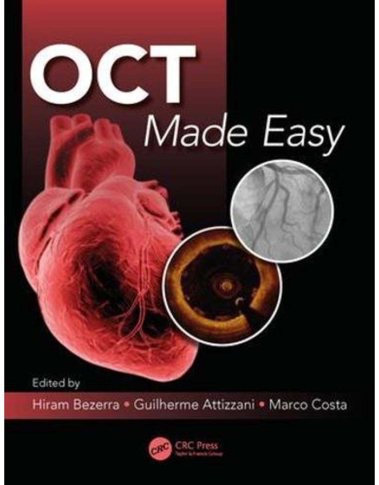
OCT Made Easy
Livrare gratis la comenzi peste 500 RON. Pentru celelalte comenzi livrarea este 20 RON.
Disponibilitate: La comanda in aproximativ 4 saptamani
Editura: CRC Press
Limba: Engleza
Nr. pagini: 190
Coperta: Paperback
Dimensiuni:
An aparitie: Septembrie 2017
Description:
This book, written by premier authors in the field of OCT intravascular imaging, covers the best practices for using OCT to provide high resolution cross-sectional viewing for atherosclerotic plaque assessment, stent strut coverage and apposition, assessment in stent restenosis evaluation, and PCI guide and optimization. Fully illustrated thorughout in a handy, cath-lab side handbook supplemented by online movie clips, OCT Made Easy includes case studies, angiography, CT correlations, and simple techniques for getting the best image.
Table of Contents:
Contributors
Chapter 1 Optical coherence tomography imaging for stent planning
1.1 Introduction
1.2 General outline
1.3 Preintervention OCT imaging
1.3.1 Assessment of coronary stenosis severity
1.3.1.1 Basic nomenclature and definitions
1.3.1.2 Considerations of quantitative measurements of vascular structures
1.3.1.3 Estimating the functional significance of coronary stenoses by quantitative OCT measurements
1.3.2 Assessment of lesion morphology, composition, and morphometry
1.3.3 Vessel measurements for stent sizing
1.3.3.1 Length measurement
1.3.3.2 Diameter measurement
1.4 Postintervention OCT imaging
1.4.1 Verification and optimization of stent expansion
1.4.2 Verification and optimization of stent apposition
1.4.3 Assessment of other qualitative parameters after stenting
1.4.3.1 Stent edge dissections
1.4.3.2 In-stent dissections, tissue prolapse, and in-stent thrombus
1.5 Potential clinical impact of OCT use for PCI planning and guidance
References
Chapter 2 OCT imaging for assessment of plaque modification
2.1 Introduction
2.2 Plain ordinary balloon and drug-eluting balloon angioplasty
2.2.1 Cutting balloon or scoring balloon angioplasty
2.2.2 Rotational atherectomy
2.2.3 Plaque modification after stenting
References
Chapter 3 OCT for plaque modification
3.1 Plaque modification before coronary artery stenting using cutting/scoring balloon
3.2 Cutting and scoring balloons
3.3 Trial results with cutting or scoring balloon plaque pretreatment
3.4 Patient’s clinical cases using cutting and scoring plaque pretreatment
3.4.1 First clinical case
3.4.2 Second clinical case
3.4.3 Third clinical case
References
Chapter 4 OCT for stent optimization
4.1 Introduction
4.2 Utilization of OCT for vessel preparation
4.2.1 Thrombectomy
4.2.2 Calcium modification using rotational atherectomy or cutting balloon
4.3 OCT for defining stent length and diameter
4.3.1 Defining lumen profile
4.3.2 Screening for unstable plaques
4.3.3 Defining the best stent landing zone
4.4 OCT for side branch protection
4.4.1 OCT after stent deployment
4.4.2 Stent malapposition
4.4.3 Stent underexpansion
4.4.4 Edge dissection
4.4.5 Incomplete lesion coverage
4.4.6 Tissue prolapse
4.5 Conclusion
References
Chapter 5 OCT for left main assessment
5.1 Introduction
5.2 Safety of FD-OCT for ULM Disease
5.3 FD-OCT Measurements for ULM Disease
5.4 Feasibility of FD-OCT for ULM Disease
5.5 Conclusions
References
Chapter 6 Optical coherence tomography for late stent failure
6.1 Introduction
6.2 Restenosis
6.2.1 Pathobiology of restenosis
6.2.2 OCT patterns of restenosis
6.2.3 Management
6.3 Case report: In-stent restenosis
6.4 Stent thrombosis
6.4.1 Prevalence and etiology of stent thrombosis
6.4.2 Role of OCT to identify intrastent pathology in late or very late stent thrombosis
6.4.3 Can OCT guide therapy in late or very late stent thrombosis?
References
Chapter 7 OCT assessment in spontaneous coronary artery dissection
7.1 Spontaneous coronary artery dissection
7.1.1 Epidemiology
7.1.2 Pathogenesis
7.1.3 Fibromuscular dysplasia
7.1.4 Angiographic features of SCAD: saw angiographic SCAD classification
7.1.5 Intracoronary imaging of SCAD
7.2 Role of OCT in the diagnosis and management of spontaneous coronary artery dissection
7.2.1 OCT guidance for PCI
7.3 Management of SCAD
7.4 Future directions
7.5 Conclusions
References
Chapter 8 Optical coherence tomography assessment for cardiac allograft vasculopathy after heart transplantation
8.1 Introduction
8.1.1 Background
8.2 Screening and diagnosing CAV
8.3 CAV Pathophysiology
8.3.1 Role of rejection
8.3.2 Traditional atherosclerosis
8.4 OCT utilization post–heart transplantation
8.4.1 Intimal hyperplasia
8.4.2 Atherosclerotic plaques
8.4.3 Correlation with cellular rejection
8.4.4 Comparison with IVUS
8.5 Treatment of CAV
8.6 Conclusion
References
Chapter 9 OCT assessment out of the coronary arteries
9.1 Introduction
9.2 History of intravascular imaging
9.3 Optical coherence tomography
9.3.1 Comparison with IVUS
9.3.2 Technique
9.4 Clinical applications of OCT in the peripheral vasculature
9.4.1 Diagnostic utilization: lesion characterization
9.4.1.1 Fibrotic and lipid-rich plaques: case 1
9.4.1.2 Calcified plaques: case 2
9.4.1.3 In-stent restenosis: case 3
9.4.1.4 Vein graft: case 4
9.4.1.5 Vasospasm: case 5
9.4.1.6 Thrombus: case 6
9.4.2 Assessment of interventional techniques
9.4.2.1 Angioplasty
9.4.2.2 Atherectomy
9.4.2.3 Stent
9.5 Future directions
References
Chapter 10 Optical coherence tomography for the assessment of bioresorbable vascular scaffolds
10.1 Introduction
10.2 Optical coherence tomography assessment before BVS implantation
10.3 Optical coherence tomography assessment after BVS implantation
10.4 Optical coherence tomography assessment for BVS follow-up
Disclosures
References
Chapter 11 ILUMIEN OPTIS Mobile and OPTIS Integrated Technology Overview
11.1 Platform overview
11.1.1 FD-OCT operating principles
11.1.2 Common system specifications
11.1.3 Catheter specifications
11.1.4 Comparison to ILUMIEN, C7xr, M3, and M2 specs
11.2 ILUMIEN OPTIS Mobile System
11.2.1 Imaging engine
11.2.2 Driver-motor and optical controller
11.2.3 Mobile console
11.3 OPTIS Integrated System
11.3.1 Architectural design
11.3.2 Control room cabinet
11.3.3 DOC holster
11.3.4 Remoting cable
11.3.5 Tableside controller
11.3.6 Installation options (boom vs. table mount)
11.4 OCT application software
11.4.1 B-Mode, L-Mode, and lumen profile displays
11.4.2 3D visualization
11.4.3 Data management
11.5 Angio coregistration software
11.5.1 Operating principle
11.5.2 Guided workflow
11.6 Conclusion
References
Chapter 12 Terumo OFDI system
12.1 Introduction
12.2 Fastview imaging catheter
12.2.1 Components and architecture
12.2.2 Clinical setup and compatibility
12.2.3 Characteristics of FastView
12.3 Lunawave OFDI imaging console
12.3.1 LUNAWAVE imaging technologies
12.3.1.1 Polygon scanner wavelength filter
12.3.1.2 Sensitivity peak shift
12.3.1.3 Laser safety mechanisms
12.3.2 Reliability
12.3.2.1 Data protection and backup system
12.3.3 Image acquisition setups
12.3.3.1 Pullback condition setting
12.3.3.2 Pullback speed and frame pitch
12.3.3.3 Automatic injector setup: flush rate and volume
12.3.4 Stent implantation supporting software
12.3.4.1 Preinterventional diagnosis
12.3.4.2 Area measurement
12.3.4.3 Length measurement
12.3.4.4 Angio coregistration
12.3.5 PCI procedure evaluation
12.3.5.1 Distance and thickness measurement
12.3.5.2 Dual-review mode
12.3.6 Evaluation of bifurcation lesion: 3D OFDI
12.3.6.1 3D reconstruction
12.3.7 File export
12.4 Case report: Usefulness of OFDI-guided PCI for the separated proximal and mid–left anterior descending artery lesions
12.5 Case report: Usefulness of 3D OFDI for bifurcation PCI
12.5.1 Introduction
12.5.2 Case
12.5.3 Discussion
Videos
Acknowledgment
Disclosures
References
Chapter 13 OCT imaging acquisition
13.1 Introduction
13.2 System of OCT
13.3 Imaging procedure
13.4 Flushing parameters: Recommended injection setting
13.5 Pitfalls and limitations
13.5.1 Incorrect calibration
13.5.2 Correction with refractive index
13.5.3 Filling of catheter sheath with contrast media
13.5.4 Inadequate flushing
13.5.5 Lesion selection
13.6 Conclusions
References
Appendix: Intravascular Bioresorbable Vascular Scaffold Optimization Technique
References
Index
| An aparitie | Septembrie 2017 |
| Autor | Guilherme F. Attizzani, Hiram G. Bezerra, Marco A. Costa |
| Editura | CRC Press |
| Format | Paperback |
| ISBN | 9781498714563 |
| Limba | Engleza |
| Nr pag | 190 |
-
1,01200 lei 94100 lei





Clientii ebookshop.ro nu au adaugat inca opinii pentru acest produs. Fii primul care adauga o parere, folosind formularul de mai jos.