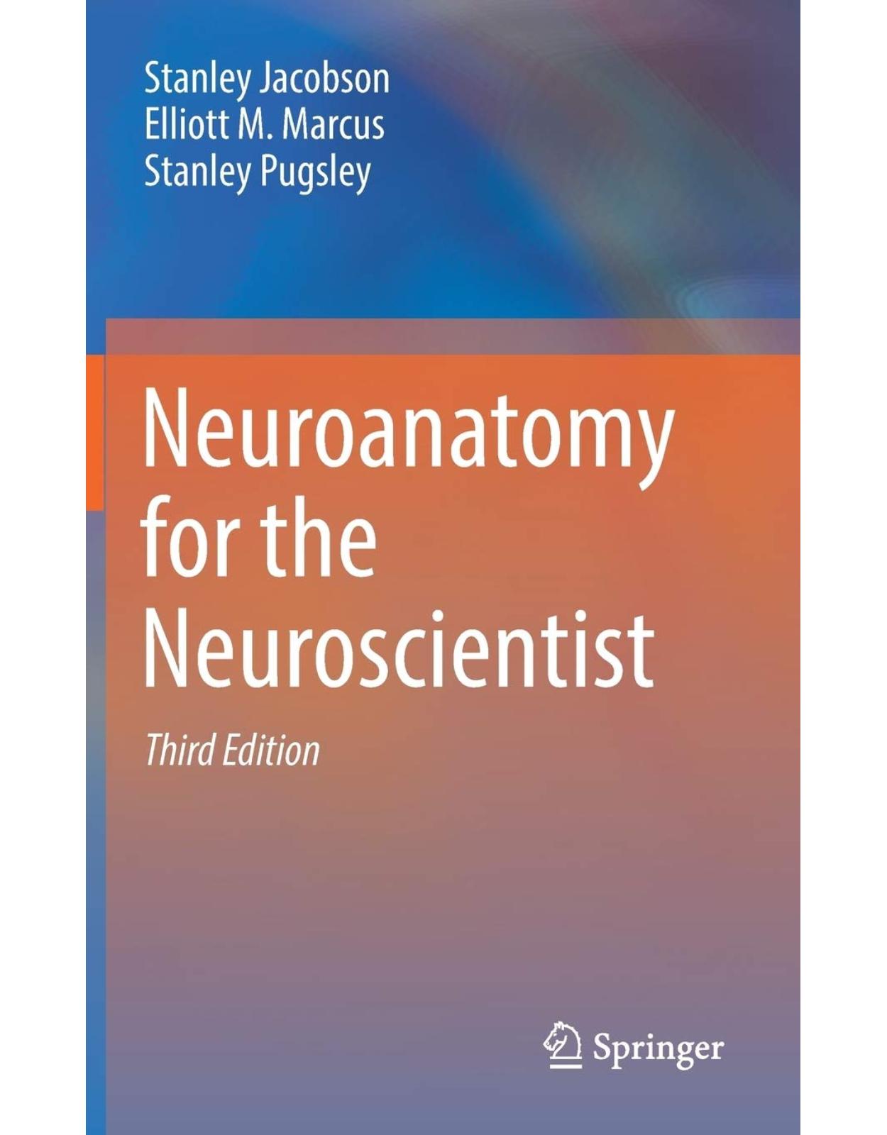
Neuroanatomy for the Neuroscientist
Livrare gratis la comenzi peste 500 RON. Pentru celelalte comenzi livrarea este 20 RON.
Description:
The purpose of this textbook is to enable a Neuroscientist to discuss the structure and functions of the brain at a level appropriate for students at many levels of study including undergraduate, graduate, dental or medical school level. It is truer in neurology than in any other system of medicine that a firm knowledge of basic science material, that is, the anatomy, physiology and pathology of the nervous system, enables one to readily arrive at the diagnosis of where the disease process is located and to apply their knowledge at solving problems in clinical situations. The authors have a long experience in teaching neuroscience courses at the first or second year level to medical and dental students and to residents in which clinical information and clinical problem solving are integral to the course.
Table of Contents:
Part I: Introduction to the Central Nervous System
Chapter 1: Introduction to the Central Nervous System
1.1 The Neuron
1.2 The Nervous System
1.2.1 Peripheral Nervous System (Fig. 1.3)
1.2.2 Central Nervous System
1.2.2.1 Spinal Cord (Fig. 1.5)
1.2.2.2 Brain
Brain Stem (Chaps. 5–7) (Fig. 1.6)
Cerebellum (Chap. 13) (Fig. 1.6)
Diencephalon (Chap. 8) (Fig. 1.6)
Cerebrum Cerebral Hemispheres (Chaps. 10–17) (Fig. 1.8)
Basal Ganglia (Fig. 1.10) (Chap. 12)
Central Nervous System Pathways
The Motor–Sensory Cortex (Fig. 1.8)
Introduction to Functional Localization Within the CNS
Bibliography
Chapter 2: Neurocytology: Cells of the CNS
2.1 The Neuron
2.1.1 Dendrites
2.1.2 Soma
2.1.3 Golgi Type I and II Neurons
2.1.4 Dendritic Spines (Fig. 2.2)
2.1.5 Nucleus
2.1.5.1 Rough Endoplasmic Reticulum: Nissl Body (Figs. 2.6 and 2.7)
2.1.5.2 Mitochondria (Figs. 2.8 and 2.12)
2.1.5.3 Neurosecretory Granules
2.1.6 Neuronal Cytoskeleton
2.1.7 Microtubules and Axoplasmic Flow
2.1.8 Neurofibrillary Tangles
2.1.8.1 Axon and Axon Origin (Axon Hillock) (Fig. 2.10a)
2.1.8.2 Myelin Sheath: The Insulator in an Aqueous Media (Fig. 2.14)
2.1.8.3 Myelination: Schwann Cell in PNS and Oligodendrocyte in CNS
2.1.8.4 Central Nervous System Pathways
2.2 Synapse
2.2.1 Synaptic Structure
2.2.2 Synaptic Types
2.2.3 Synaptic Transmission
2.2.4 Neurotransmitters (Table 2.3)
2.2.5 Modulators of Neurotransmission
2.2.6 Synaptic Vesicles (Fig. 2.16) (Table 2.4)
2.2.6.1 Excitatory Synapses
2.2.6.2 Inhibitory Synapses
2.2.6.3 Synaptic Architecture (Fig. 2.17)
2.2.7 Effectors and Receptors
2.3 Supporting Cells of the Central Nervous System
2.3.1 Astrocytes (Figs. 2.6 and 2.14; Table 2.7)
2.3.2 Oligodendrocytes (Fig. 2.9)
2.3.3 Endothelial Cells
2.3.4 Mononuclear Cells: Monocytes and Microglia
2.3.4.1 Mononuclear Cells/Mesodermal in Origin
2.3.5 Ependymal Cells (Fig. 2.20)
2.3.6 Supporting Cells in the Peripheral Nervous System
2.4 Response of the Nervous System to Injury
2.4.1 Degeneration
2.5 Regeneration
2.5.1 Peripheral Nerve Regeneration
2.5.2 Regeneration in the Central Nervous System
2.5.3 Neurogenesis in the Adult Brain Stem
2.5.4 Nerve Growth Factors (NGF)
2.5.5 Glial Response to Injury
2.6 Blood–Brain Barrier
2.6.1 Blood–Brain Barrier (Fig. 2.24)
2.6.2 Extracellular Space
Specific References
Chapter 3: Neuroembryology and Congenital Malformations
3.1 Formation of the Central Nervous System
3.2 Histogenesis
3.2.1 Repair of Damaged Nervous System
3.2.2 Growth Cone Guidance
3.2.3 Programmed Cell Death (PCD): Apoptosis
3.2.4 Neuronal Death
3.2.5 Development of Blood Vessels in the Brain
3.2.6 Ventricular System
3.2.7 Formation of Peripheral Nervous System
3.2.8 Spinal Cord Differentiation
3.3 Brain Differentiation
3.3.1 Rhombencephalon (Hindbrain) > Pons, Medulla, and Cerebellum
3.3.2 Mesencephalon > Adult Midbrain
3.3.3 Prosencephalon > Cerebral Hemispheres and Diencephalon
3.3.4 Diencephalon
3.3.5 Cranial Nerves
3.3.5.1 Cranial Nerve Innervation for Muscles of Somite Origin
3.3.5.2 Cranial Nerves Innervating the Muscles (Skeletal) and Skin in the Pharyngeal Arches (Tab
3.3.5.3 Mammals Have a Total of Six Arches (Table 3.5)
3.3.5.4 Preganglionic Parasympathetic Innervation to the Smooth Muscle
3.3.5.5 Cranial Nerves Associated with the Special Senses
3.3.6 Telencephalon
3.3.7 Primary Sulci
3.3.8 Development of the Cerebral Cortex
3.4 Prenatal Development of the Cerebral Cortex
3.5 Changes in the Cortical Architecture as a Function of Postnatal Age
3.6 Abnormal Development
3.6.1 Malformations Resulting from Abnormalities in Growth and Migration with Incomplete Develop
3.6.2 Genetically Linked Migration Disorders
3.6.2.1 Laboratory Data
3.6.3 Environmentally Induced Migration Disorder: Fetal Alcohol Syndrome
3.6.4 Malformations Resulting from Chromosomal Trisomy and Translocation
3.6.5 Malformations Resulting from Defective Fusion of Dorsal Structures
3.6.6 Malformations Characterized by Excessive Growth of Ectodermal and Mesodermal Tissue Affecti
3.6.7 Cutaneous Angiomatosis with Associated Malformations of the Central Nervous System
3.6.8 Malformations Resulting from Abnormalities in the Ventricular System
Bibliography
Chapter 4: Spinal Cord
4.1 Gross Anatomy
4.1.1 Spinal Cord: Structure and Function
4.1.2 Nerve Roots
4.1.3 Gray Matter
4.2 Interneurons
4.3 Central Pattern Generators
4.4 Segmental Function
4.4.1 Motor/Ventral Horn Cells
4.4.2 Sensory Receptors
4.4.3 Stretch Receptors
4.5 Nociception and Pain
4.5.1 Modulation of Pain Transmission
4.6 White Matter Tracts
4.6.1 Descending Tracts in the Spinal Cord
4.6.2 Ascending Tracts in the Spinal Cord
4.6.3 The Anterolateral Pathway
4.7 Upper and Lower Motor Neurons Lesions
4.7.1 Upper Motor Neuron Lesion (UMN)
4.7.2 Lower Motor Neuron Lesion
4.8 Illustrative Spinal Cord Case Histories
4.9 Illustrative Non-spinal Cord Cases with Involvement of Specific Peripheral Nerves: Case Histor
4.10 Carpal Tunnel Syndrome
Bibliography
Chapter 5: Brain Stem: Gross Anatomy
5.1 Gross Anatomical Divisions
5.1.1 Sites of Transition
5.2 Relationship of Regions in the Brain to the Ventricular System: Fig. 5.2
5.3 Gross Anatomy of Brain Stem and Diencephalon
5.3.1 Anterior Surface of Gross Brain Stem: Fig. 5.3
5.3.1.1 Anterior Surface Medulla and Pons
5.3.1.2 Anterior Surface Midbrain
5.3.2 Posterior Surface of Brain Stem and Diencephalon: Fig. 5.4
5.3.2.1 Medulla and Pons Posterior Surface (Fig. 5.4)
5.3.2.2 Midbrain Posterior Surface
5.3.2.3 Diencephalon: Ventral and Posterior Surfaces
5.4 Arterial Blood Supply to the Brain Stem and Diencephalon (Fig. 5.5)
5.4.1 Medulla
5.4.2 Pons
5.4.3 Midbrain
5.4.4 Diencephalon
Bibliography
Chapter 6: Brain Stem Functional Localization
6.1 Introduction to the Brain Stem
6.2 Differences Between the Spinal Cord and Brain Stem
6.3 Functional Localization in Brain Stem Coronal Sections and an Atlas of the Brain Stem
6.3.1 Medulla
6.3.1.1 Blood Supply Branches from the Vertebral Artery
6.3.1.2 Ventricular Zone
6.3.1.3 Lateral Zone
6.3.1.4 Medial Zone
6.3.1.5 Central Zone
6.3.1.6 Ventricular Zone
6.3.1.7 Lateral Zone
6.3.2 Pons-Blood Supply: Basilar Artery and Its Branches
6.3.2.1 Gross pons
6.3.2.2 Pons Ventricular Zone
6.3.2.3 Pons Lateral Zone
6.3.2.4 Pons Medial Zone
6.3.2.5 Pons Lateral Zone
6.3.3 Midbrain Blood Supply: Basila Arrteraynd Posterio Crerebral Arteries
6.3.3.1 Gross Anatomy Inferior Colliculus
6.4 Midbrain Tectum
6.5 Midbrain Tegmentum
6.6 Superior Colliculus
6.6.1 Midbrain Tegmentum
6.6.2 Blood Supply: Posterior Cerebral Arteries
6.7 Superior Colliculus Tectum
6.8 Superior Colliculus Tegmentum
6.8.1 Superior ColliculusVentricular Zone
6.8.1.1 Sensory Cranial Nerve Nuclei (Fig. 6.8a)
6.8.1.2 Motor Cranial Nerve Nuclei (Fig. 6.8)
6.8.1.3 Lateral Zone
6.8.1.4 Superior Colliculus/Anterolateral Column
6.8.1.5 Cerebellar Fibers
6.9 Functional Centers in the Brain Stem
6.9.1 Reticular Formation
6.9.1.1 Role of Descending Reticular Systems
6.9.1.2 Neurochemically Defined Nuclei in the Reticular Formation Affecting Consciousness
6.9.2 Respiration Centers
6.9.3 Cardiovascular Centers
6.9.4 Deglutition
6.9.5 Vomiting
6.9.6 Emetic Center
6.9.7 Coughing
6.9.8 Taste
6.10 Localiozation of Dysfunction in the Cranial Nerves Associated with the Eye (Table 6.8)
6.11 Localization of Disease Processes in the Brain Stem
6.11.1 Exercise to Identify the Tracts and Nuclei in the Brain Stem (Figs. 6.10–6.14)
Bibliography
Chapter 7: The Cranial Nerves
7.1 How the Cranial Nerves Got Their Numbers
7.2 Functional Organization of Cranial Nerves
7.3 The Individual Cranial Nerves
7.3.1 Cranial Nerve I, Olfactory (Fig. 7.4), Special Sensory/Special Visceral Afferent
7.3.2 Cranial Nerve II, Optic (Fig. 7.5), Special Somatic Sensory
7.3.3 Cranial Nerve III, Oculomotor (Fig. 7.6), Pure Motor (Somatic and Parasympathetic, Only III)
7.3.4 Cranial Nerve IV, Trochlear (Fig. 7.6), Pure Motor
7.3.5 Cranial Nerve VI, Abducens (Fig. 7.6), Pure Motor
7.3.6 Cranial Nerve V, Trigeminal (Fig. 7.7), Mixed Nerve (Sensory and Motor but No Parasympathet
7.3.7 Cranial Nerve VII, Facial (Fig. 7.8), Mixed Nerve (Sensory, Motor, Parasympathetic)
7.3.8 Cranial Nerve VIII, Vestibulocochlear (Fig. 7.9), Pure Special Somatic Sensory
7.4 Auditory Pathway
7.4.1 Cranial Nerve IX, Glossopharyngeal (Fig. 7.13), Mixed (Sensory, Motor, Parasympathetic): Nerv
7.4.2 Cranial Nerve X, Vagus (Fig. 7.14), Mixed (Sensory, Motor, Parasympathetic), and Longest Cra
7.4.3 Cranial Nerve XI, Spinal Accessory (Fig. 7.15), Pure Motor: Somatic and Visceral
7.4.4 Cranial Nerve XII, Hypoglossal (Fig. 7.16): Pure Motor Nerve
7.5 Cranial Nerve Dysfunction
7.6 Cranial Nerve Case Histories
Bibliography
Chapter 8: Diencephalon
8.1 Overview
8.2 Functional Organization of Thalamic Nuclei (Table 8.1)
8.2.1 Sensory and Motor Relay Nuclei: The Ventrobasal Complex and Lateral Nucleus
8.2.2 Limbic Nuclei: The Anterior, Medial, Lateral Dorsal, Midline, and Intralaminar Nuclei (Fig.
8.2.3 Specific Associational: Polymodal/Somatic Nuclei, the Pulvinar Nuclei (Fig. 8.5)
8.2.4 Special Somatic Sensory Nuclei: Vision and Audition, the Lateral Geniculate and Medial Geni
8.2.5 Nonspecific Associational
8.3 White Matter of the Diencephalon
8.4 Relationship Between the Thalamus and the Cerebral Cortex (Figs. 8.7 and 8.8)
8.5 Subthalamus (Fig. 8.3)
8.6 Thalamic Atlas Figs. 8.10, 8.11, and 8.12
8.7 Level: Midbrain, Diencephalic Junction (Fig. 8.10)
8.8 Level: Midthalamus (Fig. 8.11)
8.9 Level: Anterior Tubercle of Thalamus (Fig. 8.12)
Bibliography
Chapter 9: Hypothalamus, Neuroendocrine System, and Autonomic Nervous System
9.1 Hypothalamus
9.1.1 Hypothalamic Nuclei
9.1.2 Afferent Pathways
9.1.3 Efferent Pathways (Fig. 9.6)
9.1.4 Functional Stability
9.2 Neuroendocrine System, the Hypothalamus, and Its Relation to the Hypophysis
9.2.1 Hypophysis Cerebri
9.2.2 Hypothalamic–Hypophyseal Portal System
9.2.3 Hypophysiotrophic Area
9.2.4 Hormones Produced by Hypothalamus
9.2.5 Hormones Produced in Adenohypophysis (Fig. 9.12)
9.2.6 Case 9.1
9.2.6.1 Clinical Diagnosis: Pituitary Adenoma
9.2.6.2 Comments
9.2.6.3 Clinical Diagnosis: Pituitary Adenoma–Cushing Syndrome
9.2.7 Hypothalamus and the Autonomic Nervous System (Fig. 9.12)
9.2.8 Functional Localization
9.2.9 Water Balance and Neurosecretion (Fig. 9.7)
9.2.10 Hypothalamus and Light Levels: Optic Nerve Terminations in the Hypothalamus
9.2.11 Hypothalamus and Emotions
9.2.12 Hypothalamus and Light Levels
9.3 Autonomic Nervous System (Fig. 9.13)
9.4 Parasympathetic System (Craniosacral) (Fig. 9.13)
9.4.1 Cranial Nerves: III, VII, IX, and X
9.4.2 Sacral Segments S2–S4
9.5 Sympathetic System (Fig. 9.13)
9.6 Enteric Nervous System (Fig. 9.14)
Bibliography
Chapter 10: Cerebral Cortex Functional Localization
10.1 Anatomical Considerations
10.1.1 Cerebral Cortical Gray Matter
10.1.2 Cytology
10.2 Basic Design and Functional Organization of Cerebral Cortex
10.3 Fundamental Types of Cerebral Cortex
10.4 The Schema of the Fundamental Six-Layered Neocortices (Refer to Fig. 10.4)
10.5 Organization of the Neocortex (Fig. 10.6a)
10.5.1 How the Brodmann Areas Got Their Numbers
10.6 Correlation of Neocortical Cytoarchitecture and Function
10.6.1 Frontal Lobe (Figs. 10.8 and 10.9 and Table 10.1)
10.6.2 Motor Areas
10.6.3 Parietal Lobe (Figs. 10.9 and 10.10)
10.6.4 Temporal Lobe (Figs. 10.6 and 10.8)
10.6.5 Occipital Lobe (Figs. 10.8 and 10.9 and Table 10.4)
10.7 Subcortical White Matter Afferents and Efferents
10.8 Afferent Inputs and Efferent Projections of the Neocortex
10.8.1 Non-thalamic Sources of Cortical Input
10.9 Methods for the Study of Functional Localization in the Cerebral Cortex
10.9.1 How Do We Study Function?
10.9.1.1 Stimulation
10.9.1.2 Evoked Potentials (Fig. 10.14)
10.9.1.3 Lesions
10.9.2 How Do We Confirm the Location of the Pathology?
10.9.3 Neurophysiology Correlates of Cortical Cytoarchitecture and the Basis of the Electroence
General Bibliography-Cerebral Cortical Organization
Callosal References
Part II: The Systems
Chapter 11: Motor System, Movement, and Motor Pathways
11.1 Cerebral Cortical Motor Functions
11.1.1 Reflex Activity
11.2 Concept of Central Pattern Generators
11.3 Postnatal Development of Motor Reflexes
11.4 Relationship of Primary Motor, Premotor, and Prefrontal Cortex
11.4.1 Functional Overview
11.4.2 Primary Motor Cortex: Area 4 (Figs. 11.3 and 11.5)
11.4.3 Areas 6, Premotor Cortex (Areas 6 and 8; Fig. 11.5)
11.4.4 Dysfunction in the Premotor and Supplementary Motor Cortex
11.4.4.1 Effects of Metastatic Lesion to Frontal Lobe
11.4.4.2 Road Traffic Injury to frontal lobe (RTA)
11.4.4.3 Concussions-A former boxer
11.4.5 Prefrontal Cortex (Areas 9, 10, 11, 12, 13, 14, and 46, Fig. 11.5)
11.5 Disorders of Motor Development
11.6 Studies of Recovery of Motor Function in the Human
11.7 Cortical Control of Eye Movements-Frontal and Parietal Eye Fields
11.8 Major Voluntary Motor Pathways
11.8.1 Rubrospinal and Tectospinal Tracts (Fig 4.16; Table 4.2)
References
Motor System Movement, Motor Pathways General, Anatomy, Physiology and Functional Localization
Specific References Central Pattern Generators
Gait Disorders of the Elderly
Chapter 12: Motor System II: Basal Ganglia
12.1 Clinical Symptoms and Signs of Dysfunction
12.2 Specific Syndromes, Parkinson’s Disease, and the Parkinsonian Syndrome (Olanow and Tanner
12.3 Differential Diagnosis of Parkinson’s Disease
12.4 Chorea, Hemichorea, and Hemiballismus:
12.5 Hemichorea and Hemiballism
12.6 Other Movement Disorders Associated with Diseases of the Basal Ganglia
Bibliography
Specific References and Citations Parkinson Disease
Chapter 13: Motor Systems III: The Cerebellum Movement and Major Fiber Pathways of the Cerebellu
13.1 Anatomic Considerations
13.1.1 Subdivisions of the Cerebellum
13.1.2 Longitudinal Divisions
13.1.3 Transverse Divisions
13.2 Cytoarchitecture of the Cerebellum
13.3 Cerebellar Circuitry–Cerebellar Peduncles (Fig 13.4)
13.3.1 Afferents
13.3.2 Efferents
13.4 Topographic Patterns of Representation in Cerebellar Cortex
13.5 Functions of the Cerebellum and Correlations
13.5.1 Regional Functional Correlations
13.6 Effects of Disease on the Cerebellum
13.7 Major Cerebellar Syndromes
13.8 Syndrome of the Flocculonodular Lobe and Other Midline Cerebellar Tumors
13.9 Syndrome of the Anterior Lobe
13.10 Syndrome of the Lateral Cerebellar Hemispheres (Neocerebellar or Middle-Posterior Lobe Syndr
13.10.1 Syndromes of the Cerebellar Peduncles
13.11 Other Causes of Cerebellar Atrophy
13.12 Vascular Syndromes of the Cerebellum
13.13 Syndromes of Occlusion and Infarction
13.13.1 Neurological Examination
13.14 Cerebellar Degenerative Diseases
13.14.1 Gene Mechanisms
13.14.2 Olivopontocerebellar Atrophy (OPCA)
13.15 An Overview of Tremors
13.16 Major Fiber Pathways of the Cerebellum
13.16.1 Cerebellar Peduncles (Fig. 13.4)
13.16.2 Spinocerebellar Tracts (Fig. 13.11)
13.16.3 Cuneocerebellar Tract
Bibliography
Cerebellum Degenerations and Systemic Disorders
Vascular Syndromes of the Cerebellum
Cerebellum and Tremor
Gait Disorders of the Elderly
Chapter 14: Somatosensory Functions and the Parietal Lobe
14.1 Postcentral Gyrus: Somatic Sensory Cortex [Primary Sensory S-I]
14.1.1 Neurological Examination
14.2 Superior and Inferior Parietal Lobules
14.2.1 Dominant Hemisphere in the Parietal Lobules
14.2.1.1 Gerstmann’s Syndrome
14.2.2 Non-dominant Hemisphere in the Parietal Lobules
14.3 Parietal Lobe and Tactile Sensation from the Body
14.3.1 Tactile Sensation from the Body: Medial Lemniscus (Fig. 14.5)
14.3.2 Tactile Sensation from the Head: The Trigeminal Nerve (Fig. 14.6)
Bibliography
Chapter 15: Visual System and Occipital Lobe
15.1 Structure of the Eye
15.2 Photoreceptor Layer: Rods and Cones
15.2.1 Optic Nerve
15.2.2 Blind Spot
15.3 Visual Pathway (Figs. 15.4 and 15.5a–c)
15.3.1 Retina and Visual Fields
15.4 Occipital Lobe
15.4.1 Areas in the Occipital Lobe: 17, 18, and 19 (V1–V5)
15.4.2 Parallel Processing in the Visual Cortex
15.5 Perceptual Pathways: Color Vision
15.5.1 Effects of Stimulation of Areas 17, 18, and 19
15.6 Summary
15.6.1 Effects of Lesions in the Occipital Visual Areas
15.6.2 Occipital Lobe and eye Movements
15.7 Visual Field Deficits Produced by Lesions in the Optic Pathway
15.7.1 Case Histories: Examples with Lesions in the Visual System
15.7.1.1 Lesion prechiasmatic. Lesion in the optic nerve (see Fig. 15.8 #2) before the chiasm—resu
15.7.1.2 Lesion at the Optic Chiasm
15.7.2 Lesions in Occipital Cortex Result: Congruous Homonymous Hemianopsia
Bibliography
Chapter 16: The Limbic System, Temporal Lobe, and Prefrontal Cortex
16.1 Olfactory System
16.2 Limbic System
16.2.1 Subcortical Structures (Table 16.2)
16.2.2 Cortical Structures in the Limbic System
16.3 Principal Pathways of the Limbic System
16.4 Role of the Temporal Lobe in Learning and Memory
16.5 The Role of the Prefrontal Granular Areas and Emotions
16.6 Functional Neurosurgery
16.7 The Limbic Brain as a Functional System
Temporal Lobe References
Amygdala, Emotion, Autism, and Psychiatric Disorders. Specific References
Learning and Memory and the Temporal Lobe
Prefrontal Lobe General References
Chapter 17: Higher Cortical Functions
17.1 Cerebral Cortex and Disturbances of Verbal Expression
17.2 Cerebral Dominance
17.3 Aphasia: Dominant Hemispheric Functions
17.3.1 Nonfluent Aphasias
17.3.2 The Fluent Aphasias
17.4 Non-dominant Parietal Hemisphere Functions
17.5 Role of the Corpus Callosum in the Transfer of Information
General or Historical References
Part III: Neuropathology
Chapter 18: Cerebral Vascular Disease
18.1 Occlusive Cerebrovascular Disease
18.2 Clinical Correlates of Vascular Territories: Syndromes
18.3 Intracerebral Hemorrhage
18.4 Subarachnoid Hemorrhage (SAH)
18.5 Common Sites of Saccular Aneurysms (Fig. 18.7), Table 18.1
Chapter 19: Neuropathology, Nonvascular: Trauma, Neoplasms, and Communicable Diseases
19.1 Neuropathology Due to Trauma
19.1.1 Complications of Skull Fractures
19.1.2 Concussions
19.1.3 Contusions and Lacerations
19.1.4 Extradural Hematomas and Subdural Hematomas
19.1.5 Traumatic Brain Injury/TBI
19.1.6 Metastatic Lesions to the Brain
19.1.7 Meningioma: Tumors in the Coverings of the Brain
19.1.8 Glioma: Tumors Intrinsic to the Brain
19.1.9 Pituitary Tumor
19.2 Neuropathology Due to Communicable Diseases
Reference
Part IV: The Nonnervous Elements
Chapter 20: Non-nervous Elements in the CNS
20.1 Skull
20.2 Meninges: Coverings of the Brain
20.3 Blood Supply to the Brain
20.3.1 Arterial Blood Supply Figs. 20.6, 20.7, 20.8, 20.9, 20.10, and 20.11
20.4 Venous Drainage of the Brain, Head, and Neck (Fig. 20.13)
20.5 Ventricular System: Figs. 20.14 and 20.15
20.6 Cerebrospinal Fluid
20.7 The Glymphatic System and Drainage from the Brain
20.8 Glands Associated with the Brain
Reference
Chapter 21: Case History Problem Solving
Chapter 22: Movies on the Brain
22.1 Neuroanatomists, Anatomists, Neurosurgeons, and Neurologists
22.2 Developmental Disorders
22.3 Spinal Cord/Brain Stem Disorders
22.4 Disorders of Motor Systems and Motor Control
22.5 Cerebral Cortex
22.6 Limbic System
22.7 Cerebrovascular Disease
22.8 Brain Trauma
22.9 Brain Tumors and Increased Intracranial Pressure
22.10 Infections
22.11 Toxic and Metabolic Disorders
22.12 Disorders of Myelin
22.13 Seizures and Epilepsy
22.14 Coma
22.15 Memory
Part V: Atlas
Chapter 23: Descriptive Atlas
23.1 Gross Brain Sections: Coronal and Horizontal Sections
23.2 Gross Brain: Lateral Surface (Fig. 23.1)
23.3 Gross Brain: Medial Surface (Fig. 23.2)
23.4 Gross Brain Slices: Coronal (Figs. 23.3–23.9)
23.5 Gross Brain Slices Horizontal (Fig. 23.10)
Chapter 24: Myelin-Stained
24.1 Myelin-Stained Coronal Brain Sections (Figs. 24.1, 24.2, 24.3, 24.4, 24.5, 24.6, and 24.7)
24.2 Myelin-Stained Horizontal Sections (Figs. 24.8, 24.9, 24.10, and 24.11)
24.3 Myelin-Stained Sagittal Sections (Figs. 24.12 and 24.13)
| An aparitie | 2018 |
| Autor | Jacobson |
| Dimensiuni | 15.6 x 3.81 x 23.39 cm |
| Editura | Springer |
| Format | Hardcover |
| ISBN | 9783319601854 |
| Limba | Engleza |
| Nr pag | 710 |
-
68800 lei 62200 lei




Clientii ebookshop.ro nu au adaugat inca opinii pentru acest produs. Fii primul care adauga o parere, folosind formularul de mai jos.