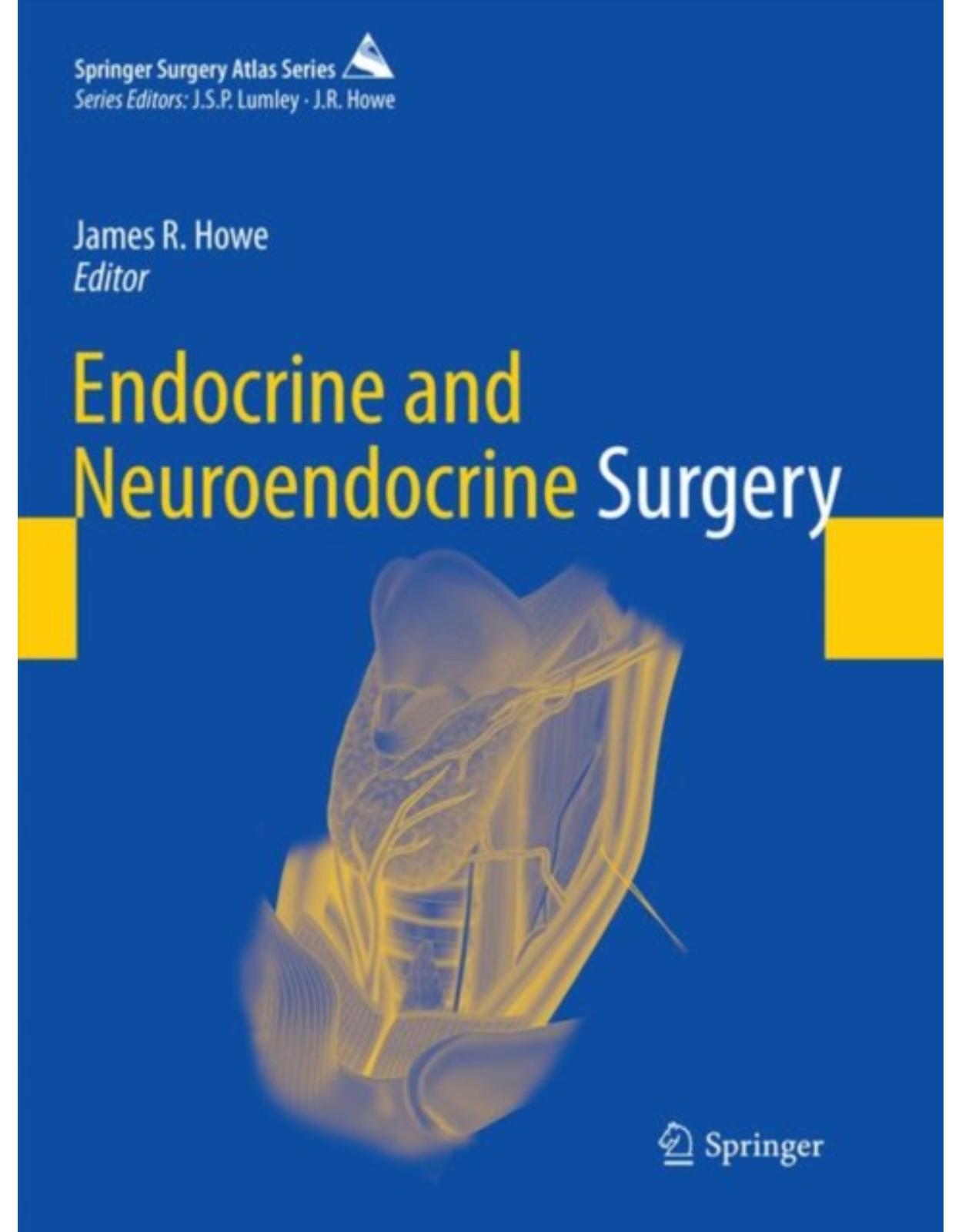
Head and Neck and Endocrine Surgery
Livrare gratis la comenzi peste 500 RON. Pentru celelalte comenzi livrarea este 20 RON.
Disponibilitate: La comanda in aproximativ 4 saptamani
Autor: Mahmoud Sakr
Editura: Springer
Limba: Engleza
Nr. pagini: 393
Coperta: Paperback
Dimensiuni: 178 x 254 x 31 mm
An aparitie: 19 Feb 2016
Description:
This book addresses a wide range of topics relating to head and neck and endocrine surgery, including: maxillofacial injuries, surgery of the scalp, surgery of the salivary glands, jaw tumors, surgery of the oral cavity (lips, tongue, floor of the mouth, and palate), swellings and ulcers of the face, inflammation in the neck, cervical lymphadenopathy, midline and lateral neck swellings, tumors of the pharynx, and endocrine surgery (thyroid gland, parathyroid glands, suprarenal glands, and neuroendocrine tumors). The aim is to clearly describe and illustrate how to diagnose and treat diverse conditions in accordance with evidence-based practice. The coverage thus extends beyond surgical indications and procedures to encompass aspects such as anatomy, clinical presentation, and imaging diagnosis. The book has been structured in such a way as to facilitate quick reference. While it is primarily intended for practitioners, it will also be suitable for upper graduate students.
Table of Contents:
Preface
Acknowledgments
Contents
Contributors
1: Maxillofacial Injuries
1.1 Introduction
1.1.1 Classification
1.1.2 General Principles of Management
1.1.2.1 First Aid Treatment
1.1.2.2 Hospital Evaluation
1.1.2.3 Primary Care
1.1.2.4 Anesthesia
1.2 Soft Tissue Injuries
1.2.1 General Principles
1.2.1.1 Preoperative Care
1.2.1.2 Anesthesia
1.2.2 Wound Preparation
1.2.3 Principles of Repair
1.2.3.1 Surgical Repair According to the Type of Injury
Abrasions
Contusions and Hematomas
Lacerations
Avulsions
Special Types of Injury
1.2.3.2 According to the Location of Injury
Lip Injuries
Nasal Injuries
Eyebrow Injuries
Eyelid Injuries (Fig. 1.3)
1.2.4 Postoperative Care
1.2.5 Complications
1.3 Injury of Specialized Structures
1.3.1 Parotid Gland and Duct Injury
1.3.1.1 Evaluation
1.3.1.2 Treatment
1.3.1.3 Complications
1.3.2 Facial Nerve Injury
1.3.2.2 Evaluation and Treatment
1.3.3 Ear Injuries
1.4 Fractures of Facial Bones
1.4.1 Surgical Anatomy
1.4.2 Nasal Fractures
1.4.2.1 Assessment of Nasal Fractures
1.4.2.2 Treatment of Nasal Fractures
Closed Reduction
Open Reduction
Postoperative Management and Complications
1.4.3 Mandibular Fractures
1.4.3.1 Anatomical Considerations
1.4.3.2 Assessment of Mandibular Fractures
1.4.3.3 Classification (According to Site)
1.4.3.4 Management of Mandibular Fractures
Fractures of the Ramus
Condylar Fractures
Dislocation of the Temporomandibular Joint (TMJ)
Temporomandibular Ankylosis
Factures of the Body of the Mandible
Dentoalveolar Fracture
1.4.4 Middle Third Fractures
1.4.4.1 Classification
1.4.4.2 Assessment of Midfacial Injuries
1.4.4.3 Treatment
Emergency Management
Definitive Treatment
Preparations and Planning
Anatomical Considerations (Facial Pillars)
Reduction and Fixation
References
2: Surgery of the Scalp
2.1 Scalp Swellings
2.1.1 Etiology
2.2 Congenital Swellings
2.2.1 Inclusion (Sequestration) Dermoid Cyst
2.2.2 Meningocele/Encephalocele
2.2.3 Cirsoid Aneurysm
2.3 Acquired Swellings
2.3.1 Traumatic
2.3.1.1 Caput Succedaneum
2.3.1.2 Cephalhematoma
2.3.2 Inflammatory
2.3.2.1 Infected Granuloma
2.3.2.2 Osteomyelitis of Skull Bones
2.3.2.3 Cock’s Peculiar Tumor
2.3.2.4 Suppuration (Abscess)
2.3.3 Sebaceous Cyst (Trichilemmal or Pilar Cyst)
2.3.4 Neoplastic
2.3.4.1 Soft Tissue Swellings
2.3.4.2 Bony Swellings
Ivory Osteoma of the Skull (Calvaria)
Multiple Myeloma
Secondaries (Metastases)
2.3.5 Pott’s Puffy Tumor
References
3: Surgery of the Salivary Glands
3.1 Introduction
3.2 Embryology and Developmental Disorders
3.3 Histology
3.4 Physiology
3.4.1 Salivary Flow Rates
3.5 Parotid Glands
3.5.1 Surgical Anatomy
3.5.1.1 Parotid Fascia
3.5.1.2 Parotid Duct (Stensen’s Duct)
3.5.1.3 Accessory Parotid Gland
3.5.1.4 Surface Anatomy of the Parotid Gland
3.5.1.5 Nerve Supply
3.5.1.6 Arterial Supply
3.5.1.7 Venous Drainage
3.5.1.8 Lymphatic Drainage
3.5.1.9 Parapharyngeal Space (PPS)
3.5.2 Evaluation of the Parotid Gland
3.5.2.1 Clinical Evaluation
History Taking
Physical Examination
3.5.2.2 Imaging
Sialography
Computed Tomography (CT)
Magnetic Resonance Imaging (MRI)
3.5.2.3 Endoscopic Examination (Sialendoscopy)
3.5.2.4 Biopsy
3.5.3 Parotid Injuries and Fistulae
3.5.3.1 Etiology
3.5.3.2 Clinical Presentation
History
Physical Examination
3.5.3.3 Treatment
Medical Therapy
Surgical Therapy
3.5.3.4 Complications
3.5.4 Parotid Calculus
3.5.5 Inflammatory Disorders (Sialadenitis)
3.5.5.1 Viral Infections
Mumps [49]
Non-Mumps
Human Immunodeficiency Virus (HIV)-Associated Sialadenitis [50, 51]
3.5.5.2 Bacterial Infections
Acute Suppurative Parotitis (Abscess) [52]
Chronic Pyogenic Parotitis
Actinomycosis
Cat-Scratch Disease
3.5.5.3 Recurrent Parotitis of Childhood
3.5.5.4 Papillary Obstructive Parotitis
3.5.5.5 Granulomatous Sialadenitis
Tuberculosis (TB)
Sarcoidosis
3.5.5.6 Autoimmune Sialadenitis
Mikulicz Disease (MD)
Sjogren’s Syndrome (SS)
3.5.5.7 Benign Lymphoepithelial Lesion (Myoepithelial Sialadenitis, MESA)
3.5.5.8 Other Autoimmune Sialadenitis
3.5.6 Sialadenosis (Sialosis)
3.5.6.1 Clinical Presentation
3.5.6.2 Etiology
3.5.6.3 Pathogenesis
3.5.6.4 Pathology
3.5.6.5 Treatment
3.5.7 Cystic Parotid Lesions
3.5.8 Tumors of the Parotid Glands
3.5.8.1 WHO Histological Classification of Tumors of Salivary Glands
Benign Epithelial Tumors
Malignant Epithelial Tumors
Soft Tissue Tumors
Hematolymphoid Tumors
Secondary Tumors
3.5.8.2 Benign Tumors (BTs)
Pleomorphic Adenoma (PA)
Warthin’s Tumor (Adenolymphoma)
Basal Cell Adenoma (BCA)
Canalicular Adenoma
Myoepithelioma
Cystadenoma
3.5.8.3 Malignant Tumors
Mucoepidermoid Carcinoma (MEC)
Adenoid Cystic Carcinoma (ACC)
Carcinoma Ex-pleomorphic Adenoma (CEPA)
True Malignant Mixed Tumor (TMMT)
Acinic Cell Carcinoma
Basal Cell Adenocarcinoma
Myoepithelial Carcinoma (Malignant Myoepithelioma)
Cystadenocarcinoma
3.5.9 Parotidectomy
3.5.9.1 Superficial Parotidectomy
Complications
Frey’s Syndrome (Gustatory Sweating)
3.5.9.2 Total Parotidectomy
3.5.9.3 Extended Total Parotidectomy
3.6 Submandibular Glands
3.6.1 Surgical Anatomy
3.6.1.1 Fascia
3.6.1.2 Submandibular Duct (Wharton’s Duct)
3.6.1.3 Lingual Nerve
3.6.1.4 Facial Artery
3.6.1.5 Venous Drainage
3.6.1.6 Lymphatic Drainage
3.6.2 Evaluation of the Submandibular Gland
3.6.2.1 Clinical Evaluation
History Taking
Physical Examination
Extraoral Examination
Intraoral Examination
Bimanual Examination
3.6.2.2 Imaging
Plain-Film Radiographs
Sialography
Computed Tomography (CT)
Magnetic Resonance Imaging (MRI)
3.6.2.3 Endoscopic Examination (Sialendoscopy)
3.6.2.4 Biopsy
3.6.3 Submandibular Sialadenitis/Sialadenosis
3.6.3.1 Acute Submandibular Sialadenitis
Etiology
Clinical Picture
Investigations (Workup of the Patient)
Treatment
Prognosis
3.6.3.2 Chronic Submandibular Sialadenitis/Sialolithiasis
Chronic Sialadenitis
Papillary Stenosis
Sialolithiasis
Clinical Picture
Investigations
Treatment
3.6.3.3 Autoimmune Sialadenitis
Sjogren’s Syndrome
Mikulicz’s Disease/Syndrome
3.6.3.4 Sialadenosis
3.6.4 Tumors of the Submandibular Gland
3.6.4.1 Benign Tumors (BTs)
3.6.4.2 Malignant Tumors (MTs)
3.6.4.3 Clinical Presentation
3.6.4.4 Differential Diagnosis
3.6.4.5 Diagnostic Imaging
3.6.4.6 Cytopathologic Diagnosis
3.6.4.7 Patterns of Spread
3.6.4.8 Management
Benign Tumors
Malignant Tumors
Factors Influencing Selection of Therapy
Resectable Tumors
Advanced Non-resectable Tumors
Management of the Neck
Chemotherapy
3.6.5 Surgical Technique: Submandibular Sialoadenectomy
3.6.5.1 Steps of the Procedure
3.6.5.2 Postoperative Complications
3.7 Sublingual Glands
3.7.1 Surgical Anatomy
3.7.2 Tumors of the Sublingual Glands
3.7.2.1 Ranula
3.7.2.2 Treatment of Sublingual Malignant Neoplasms
3.8 Minor Salivary Glands
3.8.1 Surgical Anatomy
3.8.2 Tumors of Minor Salivary Glands
3.8.2.1 Clinical Presentation
3.8.2.2 Diagnosis
3.8.2.3 Benign Tumors
3.8.2.4 Malignant Tumors
3.8.2.5 Treatment
3.8.2.6 Prognosis
References
4: Swellings of the Jaw
4.1 Introduction
4.2 Classification of Jaw Swellings
4.3 Epulides
4.3.1 Fibrous Epulides
4.3.2 Myelomatous Epulides (Giant Cell Epulides)
4.3.3 Granulomatous Epulides
4.3.4 Carcinomatous Epulides
4.3.5 Sarcomatous Epulides
4.3.6 Epulides Associated with Pregnancy
4.4 Odontomas
4.4.1 Dental (Radicular or Subapical) and Dentigerous (Follicular) Cysts
4.4.2 Ameloblastoma (Adamantinoma)
4.5 Bony Swellings
4.5.1 Inflammatory Lesions
4.5.1.1 Dental Granuloma
4.5.1.2 Alveolar (Dental) Abscess
4.5.1.3 Osteomyelitis
4.5.1.4 Cervicofacial Actinomycosis
4.5.2 Neoplastic Lesions
4.5.2.1 Carcinoma of the Maxilla
4.5.2.2 Sarcoma of the Maxilla
4.5.2.3 Burkitt’s Lymphoma
4.5.2.4 Osteoclastoma (Giant Cell Tumor)
4.6 Reconstruction of the Mandible
4.6.1 Reconstructive Stepladder
4.6.1.1 Alloplastic Devices
4.6.1.2 Corticocancellous Bone Grafts
4.6.1.3 Pedicled Soft Tissue Flaps
4.6.1.4 Free Vascularized Flaps
4.6.1.5 Tissue Engineering and Regenerative Medicine
4.6.2 Vascularization: The Key for Regeneration
4.6.2.1 Vascularization Concerns in the Mandible After Cancer Ablation
Vascular Pattern
Radiotherapy
Defect Size
4.6.2.2 Types of Vascularization
Vasculogenesis
Angiogenesis (Fig. 4.9)
Inosculation
4.6.3 Summary
References
5: Surgery of the Oral Cavity
5.1 Surgical Anatomy
5.2 Swellings of the Oral Cavity
5.2.1 Ranula
5.2.2 Sublingual Dermoid Cyst
5.2.3 Stone in Wharton’s Duct
5.2.4 Vascular Malformations
5.2.4.1 Classification
5.2.4.2 Venous Malformations
Pathology
Clinical Presentation
Treatment
5.2.4.3 Arteriovenous Fistulae
Pathology
Clinical Presentation
Treatment
5.2.4.4 Lymphatic Malformations
Pathology
Clinical Presentation
Treatment
5.3 Ulcers of the Oral Cavity
5.3.1 Etiological Classification
5.3.2 Traumatic Ulcers
5.3.2.1 Dental Ulcers
5.3.2.2 Frenular (Pertussis) Ulcer
5.3.3 Inflammatory Ulcers
5.3.3.1 Herpetic Ulcers
5.3.3.2 Tuberculous (TB) Ulcers
5.3.3.3 Syphilitic Ulcers
5.3.3.4 Chronic Superficial Glossitis
5.3.4 Dyspeptic (Aphthous) Ulcers
5.3.5 Malignant Ulcers
5.4 Neoplastic Lesions of the Oral Cavity
5.4.1 Classification
5.4.2 Benign Tumors
5.4.2.1 Fibro-epithelial Polyp
5.4.2.2 Papilloma of the Oral Cavity
5.4.2.3 Mixed Salivary Tumors of Minor Salivary Glands
5.4.2.4 Lingual Thyroid
5.4.2.5 Hemangioma
Pathology
Clinical Presentation
Treatment
5.4.2.6 Pyogenic Granuloma
5.4.3 Malignant Tumors of the Oral Cavity
5.4.3.1 Predisposing Factors
5.4.3.2 Sites in the Tongue
Anterior Two Thirds (80 %)
Posterior Third (20 %)
5.4.3.3 Pathology
Macroscopic Picture
Microscopic Picture
Spread
5.4.3.4 Clinical Presentation
5.4.3.5 Complications (Terminal Events)
5.4.3.6 Staging
Primary Tumor (T)
Regional Lymph Nodes (N)
Distant Metastasis (M)
5.4.3.7 Histological Grade (G)
5.4.3.8 Investigations
5.4.3.9 Treatment
Surgical Treatment
Radiotherapy (RT)
Chemotherapy
Management of Recurrences
5.4.3.10 Follow-Up
5.4.3.11 Prognosis
References
6: Surgery of the Face
6.1 Swellings of the Face
6.1.1 Classification
6.2 Diffuse Swellings
6.2.1 Acute Diffuse Swellings
6.2.2 Chronic Diffuse Swellings
6.2.2.1 Diffuse Hemangioma
6.2.2.2 Plexiform Neurofibromas (PNF)
6.3 Localized Swellings
6.3.1 Cystic Swellings
6.3.1.1 Sebaceous Cyst
6.3.1.2 Dermoid Cyst
6.3.1.3 Hemangioma
6.3.1.4 Lymphangioma
6.3.1.5 Cystic Swellings of the Parotid Gland
6.3.1.6 Cystic Swellings Around the Orbit
6.3.2 Solid Swellings
6.3.2.1 Keloid (Greek Crab’s Claw)
6.3.2.2 Lipoma
6.3.2.3 Neurofibroma
6.3.2.4 Other Solid Swellings
6.4 Ulcers of the Face
6.4.1 Classification
6.5 Ulcerating Infective Lesions
6.5.1 Nonspecific Ulcers
6.5.1.1 Chronic Nonspecific Ulcer
6.5.1.2 Infected Sebaceous Cyst
6.5.2 Specific Ulcers
6.5.2.1 Tuberculous (TB) Ulcers
6.5.2.2 Syphilitic ($) Ulcer
6.5.2.3 Leishmaniasis (Oriental Sore)
6.5.2.4 Actinomycosis
6.5.2.5 Anthrax
6.6 Ulcerating Tumors
6.6.1 Molluscum Sebaceum (Keratoacanthoma, KA)
6.6.2 Basal Cell Carcinoma (BCC)
6.6.2.1 Incidence
6.6.2.2 Body (Anatomic) Distribution
6.6.2.3 Etiology
6.6.2.4 Related Syndromes
Nevoid BCC Syndrome (Gorlin’s Syndrome)
Bazex Syndrome
Rombo Syndrome
6.6.2.5 Symptoms
6.6.2.6 Physical Examination
6.6.2.7 Complications
6.6.2.8 Clinic-pathological Types of BCC
Nodular (Noduloulcerative) BCC
Variants of Nodular BCC
Infiltrative BCC
Micronodular BCC
Morphea-Like (Fibrosing, or Sclerosing) BCC
Superficial BCC
Gorlin Syndrome (Basal Cell Nevus Syndrome)
Other Types of BCC
6.6.2.9 Diagnosis
Skin Biopsy
Imaging
6.6.2.10 Differential Diagnoses
6.6.2.11 Staging
High-Risk Tumors
6.6.2.12 Treatment
Approach Considerations
Surgical Modalities
Electrodesiccation and Curettage (E&C) [55]
Curettage Without Dessication [54]
Curettage with Er:YAG Laser Ablation
Laser Ablation Without Curettage
Surgical Excision
Mohs Micrographically Controlled Surgery
Cryosurgery
Immuno-cryosurgery
Radiation Therapy (RT)
Photodynamic Therapy (PDT)
Topical Medications
Oral Medications
6.6.2.13 Prognosis
6.6.3 Squamous Cell Carcinoma (SCC)
6.6.3.1 Incidence
6.6.3.2 Etiology
6.6.3.3 Symptoms
6.6.3.4 Physical Examination
6.6.3.5 Pathology
Variants (Subtypes) of SCC
6.6.3.6 Diagnosis
6.6.3.7 Marjolin Ulcer
6.6.3.8 Management
Treatment of the Primary Tumor
Treatment of Lymph Nodes
6.6.3.9 Prognosis
6.6.4 Malignant Melanoma (MM)
6.6.4.1 Embryology of Melanocytes
6.6.4.2 Incidence
6.6.4.3 Etiology and Risk Factors
6.6.4.4 Pathophysiology
6.6.4.5 Symptoms
6.6.4.6 Physical Examination
Local Examination
General Examination
6.6.4.7 Spread
6.6.4.8 Classification (Clinic-�pathological Types)
6.6.4.9 Staging of Melanoma
The Clark Scale
The Breslow Scale
The Day Scale (Modification of Breslow)
TNM and AJC (American Joint Committee) Staging System
Clinical Staging System (Number Stages of Melanoma)
6.6.4.10 Investigations
6.6.4.11 Differential Diagnosis
6.6.4.12 Prevention
6.6.4.13 Treatment
Treatment of the Primary Lesion
Local Excision
Reconstruction
Management of Regional LNs [100]
Treatment of Disseminated Disease
Palliative Surgery
Radiotherapy (RT)
Immunotherapy
Chemotherapy
6.6.4.14 Prognosis of Melanoma
6.7 Cavernous Sinus Thrombosis (CST)
6.7.1 Etiology
6.7.2 Clinical Presentation
6.7.3 Diagnosis
6.7.4 Complications
6.7.5 Treatment
6.7.5.1 Acute General Treatment
6.7.5.2 Long-Term Treatment
6.7.6 Prognosis
References
7: Deep Neck Space Infections
7.1 Introduction
7.2 Ludwig’s Angina
7.2.1 Surgical Anatomy
7.2.2 Etiology
7.2.3 Clinical Manifestations
7.2.4 Diagnosis
7.2.5 Treatment
7.2.5.1 Medical Treatment
7.2.5.2 Surgical Therapy
7.2.6 Prognosis
7.3 Peritonsillar Abscess (PTA)
7.3.1 Etiology
7.3.2 Clinical Presentation
7.3.3 Diagnosis
7.3.4 Treatment
7.3.4.1 Medical Therapy
7.3.4.2 Surgical Treatment
7.3.5 Prognosis
7.4 Parapharyngeal Abscess
7.4.1 Surgical Anatomy
7.4.2 Etiology
7.4.3 Clinical Presentation
7.4.4 Diagnosis
7.4.5 Treatment
7.5 Retropharyngeal Abscess (RPA)
7.5.1 Surgical Anatomy
7.5.2 Etiology
7.5.3 Clinical Presentation
7.5.4 Diagnosis
7.5.5 Treatment
7.5.6 Prognosis
References
8: Cervical: Lymphadenopathy
8.1 Anatomical Considerations
8.1.1 Delineation of the Levels of Cervical Lymph Nodes [3] (Fig. 8.1)
8.1.2 Neck Dissection
8.1.2.1 Historical Overview
8.1.2.2 Classification of Neck Dissection (AAO-HNS and American Head and Neck Society) [3]
Radical Neck Dissection (RND)
Modified Radical Neck Dissection (MRND) (Figs. 8.3 and 8.4)
Selective Neck Dissection (SND)
Extended Radical Neck Dissection (ERND)
8.2 Cervical Lymphadenopathy: Pathology and Clinical Pattern
8.2.1 Definition
8.2.2 Classification
8.2.2.1 Etiological
8.2.2.2 Pathological
8.2.2.3 Clinical
8.2.3 Etiology
8.2.3.1 Infections
Tuberculosis (TB)
Syphilis
Cat-Scratch Disease
Toxoplasmosis
8.2.3.2 Malignancy
8.2.3.3 Autoimmune Disease
8.2.3.4 Endocrine Disease
8.2.3.5 Drug-Induced Lymphadenopathy
8.2.3.6 Miscellaneous Causes of Cervical Lymphadenopathy
Kawasaki Disease (KD)
Sarcoidosis
Histiocytosis X
Kikuchi-Fujimoto Disease (KFD)
Castleman’s Disease
8.2.4 Neck Staging Under the “TNM Staging System” for Head and Neck Tumors
8.2.4.1 Regional Lymph Nodes (N)
8.2.4.2 Nasopharynx
8.2.4.3 Thyroid Gland
8.2.4.4 Mucosal Melanoma
8.2.4.5 Carcinoma of Unknown Primary (CUP)
8.3 Diagnostic Approach
8.3.1 History Taking
8.3.2 Physical Examination
8.3.3 Investigations
8.3.3.1 Imaging Modalities
Ultrasonography (US)
Reactive LNs
Tuberculous LNs
Lymphomatous LNs
Metastatic LNs
Computed Tomography (CT) and Magnetic Resonance Imaging (MRI)
Other Imaging Modalities
8.3.3.2 Biopsy
Excisional Biopsy
Core Needle Biopsy
8.4 Technique of Modified Radical Neck Dissection (MRND) [94, 95]
References
9: Swellings of the Neck
9.1 Surgical Anatomy
9.1.1 Triangles of the Neck
9.1.1.1 Anterior Triangle
9.1.1.2 Posterior Triangle
9.2 Introduction
9.2.1 Diagnosis of a Neck Mass
9.3 Midline Neck Swellings
9.3.1 Neck Dermoid Cysts
9.3.2 Abscess in Relation to the Mandible
9.3.3 Subhyoid Bursitis
9.3.4 Median Ectopic Thyroid Tissue
9.3.5 Tuberculous Thyroid Chondritis
9.3.6 Cyst/Nodule of the Thyroid Gland Isthmus
9.3.7 Thyroglossal Cyst
9.3.7.1 Embryology and Pathogenesis
9.3.7.2 Sites
9.3.7.3 Incidence
9.3.7.4 Pathology
9.3.7.5 Clinical Picture
9.3.7.6 Complications
9.3.7.7 Differential Diagnosis
9.3.7.8 Treatment (Sistrunk Operation, 1928)
9.3.7.9 Thyroglossal Fistula
9.4 Lateral Neck Swellings
9.4.1 Solid Swellings of the Anterior Triangle
9.4.1.1 Lymphadenopathy
9.4.1.2 Thyroid Swelling
9.4.1.3 Swelling of the Lower Pole of Parotid Gland
9.4.1.4 Paragangliomas (Glomus Tumors: Chemodectomas)
Carotid Body Tumor
Pathology
Symptoms
Clinical Examination
Investigations
Treatment
Excision of a Carotid Body Tumor
Glomus Vagale
Glomus Jugulare
Glomus Tympanicum
9.4.1.5 Schwannoma
9.4.1.6 Ganglioneuroma
9.4.1.7 Sternocleidomastoid (SCM) Tumor
Pathology
Clinical Picture
Treatment
9.4.2 Cystic Swellings of the Anterior Triangle
9.4.2.1 Pyogenic Abscess
9.4.2.2 Cold Abscess (TB Abscess)
9.4.2.3 Branchial Cyst
Embryology and Pathogenesis
Incidence
Pathology
Clinical Picture of the Second Branchial Arch Cyst
Differential Diagnosis
Complications
Treatment
Branchial Fistula
Congenital Type
Acquired Type
First Branchial Arch Anomaly (FBAA)
Third/Fourth Branchial Arch Anomaly
9.4.2.4 Laryngocele
9.4.2.5 Carotid Artery Aneurysm
9.4.3 Solid Swellings of the Posterior Triangle
9.4.3.1 Cervical Rib Syndrome
Scalene Syndrome: Superior Thoracic Aperture Syndrome
Varieties of Cervical Rib
Pathology
Symptoms
Local Examination
Investigations
Differential Diagnosis
Treatment
9.4.4 Cystic Swellings of the Posterior Triangle
9.4.4.1 Cystic Hygroma
Etiology
Pathology
Clinical Picture
Diagnosis
Complications
Treatment
Surgical Treatment
Sclerosing Therapy
Other Less Popular Methods
9.4.4.2 Pharyngeal Pouch (Zenker’s Diverticulum)
Pathogenesis
Clinical Presentation
Complications
Treatment
9.4.4.3 Pneumatocele
Pathophysiology
Epidemiology
Etiology
Clinical Picture
Complications
Investigations
Laboratory Studies
Imaging Studies
Procedures
Differential Diagnoses
Treatment
Prognosis
References
10: Tumors of the Pharynx
10.1 Tumors of the Nasopharynx
10.1.1 Benign Tumors (Nasopharyngeal Fibroma/Angiofibroma)
10.1.2 Malignant Tumors
10.2 Tumors of the Oropharynx
10.3 Tumors of the Hypopharynx
References
11: Surgery of the Thyroid Glands
11.1 Embryology
11.2 Surgical Anatomy
11.2.1 Fascia and Ligaments
11.2.2 Relation with Strap Muscles
11.2.3 Arterial Supply
11.2.3.1 Superior Thyroid Artery (STA)
11.2.3.2 Inferior Thyroid Artery (ITA)
11.2.3.3 Thyroidea Ima Artery
11.2.4 Venous Drainage
11.2.5 Lymphatics of the Thyroid Gland
11.2.6 Innervation of the Thyroid Gland
11.2.6.1 Nerves Related to the Thyroid Gland
11.2.7 The Parathyroid Glands
11.3 Histology
11.4 Physiology
11.5 Investigating the Enlarged Thyroid
11.5.1 Serological Investigations
11.5.1.1 Thyroid Function Tests
11.5.2 Imaging
11.5.2.1 Ultrasonography (US)
11.5.2.2 Magnetic Resonance Imaging (MRI) and Computed Tomography (CT)
11.5.2.3 Thyroid Scintigraphy
11.5.3 Biopsy
11.5.3.1 Fine-Needle Aspiration Cytology (FNAC): Freehand or US Guided
Indications and Aims of FNAC
Limitations of FNAC
Cytology Results of FNAC
The Nondiagnostic Smears
Accuracy of FNA in Exploring Cervical LNs in the Presence of a Thyroid Carcinoma
Core Biopsy (With or Without US Guidance)
11.5.4 Flexible Laryngoscopy
11.6 Multinodular Goiter (MNG)
11.6.1 Introduction
11.6.2 Pathogenesis
11.6.3 Clinical Assessment
11.6.3.1 Patient’s History
11.6.3.2 Physical Examination
11.6.4 Complications of MNG
11.6.5 Investigations
11.6.5.1 Laboratory Findings
11.6.5.2 Imaging Findings
Ultrasonography (US)
Scintigraphy
Radiography and Tomography
11.6.5.3 Fine-Needle Aspiration (FNA)
11.6.5.4 Airway Assessment
11.6.6 Management of MNG
11.6.6.1 Nonoperative Treatment
11.6.6.2 Surgical Treatment
Indications and Extent of Surgery
Multinodular Goiter (Benign/Thy 2)
Thyroid Nodules Associated with Hypo-/Hyperthyroidism
Dominant Nodule in MNG
Thyroid Cystic Swelling
Results of Surgical Treatment of MNG
Prophylaxis of Recurrence
Treatment of Recurrent Goiter
11.7 Retrosternal Goiter (RSG)
11.7.1 Introduction
11.7.2 Incidence
11.7.3 Anatomical Classification
11.7.4 Clinical Manifestations
11.7.4.1 Symptoms
11.7.4.2 Physical Examination
11.7.5 Diagnosis
11.7.5.1 Plain Chest X-Ray (CXR)
11.7.5.2 CT Scan and MRI
11.7.5.3 Scintigraphy
11.7.5.4 US and FNA
11.7.6 Differential Diagnosis
11.7.7 Treatment
11.7.7.1 Pharmacotherapy
11.7.7.2 Radioactive Iodine (RAI) Therapy
11.7.7.3 Surgical Therapy
Approach to RSG
Complications of RSG Surgery
11.8 Solitary Thyroid Nodule (STN)
11.8.1 Pathological Classification
11.8.2 Clinical Considerations
11.8.2.1 History Taking
11.8.2.2 Physical Examination
11.8.3 Laboratory Tests
11.8.4 Imaging Studies
11.8.4.1 Ultrasonography (US)
11.8.4.2 Scintiscan
11.8.5 Fine-Needle Aspiration Biopsy (FNAB)
11.8.6 Management
11.8.6.1 Clinically Non-palpable Incidental Nodule
11.8.6.2 Benign Nodule
11.8.6.3 Indeterminate Lesion
11.8.6.4 Suspicious Nodule
11.8.6.5 Malignant Nodule
Treatment of Differentiated Thyroid Cancer
Principles and Strategies
Treatment: Papillary Thyroid Cancer (PTC)
Treatment: Follicular Thyroid Cancer (FTC)
11.8.6.6 Treatment of Oncocytic (Hürthle Cell) Thyroid Cancer
11.8.6.7 Treatment of Medullary Thyroid Carcinoma (MTC)
11.8.6.8 Treatment of Anaplastic Carcinoma
11.8.6.9 Nondiagnostic Biopsies
11.9 Thyrotoxicosis
11.9.1 Introduction
11.9.2 Clinical Manifestations
11.9.2.1 Symptoms
11.9.2.2 Local Examination
11.9.2.3 General Examination
11.9.3 Diagnosis
11.9.4 Differential Diagnosis
11.10 Graves’ Disease
11.10.1 Pathogenesis
11.10.2 Pathology
11.10.3 Clinical Presentation
11.10.4 Diagnosis
11.10.5 Management
11.10.5.1 Radioactive Iodine (RAI)
11.10.5.2 Antithyroid Drugs (ATDs)
11.10.5.3 Surgical Treatment
11.11 Toxic Multinodular Goiter (Plummer’s Disease)
11.11.1 Diagnosis
11.11.2 Treatment
11.11.2.1 Solitary Toxic Nodule
11.11.3 Diagnosis
11.11.4 Treatment
11.12 Thyroiditis
11.12.1 Hashimoto’s Thyroiditis
11.12.2 Silent (Painless) Thyroiditis
11.12.3 Subacute Thyroiditis (de Quervain’s, Granulomatous, or Giant Cell Thyroiditis)
11.13 Iodine-Induced Thyrotoxicosis
11.14 Thyroiditis
11.14.1 Introduction
11.15 Autoimmune Thyroiditis
11.15.1 Hashimoto’s Thyroiditis (Chronic Lymphocytic Thyroiditis, Struma Lymphomatosa)
11.15.2 Focal Lymphocytic Thyroiditis (Focal Autoimmune Thyroiditis, Chronic Nonspecific Thyroiditis
11.15.3 Postpartum Thyroiditis
11.15.4 Subacute de Quervain’s Thyroiditis (Granulomatous, Pseudotuberculous, or Giant Cell Thyroi
11.15.5 Painless Thyroiditis (Subacute Lymphocytic Thyroiditis)
11.15.6 Riedel’s Fibrosing Thyroiditis
11.16 Non-autoimmune Thyroiditis
11.16.1 Acute Infectious Thyroiditis (Acute Suppurative Thyroiditis)
11.16.2 Drug-Induced Thyroiditis
11.16.3 Postoperative Necrotizing Thyroiditis
11.16.4 Radiation Thyroiditis
11.16.5 Other Causes of Nonimmune Thyroiditis
11.16.6 Indications of Surgery in Thyroiditis
11.17 Malignant Thyroid Disease: Introduction
11.17.1 Classification
11.17.1.1 World Health Organization (WHO) Classification
11.17.1.2 Pathological Classification
11.17.2 Screening
11.17.3 Risk Factors for Thyroid Carcinoma
11.17.3.1 History Taking
11.17.3.2 Clinical Examination
11.18 Well-Differentiated Thyroid Cancer
11.18.1 Staging (Tables 11.20a, 11.20b, and 11.20c)
11.18.2 Management of DTC
11.18.2.1 Surgical Treatment
11.18.2.2 Radioactive Iodine (RAI) Ablation and Treatment for DTC
Preparation for 131I Ablation or Therapy
Postoperative 131I Ablation
Indications of Remnant Ablation with 131I
Short-Term and Long-Term Side Effects of 131I Ablation Treatment
11.18.2.3 External Beam Radiotherapy (EBRT) for the Treatment of DTC
11.18.3 Follow-Up of WDTC
11.18.3.1 Voice Dysfunction
11.18.3.2 Management of Hypocalcemia
11.18.3.3 Long-Term Suppression of Serum Thyrotropin
11.18.3.4 Measurement of Serum in Long-Term Follow-Up
11.18.3.5 Role of US and Whole-Body 131I Scan (WBS) in Routine Follow-Up
11.18.4 Recurrent/Persistent DTC
11.18.4.1 Recurrence in the Thyroid Bed or Cervical LNs
11.18.4.2 Metastases in the Lungs and Other Soft Tissue Areas
11.18.4.3 Cerebral Metastases
11.18.4.4 Bone Metastases
11.18.4.5 Other Metastatic Sites
11.18.4.6 Palliative Care
11.19 Papillary Thyroid Cancer (PTC)
11.19.1 Risk Factors
11.19.2 Gross Features
11.19.3 Microscopic Features
11.19.3.1 Histological Variants of PTC
Papillary Micro-carcinoma
Encapsulated Variant
Follicular Variant
Tall Cell and Columnar Cell Variants
Diffuse Sclerosing Variant
Other Variants of PTC
11.19.4 Clinical Aspects
11.19.5 Lymphatic Spread
11.19.6 Distant Metastases
11.19.7 Evaluation of the Neck in PTC (Primary Tumor and LNs)
11.19.7.1 Physical Examination
11.19.7.2 Ultrasound (US)
11.19.7.3 Cross-Sectional Imaging (CT and MRI)
11.19.7.4 Cytology
11.19.8 Surgery for DTC
11.19.8.1 Elective Surgical Treatment
Management of the Primary Tumor (Thyroid Surgery)
Management of the Nodal Spread
Surgical Decision for Treatment of PTC
Surgery for Papillary (or Follicular) Micro-carcinoma
11.19.8.2 Emergency Surgery
11.19.8.3 Surgery for Locally Advanced Disease
11.19.9 Prognostic Indicators
11.19.9.1 Prognostic Scales/Scores
11.20 Follicular Thyroid Cancer (FTC)
11.20.1 Gross Features
11.20.2 Microscopic Appearance
11.20.3 Clinical Aspects
11.20.4 Surgical Decision for the Treatment of Follicular Carcinoma
11.20.5 Prognostic Factors
11.21 Hürthle Cell (Oncocytic) Tumors
11.21.1 Introduction
11.21.2 Clinical Presentation
11.21.2.1 Histopathological Features
11.21.3 Diagnosis
11.21.4 Management
11.21.5 Prognosis
11.22 Anaplastic Thyroid Carcinoma (ATC)
11.22.1 Clinical Aspects
11.22.2 Diagnosis
11.22.3 Gross Appearance
11.22.4 Microscopic Appearance
11.22.5 Treatment
11.23 Medullary Thyroid Carcinoma (MTC)
11.23.1 Clinical Presentation
11.23.2 Gross Features
11.23.3 Microscopic Picture
11.23.4 Investigations
11.23.4.1 Preoperative Investigations Should Include
11.23.5 Treatment
11.23.5.1 Surgical Treatment
Investigation of Persistent or Increasing Hypercalcitoninemia
11.23.5.2 Radiotherapy and Chemotherapy
11.23.6 Follow-Up
11.23.7 Molecular Genetics: Genetic Investigation of a Patient with MTC
11.23.7.1 Clinical History
11.23.7.2 Genetic Testing
Before Testing
11.23.7.3 Testing
11.23.7.4 Action Based on Results
If a Mutation Is Found
If No Mutation Is Found
11.23.8 Multiple Endocrine Neoplasia-2B (MEN-2B)
11.23.8.1 Recognition
11.23.8.2 The Child of an MEN-2B Patient
11.24 Other Rare Thyroid Malignancies
11.24.1 Poorly Differentiated Thyroid Carcinoma (PDTC) (Insular Carcinoma)
11.24.1.1 Diagnosis
11.24.2 Biological Behavior
11.24.3 Treatment
11.25 Thyroid Lymphoma
11.25.1 Clinical Presentation
11.25.2 Diagnosis
11.25.3 Treatment
11.25.3.1 Radiation Therapy/Chemotherapy
11.25.3.2 Surgical Role
11.25.4 Prognosis
11.26 Metastatic Lesions to the Thyroid
11.26.1 Clinical Presentation and Diagnosis
11.26.2 Treatment
11.26.3 Prognosis
11.27 Molecular Basis for Thyroid Carcinogenesis
11.27.1 Introduction
11.27.2 Genetic Background for FMTC
11.27.3 Genetic Background for FNMTC
11.27.4 Evolving Molecular Understanding of Sporadic Cases of Thyroid Carcinoma
11.27.5 When to Suspect a Familial Pattern
11.28 Thyroidectomy
11.28.1 Indications
11.28.2 Total Thyroidectomy (TT) for Benign Disorders
11.29 Conventional (Open) Thyroidectomy
11.29.1 Surgical Technique
11.29.1.1 Skin Incision
11.29.1.2 Dissection of Strap Muscles
11.29.1.3 Mobilization of Thyroid Gland and Identification of Upper PTGs
11.29.1.4 Identification of RLNs and Lower PTGs
11.29.1.5 Mobilization of Pyramidal Lobe
11.29.1.6 Thyroid Resection
11.29.1.7 Drainage
11.29.1.8 Closure
11.29.2 Postoperative Care
11.30 Flapless Conventional Thyroidectomy
11.31 Minimal Invasive Thyroid Surgery (MITS)
11.31.1 Introduction
11.31.2 Advantages and Disadvantages of MITS
11.31.3 Direct Access MITS (Mini-incision)
11.31.4 Minimally Invasive Video-Assisted Thyroidectomy (MIVAT)
11.31.5 Pure Endoscopic Techniques of Thyroidectomy
11.32 Complications of Thyroidectomy
11.32.1 Bleeding
11.32.2 Respiratory Distress
11.32.3 Recurrent Laryngeal Nerve (RLN) Injury
11.32.4 External Branch of the Superior Laryngeal Nerve (EBSLN) Injury
11.32.5 Other Nerve Injuries
11.32.6 Hypoparathyroidism
References
12: Surgery of Parathyroid Glands
12.1 Introduction
12.2 Anatomy
12.3 Physiology
12.4 Pathology
12.5 Clinical Presentation
12.6 Investigations
12.7 Treatment
12.7.1 When to Operate and What Benefits to Expect
12.7.2 Surgery
12.7.2.1 Bilateral Neck Exploration (BNE)
12.7.2.2 Minimally Invasive Parathyroidectomy (MIP)
12.7.2.3 Intraoperative Techniques Aiding the Localization and Confirmation of Cure
12.7.3 Special Situations
References
13: Surgery of the Suprarenal Gland
13.1 Surgical Anatomy
13.1.1 Vascular Supply
13.1.2 Lymphatic Drainage
13.1.3 Innervation
13.2 Applied Physiology
13.2.1 Glucocorticoids
13.2.2 Mineralocorticoids
13.2.3 Adrenal Sex Steroids
13.2.4 Catecholamines
13.3 Cortical Tumors (Adenoma-Carcinoma)
13.3.1 Primary Hyperaldosteronism (Conn’s Syndrome)
13.3.2 Cushing’s Syndrome
13.3.3 Adrenocortical Cancer (ACC)
13.4 Pheochromocytoma
13.5 Incidentaloma
13.6 Bilateral Adrenalectomy
13.6.1 Technical Aspects of Adrenalectomy
References
14: Pancreatic Neuroendocrine Tumors
14.1 Introduction
14.2 Insulinoma
14.2.1 Historical and Epidemiological Aspects
14.2.2 Genetic Basis
14.2.3 Pathological Features
14.2.4 Pathophysiology and Clinical Features
14.2.5 Biochemical Diagnosis
14.2.6 Imaging Localization
14.2.7 Management
14.3 Gastrinoma
14.3.1 Historical and Epidemiological Aspects
14.3.2 Genetic Basis
14.3.3 Pathological Features
14.3.4 Pathophysiology and Clinical Features
14.3.5 Biochemical Diagnosis
14.3.6 Imaging Localization
14.3.7 Management
14.4 Glucagonoma
14.4.1 Historical and Epidemiological Aspects
14.4.2 Pathological Features
14.4.3 Pathophysiology and Clinical Features
14.4.4 Biochemical Diagnosis
14.4.5 Imaging Localization
14.4.6 Management
14.5 VIPoma
14.5.1 Historical and Epidemiological Aspects
14.5.2 Pathological Features
14.5.3 Pathophysiology and Clinical Features
14.5.4 Biochemical Diagnosis and Imaging Localization
14.6 Somatostatinoma
14.6.1 Historical and Epidemiological Aspects
14.6.2 Pathological Features
14.6.3 Pathophysiology and Clinical Features
14.6.4 Biochemical Diagnosis and Imaging Localization
14.6.5 Management
14.7 Nonfunctional Pancreatic NETs
References
15: Minimally Invasive Surgery in the Head and Neck
15.1 Introduction
15.2 Classification of Endoscopic Neck Surgery
15.2.1 Gasless Skin Lifting Methods
15.2.2 Carbon Dioxide (CO2) Insufflation Method
15.3 Benefits of Endoscopic Neck Surgery
15.4 Obstacles of Endoscopic Neck Surgery
15.5 Video-Assisted Neck Surgery (VANS)
15.5.1 Surgical Technique
15.5.2 Transcervical Approach
15.5.3 Infraclavicular Approach
15.6 Endoscopic Nodal Neck Dissection
15.7 Assessment of Minimally Invasive Neck Surgery
15.7.1 Operative Time
15.7.2 Blood Loss
15.7.3 Complications
15.8 Conclusions
| An aparitie | 19 Feb 2016 |
| Autor | Mahmoud Sakr |
| Dimensiuni | 178 x 254 x 31 mm |
| Editura | Springer |
| Format | Paperback |
| ISBN | 9783319275307 |
| Limba | Engleza |
| Nr pag | 393 |

