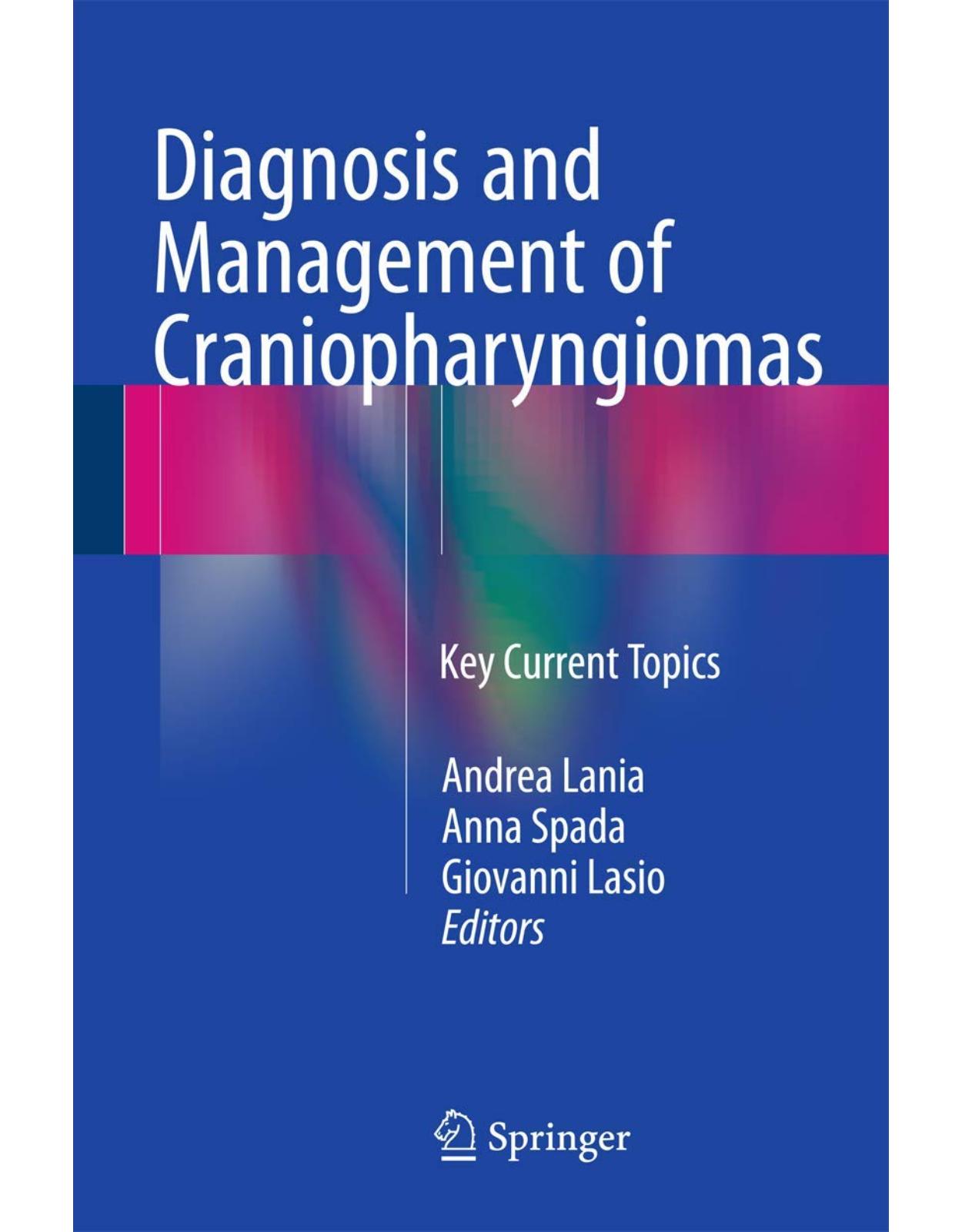
Diagnosis and Management of Craniopharyngiomas
Livrare gratis la comenzi peste 500 RON. Pentru celelalte comenzi livrarea este 20 RON.
Description:
This book provides up-to-date coverage of the most relevant topics in the diagnosis and management of craniopharyngiomas. After introductory discussion of natural history and clinical presentation, individual chapters are devoted to pathological and molecular aspects, use of diagnostic imaging techniques, the surgical approach to craniopharyngiomas, radiotherapy and radiosurgery, and associated endocrine disturbances. A particular feature of the book is the detailed attention devoted to the metabolic consequences of the disease and related treatments, including obesity and electrolyte disturbances, and to cognitive alterations. This book will be of value to oncologists, neurosurgeons, and endocrinologists by assisting in diagnostic workup, delivery of appropriate treatment, and management of the serious metabolic and endocrine consequences.
Table of Contents:
1: Craniopharyngiomas: Natural History and Clinical Presentation
1.1 Introduction
1.2 History
1.3 Epidemiology
1.4 Presentation (Clinical and Hormonal Manifestations)
References
2: Craniopharyngioma: Pathological and Molecular Aspects
2.1 An Historical Note
2.2 Adamantinous Craniopharyngioma
2.2.1 Macroscopic Features
2.2.2 Microscopic Features
2.2.3 Electron Microscopy
2.3 Papillary Craniopharyngioma
2.3.1 Macroscopic Features
2.3.2 Microscopic Features
2.3.3 Electron Microscopy
2.4 The Immunoprofile of Craniopharyngioma
2.5 Can Pathology Predict Recurrence?
2.5.1 Invasion in Craniopharyngioma and Its Implications
2.6 Ectopic Locations
2.7 Metastatic Craniopharyngioma
2.8 Malignancy in Craniopharyngioma
2.9 Differential Diagnosis: Lesions Mimicking Craniopharyngioma
2.10 Craniopharyngioma Can Occur in Association with Other Sellar Lesions
2.11 Molecular Aspects and Pathogenesis
2.11.1 Molecular Aetiology of Adamantinous Craniopharyngioma
2.11.2 Stem Cells Play a Critical Role in ACP Tumorigenesis
2.11.3 The EGFR and SHH Pathways Are Upregulated in Mouse and Human ACP
2.11.4 Molecular Aetiology of Papillary Craniopharyngioma
References
3: Neuroradiological Diagnosis of Craniopharyngiomas
3.1 Introduction
3.2 Imaging Technique
3.3 General Features
3.4 Adamantinous Subtype
3.5 Papillary Subtype
3.6 Differential Diagnosis
References
4: Transsphenoidal Approaches to Craniopharyngiomas
4.1 Introduction
4.2 Transsphenoidal Approaches
4.2.1 Microscopic Transsphenoidal Approaches
4.2.1.1 Sublabial
4.2.1.2 Endonasal
4.2.2 Transsphenoidal Transsellar Transdiaphragmatic Approach
4.2.3 Transsphenoidal Transtuberculum Sellae Approach
4.2.3.1 Cystic Suprasellar Craniopharyngiomas
4.2.3.2 Endoscopic Endonasal
4.2.4 Anterior Skull Base Endoscopic Approach
4.2.5 Combined Approaches
4.3 Endonasal Transsphenoidal Operative Technique
4.3.1 Preoperative Phase
4.3.2 Operating Room Layout and Patient Positioning
4.3.3 Surgical Procedure
4.3.3.1 Nasal Phase (Single Surgeon/Two Hands)
4.3.3.2 Sphenoidal Phase (Two Surgeons, Three or Four Hands)
4.3.3.3 Sellar Phase
4.3.4 Closure
4.4 Adjuvant Therapy
4.4.1 Intracavitary Radioisotopes
4.4.2 Intracavitary Chemotherapy
4.4.3 Interferon Therapy
4.5 Outcomes
Conclusion
References
5: Surgical Approach to Craniopharyngiomas: Transcranial Routes
5.1 Introduction
5.2 Anatomic Consideration for Surgical Planning
5.3 Surgical Indications
5.4 Surgical Anatomy
5.5 Technique
5.5.1 FOZ
5.5.2 CISTA
5.6 Clinical Series
5.6.1 Patient Population
5.6.2 Early Postoperative Results
5.6.3 Tumor Recurrence
5.6.4 Further Treatments and Disease Status at Last Follow-Up
References
6: Radiotherapy and Radiosurgery for Craniopharyngiomas
6.1 Introduction
6.2 Radiotherapy and Radiosurgery
6.2.1 Fractionated Stereotactic Radiotherapy
6.2.2 Radiosurgery
Conclusions
References
7: Endocrine Consequences: Diagnostic Workout and Treatment
7.1 GH Deficiency
7.1.1 Diagnosis
7.1.2 Treatment
7.2 Central Hypogonadism
7.2.1 Diagnosis
7.2.2 Treatment
7.2.2.1 Prepubertal Patients
Adults Patients
7.3 Central Hypoadrenalism
7.3.1 Diagnosis
7.3.2 Treatment
7.4 Central Hypothyroidism
7.4.1 Diagnosis
7.4.2 Treatment
References
8: Metabolic Consequences: Obesity and Energy Expenditure, Can They Be Treated?
8.1 Introduction
8.2 Lifestyle Therapy
8.3 Pharmacotherapy
8.4 Surgery
Conclusions
References
9: Metabolic Consequences: Electrolyte Disturbances
9.1 Introduction
9.2 CP and Presurgical Electrolyte Alterations
9.3 Postsurgical Electrolyte Alterations
9.4 Radiotherapy and Electrolyte Alterations
9.5 Diagnosis
9.6 Treatment
Conclusions
References
| An aparitie | 2016 |
| Autor | Lania |
| Dimensiuni | 15.6 x 1.45 x 24.21 cm |
| Editura | Springer |
| Format | Hardcover |
| ISBN | 9783319222967 |
| Limba | Engleza |
| Nr pag | 161 |






Clientii ebookshop.ro nu au adaugat inca opinii pentru acest produs. Fii primul care adauga o parere, folosind formularul de mai jos.