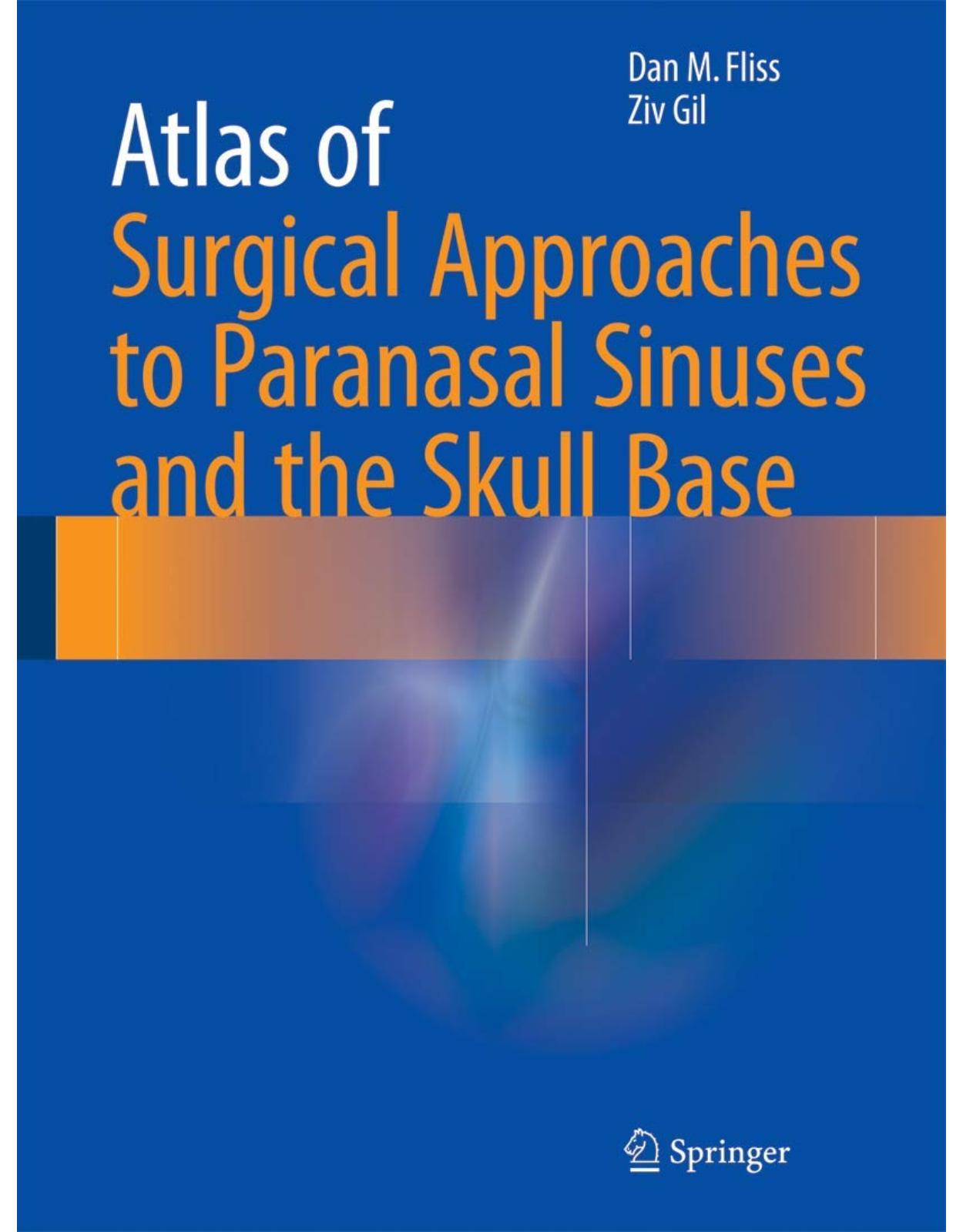
Atlas of Surgical Approaches to Paranasal Sinuses and the Skull Base
Livrare gratis la comenzi peste 500 RON. Pentru celelalte comenzi livrarea este 20 RON.
Description:
This richly illustrated atlas, compiled by authors with extensive experience in the field, offers a step-by-step guide to the surgical treatment of tumors, and congenital diseases of the skull base and nasal sinuses. Particular attention is devoted to the various techniques employed for extirpation of tumors and reconstruction of the skull base and Paranasal Sinuses. In order to facilitate understanding of the different approaches, clear surgical illustrations are presented alongside the high-quality intraoperative photographs. Whenever appropriate, technical tips are provided and briefly discussed. This atlas will appeal to a broad audience of residents, fellows, and consultants in different fields of medicine, including surgeons (head and neck, neurosurgery, otolaryngology, plastic and reconstructive surgery, ophthalmology, maxillofacial surgery) and oncologists.
Table of Contents:
1: The Cranial Base
1.1 Overview
1.2 Anterior Cranial Base
1.2.1 Endocranial Surface
1.2.2 Exocranial Surface
1.2.3 Endoscopic Endonasal Anatomy
1.3 Middle Cranial Base
1.3.1 Endocranial Surface
1.3.2 Exocranial Surface
1.3.3 Endoscopic Endonasal Anatomy
1.3.3.1 Sellar and Parasellar Areas
1.3.3.2 Cavernous Sinus and Meckel Cave
1.3.3.3 Infratemporal Fossa and Parapharyngeal Space
1.4 Posterior Cranial Base
1.4.1 Endocranial Surface
1.4.2 Exocranial Surface
1.4.3 Endoscopic Endonasal Anatomy
1.4.3.1 Clivus
1.4.3.2 Foramen Magnum and C1 and C2
1.5 Discussion
Selected Reading
2: Anterior Skull Base: Imaging
2.1 Imaging Modalities
2.1.1 Imaging Approach and Protocols
2.1.2 Computed Tomography
2.1.3 Magnetic Resonance Imaging
2.1.4 Cross-Sectional Angiographic Imaging
2.1.5 Positron Emission Tomography
2.2 Imaging for Initial Diagnosis and Staging
2.2.1 CT Scans
2.2.2 MR Imaging
2.2.3 PET/CT
2.2.4 MRI (Standard Imaging)
2.3 Dural, Perineural, and Venous Sinus Invasion by Skull Base Tumors
2.3.1 Dural Invasion
2.3.2 Direct Extension and Distant Metastasis
2.4 Pathology
2.4.1 Infection and Inflammation
2.4.2 Tumors: Sinonasal Masses
2.4.3 Meningiomas
2.4.4 Primary Osseous Tumors
2.4.5 Imaging During Follow-Up
Suggested Reading
3: Operating Room Setup in Skull Base Surgery
3.1 Introduction
3.2 Preoperative Evaluation and Anesthesia
3.3 Patient Positioning
3.3.1 Open Approaches
3.3.2 Endoscopic Approaches
4: Open Surgical Approaches to the Paranasal Sinuses
4.1 Preoperative Evaluation and Anesthesia
4.2 Surgical Technique
4.2.1 Transfacial Approaches
4.2.1.1 Lateral Rhinotomy Incision
4.2.1.2 Dieffenbach and Subciliary Incisions
4.2.1.3 Combined Lateral Rhinotomy and Dieffenbach Incisions
4.2.1.4 Weber-Ferguson Incision
4.2.1.5 Lynch Incision
4.2.2 Transfacial Subtotal Maxillectomy
4.2.3 Midfacial Degloving Approach
4.2.4 Suprastructure Maxillectomy
4.2.5 Suprastructure Maxillectomy with Orbital Exenteration
4.2.6 Total Maxillectomy and Orbital Exenteration
4.2.7 Combined Total Maxillectomy with Infratemporal Fossa Approach
4.2.8 Maxillary Swing Approach
4.2.9 Le Fort I Down-Fracture Osteotomy
4.3 Reconstruction
4.4 Postoperative Treatment
4.5 Highlights: Open Surgical Approaches to the Paranasal Sinuses
Suggested Reading
5: The Subcranial Approach to the Anterior Skull Base
5.1 Preoperative Evaluation and Anesthesia
5.1.1 Operative Room Setting
5.2 Surgical Technique
5.3 Reconstruction Following Subcranial Surgery
5.4 Postoperative Treatment
5.5 Highlights
Suggested Reading
6: Surgical Approaches to the Lateral Skull Base
6.1 Introduction
6.2 Preoperative Evaluation and Anesthesia
6.2.1 Physical Examination
6.2.2 Indications for Surgery
6.2.3 Contraindications
6.2.4 Imaging Studies
6.2.5 Tissue Diagnosis
6.3 Surgical Technique
6.3.1 Operating Room Setting
6.3.2 Skin Incision
6.3.3 Vascular Control and Identification of Cranial Nerves
6.3.4 Pterional Approach
6.3.5 Infratemporal Fossa Approach
6.3.6 Orbitozygomatic Approach
6.4 Postoperative Treatment
6.5 Highlights
Suggested Reading
7: Approaches to the Parapharyngeal Space
7.1 Introduction
7.2 Preoperative Evaluation and Anesthesia
7.2.1 Imaging Studies
7.2.2 Tissue Diagnosis
7.3 Surgical Technique
7.3.1 The Transcervical Approach for Resection of PPS Tumor
7.3.2 The Transparotid Approach for Resection of PPS Tumor
7.3.3 The Transcervical-Transparotid Approach
7.3.4 The Transmandibular Approach
7.3.5 Transoral Robotic Surgery
7.3.6 Transcervical Endoscopic
7.4 Postoperative Treatment
7.5 Highlights
Suggested Reading
8: The Translabyrinthine Approach to the Internal Auditory Canal and Cerebellopontine Angle
8.1 Introduction
8.2 Indications for the Procedure
8.2.1 Vestibular Schwannomas: Epidemiology, Presentation, and Diagnosis
8.2.2 The Place of the TL Approach in VS Treatment
8.3 Surgical Technique: Translabyrinthine Approach for Resection of Vestibular Schwannoma
8.3.1 Perioperative Preparation in the Operating Room
8.3.2 Soft-Tissue Incisions and Flaps
8.3.3 A Wide Mastoidectomy (Canal Wall-Up or Canal Wall-Down)
8.3.4 Removing All Bone over the Posterior and Middle Fossa Dura
8.3.5 Systematic Labyrinthectomy
8.3.6 Removing the Bone Surrounding the IAC
8.3.7 Opening the IAC and CPA Dura
8.3.8 Tumor Removal
8.3.9 Obliterating the Surgical Defect
8.3.10 Closure
8.4 Postoperative Care
8.5 Complications
8.5.1 Facial Nerve Dysfunction
8.5.2 Cerebrospinal Fluid Leakage
8.5.3 Meningitis
8.5.4 Intracranial Hemorrhage
8.5.5 Headaches
8.6 Outcome and Follow-Up
8.7 Highlights
References
9: Cancer of the Temporal Bone
9.1 Introduction
9.2 Evaluation
9.3 Staging
9.4 Treatment
9.4.1 Surgery
9.4.1.1 Lateral Temporal Bone Resection
9.4.2 Reconstruction
9.4.3 Radiation Therapy
9.4.4 Hearing Loss
9.5 Outcome
Conclusion
9.6 Highlights
References
10: Endoscopic Approaches in Skull Base Surgery and Their Indications
10.1 Introduction
10.1.1 The Inferior and Superior Limits of the Endonasal Approach
10.2 Surgical Technique
10.2.1 The Expanded Endoscopic Approach
10.2.2 Approach to the Sphenoid Sinus and Planum
10.2.2.1 Surgery of the Sella and Posterior Sphenoid Sinus Wall
10.2.2.2 Sellar and Suprasellar Craniotomy
10.2.3 The Transplanum, Perichiasmatic Approach
10.2.4 The Endoscopic Transnasal Transcribriform Approach
10.2.5 The Endoscopic Transmaxillary/Retromaxillary Approach
10.2.6 The Endoscopic Transclival Approach
10.2.6.1 Identification of the Internal Carotid Artery at the Paraclival Area
10.2.6.2 Approaches to the Clivus and Cervical Spine
10.2.7 Endonasal Reconstruction of the Skull Base
10.2.7.1 The Nasal Septal Flap
10.2.7.2 The Inferior Turbinate Flap
10.2.7.3 Pericranial and Temporoparietal Fascia Flaps
References
11: Combined Approaches to Complex Skull Base Tumors
11.1 Introduction
11.2 Surgical Technique
11.2.1 Combined Transorbital–Transcervical–Infratemporal Fossa Approach
11.2.2 Combined Transfascial–Transorbital–Pterional Approach
11.2.3 Combined Transmandibular–Infratemporal Fossa Approach
11.2.4 Combined ITF–Transcervical Approach
11.2.5 Combined Temporal Bone Resection and Far Lateral Approach
11.3 Reconstruction
11.3.1 Postoperative Treatment
11.4 Special Considerations for Combined Approaches
Suggested Reading
12: Microsurgical Reconstruction of Skull Base Defects
12.1 Introduction
12.2 Anatomical Considerations
12.3 Reconstruction Method
12.3.1 Analysis of the Defect and Considerations
12.3.2 Flap Variations
12.3.2.1 Anterolateral Thigh Flap
12.3.2.2 Radial Forearm Free Flap
12.3.2.3 Rectus Abdominis Myocutaneous Free Flap
12.3.2.4 Additional Flaps
12.3.3 Recipient Vessels
12.4 Postoperative Protocol and Flap Monitoring
12.5 Complications
12.5.1 Surgical Site Complications
12.5.2 Donor-Site Complications
12.5.3 Medical Complications
12.6 Specific Considerations for Microsurgical Reconstruction of Skull Base Defects
12.6.1 Anterior Cranial Fossa Reconstruction
12.6.2 Middle Cranial Fossa Defect Reconstruction
12.6.3 Posterior Cranial Fossa Defects
Conclusions
Suggested Reading
13: Complications After Skull Base Surgery
13.1 Introduction
13.2 Risk of Complications after Anterior Skull Base Tumor Resection
13.3 Specific Complications
13.4 How to Avoid Complications
13.5 Highlights
Suggested Reading
| An aparitie | 2016 |
| Autor | Fliss |
| Dimensiuni | 21.23 x 2.26 x 28.7 cm |
| Editura | Springer |
| Format | Hardcover |
| ISBN | 9783662486306 |
| Limba | Engleza |
| Nr pag | 319 |

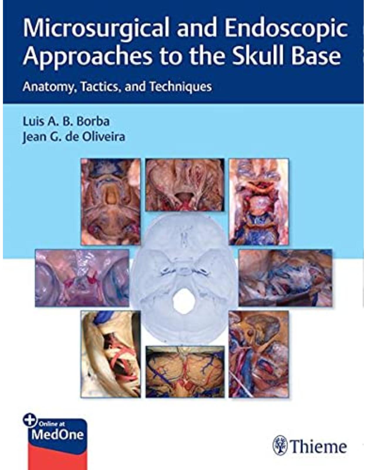

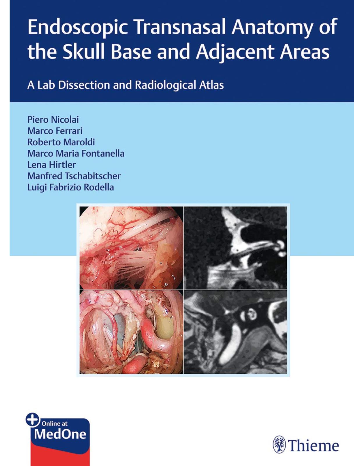


Clientii ebookshop.ro nu au adaugat inca opinii pentru acest produs. Fii primul care adauga o parere, folosind formularul de mai jos.