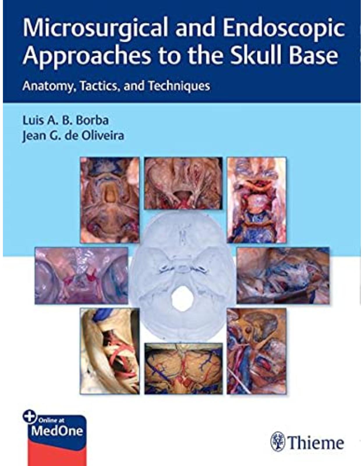
Microsurgical and Endoscopic Approaches to the Skull Base: Anatomy, Tactics, and Techniques
Livrare gratis la comenzi peste 500 RON. Pentru celelalte comenzi livrarea este 20 RON.
Disponibilitate: La comanda in aproximativ 4-6 saptamani
Autor: Jean de Oliveira
Editura: Thieme
Limba: Engleza
Nr. pagini: 550
Coperta: Hardcover
Dimensiuni: 216 x 279 mm
An aparitie: 30 Aug. 2021
Description:
The one-stop guide to microsurgical and endoscopic treatment of skull base lesions from global experts
A deep knowledge of regional anatomy, improved understanding of pathologies and their behaviors, technological advances, and multidisciplinary collaboration have led to more effective treatments for once inoperable skull base lesions. Microsurgical and Endoscopic Approaches to the Skull Base: Anatomy, Tactics, and Techniques by renowned skull base neurosurgeons Luis A. B. Borba and Jean G. de Oliveira presents a balanced, anatomy-based perspective on microsurgical and endoscopic approaches to manage these highly challenging lesions. The text leverages the best current scientific literature on this topic and insights from global skull base surgery experts.
Organized into 9 sections and 52 chapters, the book starts with discussion of microsurgical and endoscopic instrumentation and neurophysiological monitoring. The subsequent sections cover diverse approaches for skull base lesions involving the sphenoid and parasellar, orbit, anterior fossa, cavernous sinus, temporal bone and jugular foramen, and foramen magnum regions. Each of these sections starts with an introduction, followed by a microsurgical description of the anatomy of the impacted region.
Key Highlights
Contributions from an impressive group of internationally renowned neurosurgeons and otolaryngologists specializing in skull base pathologies
Indications, preoperative and postoperative concerns, nuances, pitfalls, tactics, techniques, and references for further reading provide a comprehensive guide to treatment
A stepwise description of the approach, high-quality four-color drawings, and illustrative cases facilitate acquisition and retention of knowledge
High-quality figures provide greater visual insights and step-by-step guidance on how to perform specific procedures
This unique textbook will help residents, fellows, and practitioners in neurosurgery and otolaryngology make an evidenced-based decision on using the most effective microsurgical and/or endoscopic approach to achieve the best outcomes in patients with skull base lesions.
Table of Contents:
Section I: Instrumentals and Equipment
1 Instrumentation for Endoscopic Skull Base Surgery
1.1 Introduction
1.2 Special Requirements for Endoscopic Approaches
1.3 Patient Positioning and Operating Room Setup
1.3.1 Positioning
1.3.2 Operating Room Setup
1.4 Endoscopes and Video Systems
1.4.1 Types of Endoscopes: Function and Management
1.4.2 Irrigation Sheaths and Pumps
1.4.3 Pneumatic Holding Arms
1.4.4 Light Sources
1.4.5 High-Definition Digital Cameras and Monitors
1.5 Surgical Instruments
1.5.1 Microdrills Handpieces and Burrs
1.5.2 Microinstruments, Dissectors, and Micro-Ultrasonic Aspirator (microCUSA)
1.6 Neuromonitoring and Neuronavigation in Endoscopic Skull Base Surgery
1.7 Conclusion
References
2 Neurosurgical Instrumentation
2.1 Operating Room
2.2 Microscope
2.3 Neuroendoscopy
2.4 Patient Positioning
2.5 Instruments
2.5.1 Bipolar Coagulator
2.5.2 Scissors
2.5.3 Dissectors
2.5.4 Needle, Suture, and Needle Holder
2.5.5 Brain Retractor
2.5.6 Suction
2.5.7 Drills
2.5.8 Tissue Forceps
2.5.9 Aneurysm and Vascular Clips
References
3 Neurophysiological Monitoring in Skull Base Surgery
3.1 Introduction
3.2 Electromyography (EMG)
3.2.1 Background
3.2.2 Cranial Nerves III, IV, and VI
3.2.3 Cranial Nerve V
3.2.4 Cranial Nerve VII
3.2.5 Cranial Nerves IX, X, and XII
3.2.6 Cranial Nerve XI
3.3 Brainstem Auditory Evoked Potential (BAEP)
3.4 Visual Evoked Potential (VEP)
3.5 Cases
3.5.1 Case 1
3.5.2 Case 2
3.5.3 Case 3
3.6 Conclusion
References
Section II: Anterior Approaches Transnasal and Transoral
4 Anatomical Landmarks of Anterior Approaches: An Endoscopic View
4.1 Introduction
4.2 Nasal Cavity
4.3 Anatomy of the Transshpenoidal, Transtuberculum, and Transplanum Approaches
4.4 Anatomy of the Transethmoidal Transcribriform Approach
4.5 Anatomy of the Transclival and Craniovertebral Junction Approaches
4.6 Anatomy of the Transpterygoid and Infratemporal Approaches
References
5 Direct Microscopic Transsphenoidal Surgery with Hybrid Use of Endoscopy
5.1 Introduction
5.2 Direct Endonasal Microscopic TSS with Hybrid Use of Endoscopy
5.2.1 Patient Preparation
5.2.2 Endonasal Stage I
5.2.3 Sphenoid Sinus Stage II
5.2.4 Intrasellar Stage III
5.2.5 Suprasellar Stage IV
5.2.6 Hybrid Use of Endoscopy
5.2.7 Closure
5.3 Conclusion
References
6 The Extended Transsphenoidal Approach (Mid-maxillotomy) to the Clivus
6.1 Introduction
6.2 Anatomical Background
6.3 Indications
6.4 Surgical Technique
6.5 Patient Positioning
6.6 Incision and Soft Tissues Dissection
6.7 Osteotomy
6.8 Nasal Dissection and Clivus Stage
6.9 Closure
References
7 The Microsurgical Transoral Approach
7.1 Introduction
7.2 Anatomy of the Craniovertebral Junction
7.3 Indications
7.4 Preoperative Evaluation
7.5 Operative Technique
7.6 Endoscopic Endonasal Approach
7.7 Postoperative Care and Complications
7.8 Conclusion
References
8 Elongated and Pediculated Pericranial Flap for Endonasal Reconstruction of the Entire Ventral Skull Base
8.1 Background
8.2 Introduction
8.3 Objective
8.4 Methods
8.5 Surgical Technique
8.6 Results
8.7 Discussion
8.8 Conclusion
References
9 Expanded Endoscopic Endonasal Approaches to the Sellar and Parasellar Regions
9.1 Introduction
9.2 Endoscopic Anatomy of the Sellar and Parasellar Regions
9.2.1 Anterior Skull Base
9.2.2 Sellar and Parasellar Regions
9.2.3 Clival Region and Posterior Skull Base
9.3 Surgical Technique, Technical Nuances, and Complications
9.3.1 Trans-sellar Approach
9.3.2 Transtuberculum Transplanum
9.3.3 Trans-cavernous and Trans-clival (Superior Third)
9.4 Illustrative Cases
9.4.1 Case 1
9.4.2 Case 2
9.5 Conclusion
References
10 The Endoscopic Transpterygoid Approach to the Parasellar and Infratemporal Fossa
10.1 Introduction
10.2 Anatomical Landmarks and Surgical Strategies
10.2.1 Zone 1: Medial Petrous Apex (Infrapetrous)
10.2.2 Zone 2: Petroclival Approaches (Infrapetrous)
10.2.3 Zone 3: Inferior Cavernous Sinus and Quadrangular Space (Suprapetrous)
10.2.4 Zone 4: Superior Portion of the Cavernous Sinus (Suprapetrous)
10.2.5 Zone 5: Transpterygoid Infratemporal Approach (Suprapetrous)
10.3 Limits of the Approach
10.4 Conclusion
References
11 Endoscopic Endonasal Transclival Approach to the Posterior Fossa
11.1 Introduction
11.1.1 Extradural Lesions
11.1.2 Intradural Lesions
11.2 Anatomy
11.3 Tactics and Techniques
11.3.1 Imaging
11.3.2 Technical Approach
11.3.3 Skull Base Reconstruction
11.4 Conclusions
References
Section III: Surgical Approaches to the Orbit
12 Microsurgical Anatomy of the Orbit
12.1 Historical Account
12.2 The Rule of Seven of the Orbit
12.3 Orbital Bones
12.4 Orbital Muscles and Vessels
12.5 Orbital Nerves
12.6 Topographical Relationships and Orbital Surgical Approaches
12.7 Conclusion
References
13 Lateral Orbitotomy Approach to the Orbit and Cavernous Sinus
13.1 Historical Background
13.2 Relevant Surgical Anatomy
13.3 Indications
13.4 Surgical Technique
13.5 Advantages and Disadvantages
13.6 Surgical Outcome and Complications
13.7 Illustrative Cases
13.7.1 Case 1: Cavernous Sinus Lesion Biopsy
13.7.2 Case 2: SOF Lesion Removal
13.7.3 Case 3: Temporal Pole Tumor Removal
13.8 Conclusion
References
14 Transcranial Approaches to the Orbit
14.1 Historical Background
14.2 Relevant Surgical Anatomy of the Orbit
14.2.1 Structure of the Orbit
14.2.2 Nerves of the Orbit
14.2.3 Blood Supply to the Orbit
14.2.4 Muscles of the Orbit
14.2.5 Lacrimal Gland
14.3 Indications for Transcranial Approaches to the Orbit
14.4 Surgical Techniques
14.4.1 Preoperative Preparation
14.4.2 The Subfrontal Route
14.4.3 The Supraorbital Route
14.4.4 The Frontotemporal Approach
14.4.5 The Superolateral Approach
14.5 General Considerations to Avoid Postoperative Complications
14.5.1 Nasal Sinuses
14.5.2 Orbital Fat
14.5.3 Brain Retraction
14.5.4 Reconstruction of OrbitalWalls
14.6 Illustrative Cases
14.6.1 Case 1: Cavernous Hemangioma of the Superomedial Orbital Quadrant
14.6.2 Case 2: Recurrent Pleomorphic Adenoma of the Lacrimal Gland with Intracranial Extension
14.6.3 Case 3: Meningioma of the Lateral Orbit
14.7 Conclusion
References
15 The Medial Endoscopic Approach to the Orbit
15.1 Introduction
15.2 Materials and Methods
15.3 Surgical Technique
15.4 Illustrative Cases
15.4.1 Case 1
15.4.2 Case 2
15.5 Discussion
15.6 Conclusion
References
Section IV: Anterior Approaches to the Skull Base
16 Microsurgical Anatomy of the Anterior Skull Base Through a Cranial View
16.1 Bony Anatomy
16.2 Neurovascular Relationships
16.3 Orbit
References
17 Microsurgical Anatomy of the Anterior Skull Base as Seen Through the Endonasal
17.1 History
17.2 Guiding Principles in Ventral Skull Base Surgery
17.3 Microsurgical Anatomy of the Anterior Cranial Base
17.4 Endocranial Surface of the Anterior Cranial Base: Overview
17.5 Sinonasal Anatomy
17.5.1 The Outer Circumferential-Radial Corridor Overview
17.6 Anatomy from Frontal Sinus to Planum Sphenoidale (Transcribriform Corridor)
17.6.1 Outer Circumferential-Radial Corridor
17.6.2 Inner Circumferential-Radial Corridor (ICRC)
17.7 Anatomy from Planum to the Superior Edge of the Sella (Transtuberculum/Transplanum Corridor)
17.7.1 Outer Circumferential-Radial Corridor (OCRC)
17.7.2 Inner Circumferential-Radial Corridor (ICRC)
17.8 Anatomy of the Orbit (Transorbital Corridor)
17.8.1 Outer Circumferential-Radial Corridor (OCRC)
17.8.2 Inner Circumferential-Radial Corridor (ICRC)
17.9 Conclusion
18 Eyebrow Supraorbital Approach for Skull Base Lesions
18.1 History of Supraorbital Craniotomy
18.2 Relevant Surgical Anatomy
18.3 Anatomical Limitations and Surgical Applications
18.4 Surgical Technique
18.4.1 Positioning and Anesthesia
18.4.2 Skin Incision
18.4.3 Craniotomy
18.4.4 Intradural Dissection
18.4.5 Closure
18.5 Advantages and Disadvantages
18.6 Illustrative Cases
18.6.1 Case 1
18.6.2 Case 2
18.7 Conclusion
References
19 Transbasal/Transcranial Microsurgical Approaches to Anterior Fossa Meningiomas
19.1 Introduction
19.2 Incidence
19.3 Clinical Features
19.3.1 Tuberculum Sellae Meningiomas
19.3.2 Olfactory Groove Meningiomas
19.3.3 Anterior Fossa Floor Meningiomas
19.4 Diagnostic Imaging
19.4.1 Skull Radiography
19.4.2 Digital Subtraction Angiography of the Internal Carotid Artery
19.4.3 Computed Tomography (CT)
19.4.4 Magnetic Resonance Imaging (MRI)
19.5 Surgical Treatment
19.6 Surgical Approaches
19.7 Preoperative Assessment
19.8 Anesthesia
19.9 Surgical Technique for the Approach of Tuberculum Sellae Meningiomas
19.9.1 Positioning
19.9.2 Frontotemporal Craniotomy with Resection of the Supraorbital Rim
19.9.3 Lateral Fissure Splitting
19.9.4 Tumor Resection
19.9.5 Optic Nerves and Chiasm
19.9.6 Arterial Dissection
19.9.7 Pituitary Stalk
19.9.8 Invasion of the Optic Canal and the Cavernous Sinus
19.9.9 Tumor Implantation
19.9.10 Closure
19.10 Surgical Technique for the Approach to Olfactory Groove Meningiomas
19.10.1 Positioning
19.10.2 Bifrontal Craniotomy
19.10.3 Dura Mater Incision
19.10.4 Tumor Resection
19.10.5 Arterial Dissection
19.10.6 Optic Nerves and Chiasm
19.10.7 Tumor Implantation
19.10.8 Closure
19.10.9 Subcranial Approach
19.11 Surgical Technique for the Approach to Meningiomas of the Anterior Fossa Floor
19.12 Postoperative Care
19.13 Results
19.14 Visual Outcome
19.15 Complications
19.16 Damage to the Pituitary Stalk
19.17 Surgical Approach
19.18 Retraction of Brain Tissue
19.19 Damage to Vessels Close to the Tumor
19.20 Mortality
References
20 Orbitocranial Approach
20.1 Introduction
20.1.1 Historical Background
20.1.2 Indications
20.2 Surgical Technique
20.2.1 Position
20.2.2 Skin Incision
20.2.3 Scalp Elevation
20.2.4 Craniotomy
20.2.5 Removal of the Orbital Rim
20.2.6 Reconstruction
20.3 Cases
20.3.1 Case 1
20.3.2 Case 2
References
21 The Extended Endoscopic Endonasal Approach to the Anterior Fossa
21.1 Introduction
21.2 Surgical Approachs
21.2.1 Transplanum Transtuberculum Approach
21.2.2 Surgical technique – transplanum transtuberculum approach
21.2.3 The Transcribriform Approach
21.2.4 Surgical technique transcribriform approach
21.2.5 Reconstruction
21.3 Clinical Cases
21.4 Conclusions
References
Section V: Anterolateral Approaches
22 Microsurgical Anatomy of the Cavernous Sinus
22.1 Introduction
22.2 The Sinus
22.3 Osseous Relationships
22.4 Nerves
22.5 Ligaments
22.6 Internal Carotid Artery and Branches
22.7 Venous Compartments
22.8 Cavernous Sinus Triangles
22.8.1 Clinoidal Triangle
22.8.2 Oculomotor Triangle
22.8.3 Supratrochlear Triangle
22.8.4 Infratrochlear Triangle (Parkinson’s Triangle)
22.9 Middle Fossa and Paraclinoid Triangles
22.9.1 Anteromedial Triangle
22.9.2 Anterolateral Triangle
22.9.3 Posterolateral Triangle (Glasscock’s Triangle)
22.9.4 Posteromedial Triangle (Kawase’s Triangle)
22.9.5 Paraclival Triangles
References
23 Transcavernous Approach
23.1 Introduction
23.1.1 Anatomy of the Cavernous Sinus and Temporal Fossa
23.1.2 Triangles of the Cavernosus Sinus
23.1.3 Triangles of the Middle Fossa
23.2 The Pretemporal Craniotomy
23.2.1 Positioning
23.2.2 Trichotomy
23.2.3 Incision
23.2.4 The Temporal Muscle
23.3 Craniotomy
23.3.1 The Exposure of the LateralWall of the Cavernous Sinus
23.3.2 The Anterior Clinoid
23.3.3 The Meckel’s Cave
23.3.4 Anterior Petrosectomy
23.3.5 Posterior Clinoid
23.4 Discussion
References
24 Microsurgical Anatomy of the Middle Fossa and Petrous Apex
24.1 Background
24.2 Anatomy .
24.2.1 Endocranial Surface and Its Limits
24.2.2 The Foramens of the Middle Fossa
24.2.3 Petrous Apex
24.3 The Middle Fossa as a Surgical Corridor
24.4 Cases
24.4.1 Case 1
24.4.2 Case 2
24.4.3 Case 4
24.5 Conclusions
References
25 Pterional Approach
25.1 Introduction
25.2 Historical Landmarks
25.3 Indications
25.4 Step by step procedure
25.4.1 Positioning
25.4.2 Trichotomy
25.4.3 Skin Incision
25.4.4 Interfascial dissection
25.4.5 Temporal muscle dissection
25.4.6 Craniotomy
25.4.7 Sphenoid wing drilling
25.4.8 Dural opening
25.4.9 Neurosurgical routes
25.4.10 Dural closure
25.4.11 Cranioplasty
25.4.12 Muscle reconstruction
25.4.13 Variations
25.5 Conclusion
25.6 Illustrative Cases
References
26 Anterolateral Skull Base: Anatomy, Surgical Technique, and Nuances
26.1 Surgical Anatomy
26.1.1 Sagittal and Axial Unlocking: A Wider Exposure to Skull Base
26.2 Extended Skin Incision and Craniotomy
26.3 Orbitomeningeal Band Dissection
26.4 Pericavernous Dissection Techniques
26.5 Anterior Clinoidectomy
26.6 Posterior Clinoidectomy
26.7 Petrous Anatomy and Anterior Petrosectomy
26.8 Carotid Anatomy and Relations
References
27 Pretemporal Approach
27.1 Introduction
27.2 Advantages of the Pretemporal Approach
27.3 Anatomical Exposure and Surgical Access
27.4 Indications
27.5 Step-by-Step Procedure
27.5.1 Patient Positioning
27.5.2 Skin Incision
27.5.3 Preservation of the STA
27.5.4 Facial Nerve Branches Preservation
27.5.5 Mobilization of the Temporal Muscle
27.5.6 Craniotomy
27.5.7 Variations and Alternative Routes
27.6 Microsurgical Access to the Interpeduncular Fossa Region
27.7 Conclusion
27.8 Illustrative Cases
References
28 Cranio-Orbit-Zygomatic (COZ) Approach
28.1 Background
28.2 Indications
28.3 Technique
28.3.1 Positioning
28.3.2 Skin Incision and Pericranial Flap
28.3.3 Subfascial Dissection
28.3.4 Zygomatic Osteotomy
28.3.5 Mobilizing the Temporal Muscle
28.4 Craniotomy
28.4.1 Cranial Flap
28.4.2 Orbital Osteotomy
28.5 Closure
28.6 Limitations
28.7 Complications and Avoidance
28.8 Illustrative Cases
28.9 Conclusions
References
29 Anterior Petrosal Approach
29.1 Introduction
29.1.1 Anatomical Background
29.1.2 Indications
29.2 Surgical Technique
29.2.1 Patient Positioning
29.2.2 Skin Incision and Soft Tissue Dissection
29.2.3 Zygomatic Osteotomy
29.2.4 Temporalis Muscle Reflection
29.2.5 Craniotomy
29.2.6 Middle Fossa Dissection
29.2.7 Closure
29.2.8 Complications
References
30 Endoscopic Approach to the Cavernous Sinus Area: Surgical Technique
30.1 Introduction
30.2 Technical Equipment and Surgical Supplies
30.3 Preoperative Preparation Stage
30.3.1 Setup of the Patient and the Surgeon
30.4 Surgical Technique
30.4.1 Stages of the Surgical Procedure
30.5 A Clinical Case
30.6 Perspectives
References
Section VI: Posterolateral Approaches
31 Temporal Bone Anatomy
31.1 Introduction
31.2 Surgical Anatomy
31.2.1 The Squamous Portion
31.2.2 The Tympanic and Styloid Portions
31.2.3 The Mastoid Portion
31.2.4 The Petrous Portion
31.3 Craniotopographic Relationships
Suggested Readings
32 Microsurgical Anatomy of the Jugular Foramen
32.1 Introduction
32.2 Anatomy
32.3 Bone and Osseous Relations
32.4 Meningeal Structure and Cranial Nerves
32.5 Vascular Structures Related to the Jugular Foramen
32.6 Muscular Relationships
32.7 Conclusions
References
33 Anatomy of the Cerebellopontine Angle: A Microscopic Perspective
33.1 A Brief Historical Review
33.2 A Brief Introduction to the Anatomy of the CPA: A Detailed Look
33.3 Surgical Anatomy of the CPA Region
33.3.1 Continent:Walls of the CPA
33.3.2 Content: The Neurovascular Complexes.
References
34 The Posterior Retrolabyrinthine Presigmoid Approach
34.1 Background
34.2 Indications
34.3 Surgical Technique
34.3.1 Patient Positioning
34.3.2 Soft TissueWork
34.3.3 BonyWork
34.3.4 Closure
34.4 Limitations
34.5 Complications and Avoidance
34.6 Illustrative Cases
34.7 Conclusions
References
35 Retrosigmoid Approach
35.1 Introduction
35.1.1 Anatomical Background
35.1.2 Indications
35.2 Surgical Technique
35.2.1 Patient Positioning
35.2.2 Skin Incision and Soft Tissue Dissection.
35.2.3 Craniotomy
35.2.4 Dural Opening and Intradural Steps
35.2.5 Closure
35.2.6 Complications
References
36 Endoscopic Assisted Approach to the Cerebellopontine Angle
36.1 History
36.2 CPA Anatomy
36.3 Advantages/Disadvantages
36.4 Indications
36.5 Vestibular Schwannoma
36.5.1 Vestibular Schwannoma Endoscopic Assisted Approach Technique
36.6 Trigeminal Schwannomas
36.6.1 Trigeminal Schwannoma Endoscopic Assisted Approach Technique
36.7 Epidermoid Tumors
36.8 Meningiomas
36.9 Other Tumors
References
37 Infratemporal Fossa Approach to the Jugular Foramen
37.1 Introduction
37.2 Anatomy
37.3 Technique and Tactics
37.3.1 Positioning
37.3.2 Incision and Superficial Dissection
37.3.3 Muscle and Cervical Region Dissection
37.3.4 Mastoidectomy and Craniotomy
37.3.5 Tactics
37.4 Closure
37.5 Conclusion
References
38 The Facial Reanimation: Multidisciplinary Approaches
38.1 Introduction
38.2 Different Facial Nerve Reconstruction Techniques
38.2.1 Direct Neurorrhaphy
38.2.2 Hypoglossal-Facial Nerve Neurorrhaphy
38.2.3 Alternatives to Classic Hypoglossal-Facial Neurorrhaphy
38.2.4 Masseteric-Facial Nerve Neurorrhaphy
38.2.5 Accessory-Facial Nerve Neurorrhaphy
38.3 Transfacial Anastomosis (Cross-Face Facial Nerve Anastomosis)
38.4 Neuromuscular Transplant
38.5 Conclusion
References
Section VII: Posterior and Lateral Approaches to the Craniocervical Junction
39 Microsurgical Anatomy of the Foramen Magnum
39.1 Brief Historical Review
39.2 Introduction
39.3 Surgical Anatomy of the FM Region
39.4 Extradural Stage
39.4.1 Nuchal Muscles and V3 Segment
39.5 Anatomy of the VA
39.5.1 Vertebral Venous Plexus
39.5.2 Nuchal Lines Method
39.6 Bone Structure and Articulations
39.6.1 Occipital Bone
39.6.2 Occipital Condyles
39.6.3 Atlanto-occipital Joints
39.6.4 Atlanto-axial Joints
39.7 Dural Stage
39.7.1 Dural Venous Sinuses
39.7.2 Surgical Tips
39.8 Intradural Stage
39.8.1 Neural Elements
39.8.2 Denticulate Ligament
39.8.3 Cerebellum
39.8.4 Nerves
39.8.5 Vascular Elements
References
40 Far-Lateral Approach and Its Variants
40.1 Background
40.2 Indications
40.3 Surgical Technique
40.3.1 Patient Positioning
40.3.2 Skin Incision
40.3.3 Intradural versus Extradural Approach
40.3.4 Soft Tissue Dissection
40.3.5 Bone Flap and Dura Opening
40.3.6 Condyle Removal: The Transcondylar Approach
40.3.7 Closure
40.4 Complications and Avoidance
40.5 Illustrative Cases
40.5.1 Case 1
40.5.2 Case 2
40.6 Conclusions
References
41 Surgical Treatment for Atlantoaxial Instability
41.1 Introduction
41.2 Atlantoaxial Dislocation
41.3 Basilar Invagination
41.4 Chiari 1 Malformation and Syringomyelia
41.5 Central or Axial Atlantoaxial Facetal Instability
41.6 Investigations
41.7 Surgery
41.7.1 Operative Technique for Lateral Mass Plate (or Rod) and Screw (Monoaxial or Polyaxial) Fixation (Goel and Laheri)
41.8 Complication Avoidance
41.9 Craniovertebral Realignment for Group A Basilar Invagination: Role of Interarticular Cages
41.10 Technique
41.11 Atlantoaxial Fixation for Group B Basilar Invagination
References
42 Endoscopic Approach to the Craniocervical Junction
42.1 Introduction
42.2 The Endoscopic Endonasal Approach
42.3 Surgical Technique and Anatomic Landmarks
42.4 The Endoscopic Transoral Approach
42.5 The Endoscopic Transcervical Approach
42.6 Surgical Technique
42.7 Approach Selection
42.7.1 Preoperative Imaging
42.8 Patient Selection
42.9 Limitations to the Endoscopic Approaches
42.10 Conclusion
References
Section VIII: Approaches to the Posterior Incisural Space
43 Anatomy of the Posterior Incisural Space: Microscopic Perspective
43.1 Introduction
43.2 Objective
43.3 Microsurgical Anatomy
43.4 Overview
43.4.1 The Posterior Incisural Space
43.4.2 The Inferior and Middle Surface of the Posterior Temporal Lobe and the Occipital Lobe
43.4.3 The Tentorium
44 Anatomy of the Posterior Incisural Space: Endoscopic Perspective
44.1 Introduction
44.2 History
44.3 Anatomy
44.4 Endoscopic-Assisted Microsurgery
44.5 Conclusion
References
45 The Occipito-polar Approach: Study of the Occipital lnterhemispheric Supratentorial Transtentorial Transfalcine Approach
45.1 Introduction
45.2 Patient Series, Anatomical Study, and Methods
45.2.1 Anatomical Study and Patient Series
45.2.2 Surgical Technique
45.3 Illustrative Surgical Cases
45.3.1 Case 1
45.3.2 Case 2
45.4 Evaluation of Surgical Outcomes of the Patients
45.5 Results
45.5.1 Anatomical Study of the Occipito-polar Approach
45.5.2 Surgical Cases
45.6 Discussion
45.7 Conclusions
45.8 Disclosures
References
46 Supracerebellar Infratentorial Approach: Microsurgical Perspective
46.1 Introduction
46.2 Preoperative Evaluation
46.3 Surgical Approach and Operative Techniques
46.4 Median SCIT Approach
46.5 Paramedian SCIT Approach
46.6 Lateral and Extreme Lateral SCIT Approach
46.7 Contralateral SCIT Approach
46.8 Intraoperative Techniques
46.9 Postoperative Management
References
47 Supracerebellar Infratentorial Approach: Endoscopic Perspective
47.1 Introduction
47.2 Evolution of a Surgical Approach
47.2.1 Supracerebellar Infratentorial Approach
47.3 Optimization of Gravity Retraction: Sitting Position
47.4 Use of the Endoscope in Neurosurgery
47.5 Nuances of the Surgical Technique
47.6 Sitting Position
47.7 Off-Midline, Minimally Invasive Craniotomy
47.8 Selection of Craniotomy Location
47.9 Representative Cases
47.9.1 Case 1
47.9.2 Case 2
47.9.3 Case 3
References
48 Supracerebellar Transtentorial Approach
48.1 Introduction
48.2 Historical Landmarks
48.3 Indications
48.4 Preoperative Assessment
48.5 Surgical Technique
48.5.1 Anesthesia and Intraoperative Neurophysiological Monitoring
48.5.2 Positioning
48.5.3 Skin Incision and Muscle Management.
48.5.4 Craniotomy
48.5.5 Dural Opening
48.5.6 Microsurgical Dissection
48.5.7 Tentorial Opening/Resection
48.5.8 Microsurgical Anatomy
48.5.9 Closure
48.6 Conclusion
48.7 Illustrative Cases
References
Section IX: Cerebrovascular Management for Skull Base Surgery
49 STA-MCA Bypass
49.1 Introduction
49.2 Historical Landmarks
49.3 Indications
49.4 Surgery
49.4.1 Preoperative Preparation
49.4.2 Anaesthesia and Monitoring
49.4.3 The Technique
49.4.4 Closure
49.4.5 Postoperative Care
49.4.6 Follow-up
49.5 Conclusion
References
50 High-Flow Cervical Carotid Artery to Middle Cerebral Artery Bypass
50.1 Introduction
50.2 Indications and Preoperative Management
50.3 Microsurgical Anatomy
50.4 Interposition Graft
50.5 Recipient Site
50.6 Donor Site
50.7 Technical Nuances
50.8 Bypass Patency and Intraoperative Troubleshooting
50.9 Conclusion
References
51 Intracranial–Intracranial Bypass for Treatment of Complex Vascular Lesions and Tumors
51.1 Introduction
51.2 Intracranial-Intracranial (IC-IC) Bypass
51.2.1 Single
51.2.2 With Graft Interposition
51.2.3 Combined
51.3 Preoperative Assessment Study
51.4 Surgical Procedure
51.5 Discussion
51.6 Conclusion
References
52 Endoscopic Endonasal Approaches for the Treatment of Vascular Lesions
52.1 Introduction
52.2 Endoscopic Anatomy during Treatment of Vascular Lesions
52.2.1 Vascular Anatomy
52.2.2 Neural Anatomy
52.2.3 Bony Anatomy
52.2.4 Dural Anatomy
52.3 Intraoperative Fluorescence and Imaging Tools in EEA for Vascular Lesions
52.3.1 Endoscopic Indocyanine Green (endoICG)
52.3.2 Sodium Fluorescein
52.3.3 Intraoperative 3D Angiography (3D-DSA)
52.4 Reconstructive Techniques in EEA
52.4.1 Nasal Phase
52.4.2 Sphenoidal Phase
52.4.3 Sealing/Packing Phase
52.5 Operative Nuances
52.6 Aneurysms
52.6.1 Anterior Circulation Aneurysms
52.6.2 Posterior Circulation Aneurysms
52.7 Complications
52.8 Cavernous Malformations
52.8.1 Technical Nuances
52.8.2 Surgical Technique
52.8.3 World Experience
52.9 Vascular Tumors of the Skull Base
52.9.1 Preoperative Evaluation
52.9.2 Intraoperative Technical Nuances
52.10 Conclusion
References
Index
Additional MedOne Access Information
| An aparitie | 30 Aug. 2021 |
| Autor | Jean de Oliveira |
| Dimensiuni | 216 x 279 mm |
| Editura | Thieme |
| Format | Hardcover |
| ISBN | 9781626239661 |
| Limba | Engleza |
| Nr pag | 550 |


Clientii ebookshop.ro nu au adaugat inca opinii pentru acest produs. Fii primul care adauga o parere, folosind formularul de mai jos.