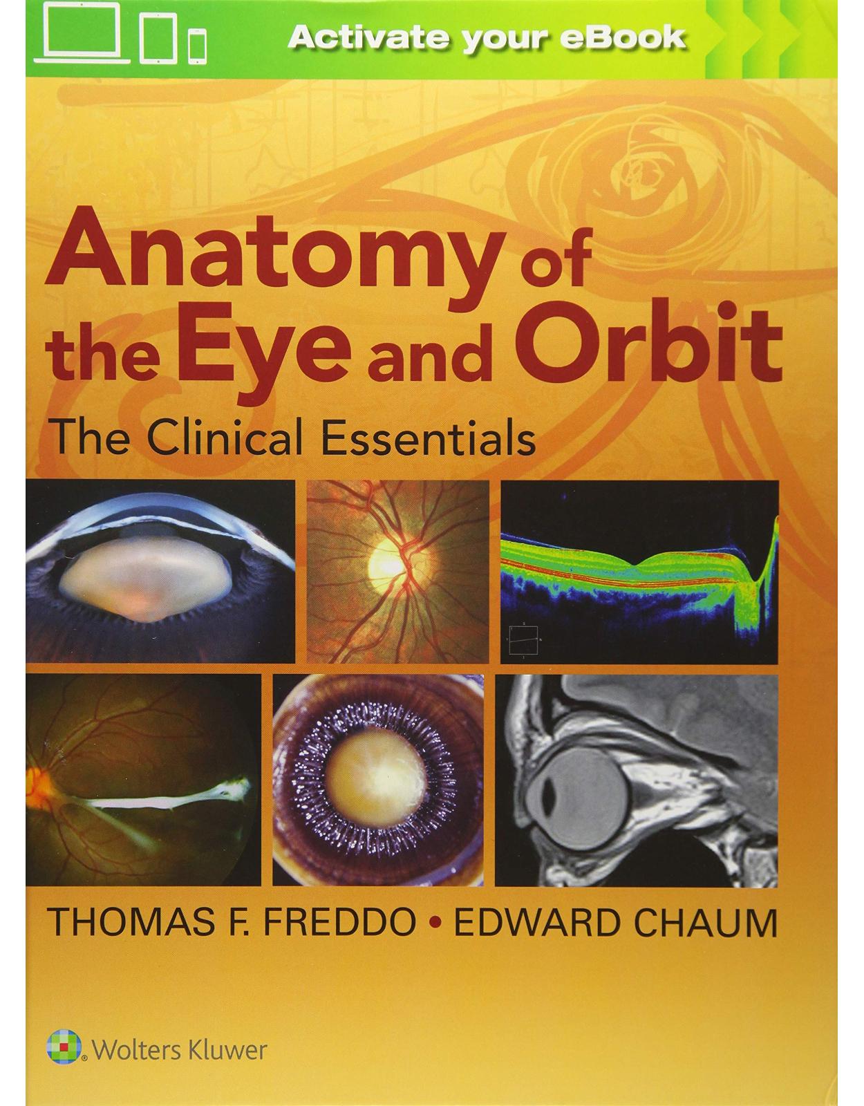
Anatomy of the Eye and Orbit: The Clinical Essentials
Livrare gratis la comenzi peste 500 RON. Pentru celelalte comenzi livrarea este 20 RON.
Disponibilitate: La comanda in aproximativ 4-6 saptamani
Editura: LWW
Limba: Engleza
Nr. pagini: 512
Coperta: Hardcover
Dimensiuni: 21.84 x 2.03 x 27.94 cm
An aparitie: 25 July 2017
Description:
Master the Clinical Essentials of ocular and orbital anatomy for clinical practice! The eye is an organ of great complexity. Anatomy of the Eye and Orbit: The Clinical Essentials achieves the impressive task of presenting all the ocular anatomy that ophthalmology residents, optometry residents, and optometry students need to know – in a single accessible, high-yield volume. It emphasizes the aspects of eye and orbit anatomy that are most relevant to clinicians in training, providing the practical, real-world foundation necessary for practice.
Table of Contents:
SECTION I: ORBITS, LIDS, AND ADNEXA
Chapter 1: The Orbits
Overview
Bones of the Orbit
Relationship between the Orbits and the Paranasal Sinuses
The Skeletal Muscles of the Orbit
Principal Fascial Compartments of the Orbit and Adnexa
The Muscle Pulley System Modulating Extraocular Muscle Movement
Neural Pathways within the Orbit
Cranial Nerves Entering the Orbit via the Optic Canal
Cranial Nerves Entering the Orbit via the Superior Orbital Fissure
Inferior Orbital Fissure
Pathway of Cranial Nerves through the Cavernous Sinus to the Superior Orbital Fissure
Pathways of the Ophthalmic and Maxillary Divisions of CN V and their Branches within the Orbit
Vascular Supply
Arterial Anatomy
Venous Anatomy
Lymphatic Anatomy
Lacrimal Gland
Acknowledgment
Chapter 2: The Eyelids and Adnexa
Overview
Surface Landmarks
Gross Anatomical Organization of the Lids
The Glands of the Eyelids
Vascular Supply
Innervation
Lymphatic Drainage
The Lacrimal Drainage System
SECTION 2: OVERVIEW OF THE EYE
Chapter 3: Overview of the Eye
Overview
Shape and Dimensions of the Eye
External Landmarks
Axes, Angles, and Sections of the Eye
Definition of Planes and Sections of the Eye
Layers of the Eye
Chambers of the Eye
Overview of the Ocular Vasculature
Venous Drainage of the Eye
Innervation
Lymphatic Drainage
Acknowledgment
SECTION 3: ANTERIOR SEGMENT OF THE EYE
Chapter 4: The Cornea
Overview
Macroscopic and Clinical Anatomy of the Cornea
Chapter 5: The Conjunctiva and the Limbus
Overview
Macroscopic and Clinical Anatomy of the Conjunctiva
Histology
Macroscopic and Clinical Anatomy of the Limbus
Acknowledgments
Chapter 6: The Sclera, Episclera, and Tenon’s Capsule
Overview
Macroscopic and Clinical Anatomy of the Sclera
Histology of the Sclera
Macroscopic and Clinical Anatomy of Tenon’s Capsule and the Episclera
Chapter 7: The Iris
Overview
Macroscopic and Clinical Anatomy of the Iris
Histology
Chapter 8: The Ciliary Body
Overview
Macroscopic and Clinical Anatomy of the Ciliary Body
Layers of the Ciliary Body
Histology of the Layers of the Ciliary Body
The Anatomic Basis of the Blood–Aqueous Barrier in the Ciliary Body
Chapter 9: Anatomy of the Aqueous Outflow Pathways
Overview
The Conventional or Trabecular Outflow Pathway
Anatomic Relationships between the Ciliary Muscle and the Structures of the Outflow Pathways
Anatomy of the Uveoscleral Outflow Pathway
Chapter 10: Crystalline Lens and Zonules
Overview
Macroscopic and Clinical Anatomy of the Crystalline Lens
Histology
Macroscopic and Clinical Anatomy of the Zonules
SECTION 4: POSTERIOR SEGMENT OF THE EYE
Chapter 11: The Vitreous
Overview
Development of the Vitreous
Macroscopic and Clinical Anatomy of the Vitreous
Histology and Ultrastructure of the Vitreous Gel
Physical Chemistry of the Vitreous
Degenerative Changes in the Vitreous Gel with Age
Chapter 12: Choroid and Choroidal Circulation
Overview
Macroscopic and Clinical Anatomy of the Choroid and Choroidal Vasculature
Histology of the Choroid
Innervation of the Choroid
Chapter 13: The Retinal Pigment Epithelium
Overview
Histology of the Retinal Pigment Epithelium
Polarized RPE Cell Anatomy and Function
Cytoplasmic Organelles and Inclusions
The Interphotoreceptor Matrix
Chapter 14: The Neurosensory Retina
Overview
Macroscopic and Clinical Anatomy of the Retina
Histology of the Retina
Introduction to the Layers of the Neurosensory Retina
Anatomical and Functional Details of Neurosensory Layers of the Retina
Histology of the Macula and Macula Lutea
Histology of the Fovea and Foveola
The Retinal Circulation
Histology of the Retinal Vessels
The Blood-Retinal Barrier
Chapter 15: The Optic Nerve and Visual Pathways
Overview
Macroscopic and Clinical Anatomy of the Optic Nerve
Histology of the Optic Nerve Head
Zones of the Optic Nerve Head
Blood Supply to the Optic Nerve
Retinotopic Organization of Axons at the Optic Nerve Head
Overview of Visual Pathways Distal to the Optic Nerves
The Lateral Geniculate Nucleus
Additional Neural Connections
The Optic Radiations and Occipital Cortex
Functional Correlates of Injuries to the Optic Nerve and Radiations
SECTION 5: EMBRYOLOGY OF THE EYE AND ORBIT
Chapter 16: Embryology of the Eye and Orbit
Overview
Early Embryogenesis
Early Development of Ocular Tissues from the Neuroectoderm
Early Development of Ocular Tissues from the Surface Ectoderm
Early Development of Ocular Tissues from the Neural Crest Cells
Early Development of Ocular and Periocular Tissues from Mesoderm
Summary of Development in the First Trimester
Maturation of the Eye after the Third Month
Genetic Programming of Eye Development
SECTION 6: OCULAR ANATOMY BY OPTICAL COHERENCE TOMOGRAPHY
Chapter 17: Ocular Anatomy by Optical Coherence Tomography
General Principles of Optical Coherence Tomography (OCT)
OCT Device Evolution
OCT Clinical Acumen and Sources of Misinterpretation
OCT Ocular Anatomy and Histology
New Frontiers in OCT Ocular Anatomy
Summary
Index
Coming soon: Serial MRI slices/images!
| An aparitie | 25 July 2017 |
| Autor | Thomas F Freddo, Edward Chaum |
| Dimensiuni | 21.84 x 2.03 x 27.94 cm |
| Editura | LWW |
| Format | Hardcover |
| ISBN | 9781469873282 |
| Limba | Engleza |
| Nr pag | 512 |

