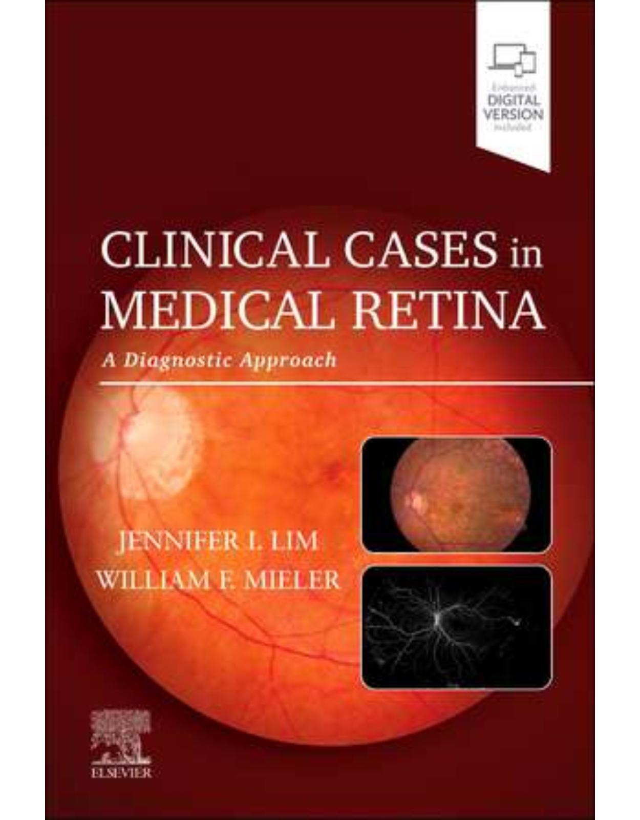
Clinical Cases in Medical Retina: A Diagnostic Approach
Livrare gratis la comenzi peste 500 RON. Pentru celelalte comenzi livrarea este 20 RON.
Disponibilitate: La comanda in aproximativ 4 saptamani
Editura: Elsevier
Limba: Engleza
Nr. pagini: 400
Coperta: Paperback
Dimensiuni: 152 x 229 mm
An aparitie: 28 iunie 2024
Medical retina is a complex subspecialty with a steep learning curve, requiring vast and diverse knowledge in basic science, diagnostic imaging, medical treatment, and surgical techniques. Clinical Cases in Medical Retina: A Diagnostic Approach provides highly visual, case-based guidance on the challenging process of gathering patient information, ordering appropriate testing, and arriving at an accurate diagnosis and effective treatment plan. In one convenient volume, it exposes retina fellows and specialists, ophthalmology residents, and other eye care clinicians to a wide variety of patient presentations and scenarios, including rare conditions and special populations.
Table of Contents:
Section 1. Hereditary Macular Conditions
1. Yellow macular spots in a child
History of present illness
Questions to ask
Assessment
Differential diagnosis
Working diagnosis
Multimodal testing and results
Management
Follow-up care
Algorithm 1.1: Algorithm for differential diagnosis for best disease
Key points
References
2. Long-standing photophobia, reduced visual acuity, myopia, and dyschromatopsia in a young adult male patient
History of present illness
Questions to ask
Assessment
Differential diagnosis
Working diagnosis
Multimodal testing and results
Management
Follow-up care
Algorithm 2.1: Algorithm for differential diagnosis for BED/BCM/X-linked cone dystrophy
Key points
References
3. Bilateral progressive severe loss of vision and obesity
History of present illness
Questions to ask
Assessment
Differential diagnosis
Working diagnosis
Multimodal testing and results
Management
Follow-up care
Algorithm 3.1: Algorithm showing differential diagnosis for retinopathy
Key points
References
4. Bilateral peripheral retinal and macular schisis in a young boy
History of present illness
Questions to ask
Assessment
Differential diagnosis
Workup and diagnosis
Multimodal testing and results
Management
Follow-up care
Key points
Algorithm 4.1: Diagnostic flow chart X-linked retinoschisis (XLR)
References
5. Congenital blindness and retinopathy in a young girl
History of present illness
Questions to ask to aid diagnosis
Assessment
Differential diagnosis
Working diagnosis
Multimodal testing and results
Management
Follow-up care
Algorithm 5.1: Algorithm for differential diagnosis of congenital blindness and retinopathy
Key points
References
6. Progressive nyctalopia and tunnel vision in a young man
History of present illness
Questions to ask to aid diagnosis
Assessment
Differential diagnosis
Working diagnosis
Multimodal testing and results
Management
Follow-up care
Surgical procedure for subretinal gene therapy
Algorithm 6.1 Algorithm of various inherited conditions causing nyctalopia with poor visual acuity in children
Key points
References
7. Rapid progression of vision loss in a child with pigmentary retinopathy
History of present illness
Questions to ask
Assessment
Differential diagnosis
Working diagnosis
Investigations
Follow-up care
Management
Follow-up care
Key points
Algorithm 7.1: Algorithm for differential diagnosis of a child with pigmentary retinopathy and progressive vision loss
References
8. Unilateral tractional retinal detachment in a 6-month-old female infant with erythematous skin lesions
History of present illness
Questions to ask
Assessment
Differential diagnosis
Working diagnosis
Multimodal testing and results
Management
Follow-up care
Algorithm 8.1: Algorithm for differential diagnosis for retinal nonperfusion in infants
Key points
References
9. Bilateral peripheral pigmentary changes in a woman
History of present illness
Questions to ask
Assessment
Differential diagnosis
Working diagnosis
Multimodal testing and results
Management
Follow-up care
Algorithm 9.1: Algorithm for bilateral peripheral pigmentary changes
Key points
References
10. I was never good at ghosts in the graveyard
History of present illness
Questions to ask
Assessment
Differential diagnosis
Working diagnosis
Genetic testing
Multimodal testing and results
Diagnosis
Enhanced S-cone syndrome
Management
Follow-up care
Algorithm 10.1: Algorithm showing diagnosis for enhanced s-cone syndrome
Key points
References
11. Bilateral perifoveal degeneration in a woman
History of present illness
Questions to ask
Assessment
Differential diagnosis
Working diagnosis
Multimodal testing and results
Management
Follow-up care
Algorithm 11.1: Algorithm for differential diagnosis for bilateral perifoveal degeneration
Key points
References
12. Autosomal dominant radial drusen
History of present illness
Questions to ask
Assessment
Differential diagnosis
Working diagnosis
Multimodal testing and results
Management
Follow-up care
Algorithm 12.1: Basic algorithm for macular dystrophies
Key points
References
Section 2. Degenerative/Deficiency
13. Bilateral asymptomatic pigmentary retinopathy
History of present illness
Questions to ask
Assessment
Differential diagnosis1
Working diagnosis
Multimodal testing and results
Management
Follow-up care
Algorithm 13.1: Algorithm for the differential diagnosis of pigmented paravenous retinochoroidal atrophy
Key points
References
14. Unilateral macular schisis with blurred vision in a woman
History of present illness
Questions to ask
Assessment
Differential diagnosis1
Working diagnosis
Multimodal testing and results
Management
Follow-up care
Algorithm 14.1: Algorithm representing myopic macular schisis
Key points
References
15. Acute vision loss in an elderly patient associated with unilateral intraretinal blot hemorrhage in the macula
History of present illness
Questions to ask
Assessment
Differential diagnosis
Working diagnosis
Multimodal imaging
Management
Follow-up care
Algorithm 15.1: Algorithm for differential diagnosis of unilateral intraretinal macular blot hemorrhage with cystoid macular edema
Key points
References
16. Bilateral macular and peripheral drusen in a young man
History of present illness
Questions to ask
Assessment
Differential diagnosis
Working diagnosis
Multimodal testing and results
Management
Follow-up care
Algorithm 16.1: Algorithm for differential diagnosis of yellowish macular deposits/lesions
Key points
References
17. Long-standing macular scars
History of present illness
Questions to ask
Assessment
Differential diagnosis and associated confirmatory tests
Working diagnosis
Multimodal testing and results
Management
Clinical pearls
Recognize the grades of NCMD
Algorithm 17.1: Algorithm showing steps involved in diagnosing and managing north carolina macular dystrophy
Acknowledgements
Key points
References
18. Bilateral presentation of bull’s eye maculopathy
History of present illness
Questions to ask
Assessment
Differential diagnosis
Working diagnosis
Multimodal testing and results
Management
Follow-up care
Algorithm 18.1: Algorithm for differential diagnosis for bull’s eye maculopathy
Key points
References
19. Late-onset nyctalopia and widespread geographical atrophy
History of present illness
Questions to ask
Assessment
Differential diagnosis
Working diagnosis
Other features
Multimodal testing and results
Management and follow-up
Algorithm 19.1: Late-onset retinal macular degeneration (LORMD) flowchart
Key points
References
20. Bilateral gradual visual decline with subtle parafoveal graying and refractile foci
History of present illness
Questions to ask
Assessment
Differential diagnosis
Working diagnosis
Multimodal testing and results
Management
Follow-up care
Algorithm 20.1: Algorithm for retinal cysts/cavitations
Key points
References
21. Transient peripheral white retinal lesions
History of present illness
Ocular examination findings
Questions to ask
Assessment
Differential diagnosis
Working diagnosis
Multimodal testing and results
Electroretinogram
Management
Follow-up care
Algorithm 21.1: Differential diagnosis and algorithm for nyctalopia
References
22. Bilateral atypical drusen and slow dark adaptation in a woman
History of present illness
Questions to ask
Assessment
Differential diagnosis
Working diagnosis
Multimodal testing and results
Management
Follow-up care
Algorithm 22.1: Algorithm for workup of reticular pseudodrusen: staging and differential diagnosis of their parent condition
Key points
References
23. Bilateral diffuse macular and peripheral yellow spots
History of present illness
Questions to ask
Assessment
Differential diagnosis
Working diagnosis
Multimodal testing and results
Management
Follow-up care
Algorithm 23.1: Differential diagnosis for cuticular drusen with vitelliform lesion
Key points
References
Section 3. Inflammatory/Autoimmune Macular Diseases
24. Night blindness in a man with a normal fundus examination
History of present illness
Questions to ask
Assessment
Differential diagnosis
Working diagnosis
Multimodal testing and results
Management
Follow-up care
Algorithm 24.1: Algorithm showing differential diagnosis for nyctalopia
Key points
References
25. Hypopyon uveitis
History of present illness
Questions to ask
Ocular exam findings
Imaging
Differential diagnosis
Working diagnosis
Multimodal testing and results
Management
Follow-up care
Algorithm 25.1: Approach to retinal whitening
Key points
References
26. Treatment-resistant bilateral neurosensory macular detachment
History of present illness
Questions to ask
Assessment
Differential diagnosis
Working diagnosis
Multimodal testing and results
Management
Follow-up care
Algorithm 26.1: Algorithm representing macular subretinal fluid in eyes without inflammation
Key points
References
27. Malignant photopsias
History of present illness
Questions to ask
Ocular examination findings
Differential diagnosis
Working diagnosis
Multimodal testing and results
Management
Follow-up care
Algorithm 27.1: Approach to photopsias
Key points
References
28. Unilateral paracentral scotoma and photopsia in a young woman with myopia
History of present illness
Questions to ask
Assessment
Differential diagnosis
Working diagnosis
Multimodal testing and results
Management
Follow-up care
Algorithm 28.1 Algorithm representing differential diagnosis for punctate inner choroidopathy (PIC)
Key points
References
29. Persistent bilateral flashes with vitreous cell and haze
History of present illness
Questions to ask
Assessment
Differential diagnosis
Working diagnosis
Multimodal testing and results
Management
Follow-up care
Key points
Algorithm 29.1: Algorithm for photopsias
References
30. Sudden-onset bilateral scotomas with punched-out, pigmented lesions
History of present illness
Questions to ask
Assessment
Differential diagnosis
Working diagnosis
Multimodal testing and results
Management
Follow-up care
Algorithm 30.1: Differential diagnosis for multifocal choroiditis
Key points
References
31. Bilateral munir-focal serous retinal detachments
History of present illness
Questions to ask
Assessment
Differential diagnosis
Working diagnosis
Multimodal testing and results
Management
Follow-up care
Algorithm 31.1: Applicable differential diagnosis for serous retinal detachment
Key points
References
32. Bilateral multifocal placoid lesions in a young woman
History of present illness
Questions to ask
Assessment
Differential diagnosis
Working diagnosis
Multimodal testing and results
Management
Follow-up care
Algorithm 32.1: Algorithm for diagnosis of acute posterior multifocal placoid pigment epitheliopathy
Key points
References
33. Bilateral progressive vision loss in an otherwise healthy man
History of present illness
Questions to ask
Assessment
Differential diagnosis
Multimodal testing and results
Management
Working diagnosis
Follow-up care
Algorithm 33.1: Algorithm for relentless placoid chorioretinitis
Key points
References
34. Flashes and floaters with a well-demarcated peripapillary lesion of the right eye
History of present illness
Questions to ask
Assessment
Differential diagnosis
Working diagnosis
Multimodal testing and results
Management
Follow-up care
Algorithm 34.1: Algorithm for differential diagnosis for acute zonal occult outer retinopathy
Key points
References
35. Acute vision loss in a pregnant woman associated with bilateral serous retinal detachment
History of present illness
Questions to ask
Assessment
Differential diagnosis
Working diagnosis
Multimodal testing and results
Management
Follow-up care
Key points
Algorithm 35.1: Algorithm for bilateral subretinal fluid in a young patient
References
Section 4. Infectious Macular Diseases
36. Unilateral vision loss in a 45-year-old woman
History of present illness
Questions to ask
Assessment
Differential diagnosis
Working diagnosis
Multimodal testing and results
Management
Follow-up care
Algorithm 36.1: Algorithm for differential diagnosis for placoid retinal lesions
Key points
References
37. Unilateral painless vision loss with retinal detachment
History of present illness
Questions to ask
Ocular exam findings and imaging
Differential diagnosis
Working diagnosis
Algorithm 37.1: General posterior uveitis differential diagnosis
Multimodal testing and results
Management
Follow-up care
Algorithms
Key points
References
38. Unilateral vitreous cell and chorioretinal lesions in an asymptomatic woman
History of present illness
Questions to ask
Assessment
Differential diagnosis
Working diagnosis
Multimodal testing and results
Management
Follow-up care
Key points
Algorithm 38.1 Algorithm for differential diagnosis of chorioretinal lesions
References
39. Bilateral chorioretinal scars and pigment mottling in a newborn
History of present illness
Questions to ask
Assessment
Differential diagnosis
Working diagnosis
Multimodal testing and results
Management
Follow-up care
Algorithm 39.1: Differential diagnosis for congenital chorioretinal scar
Key points
References
40. Unilateral floaters and vitreous cells
History of present illness
Questions to ask
Assessment
Differential diagnosis
Working diagnosis
Examination/multimodal testing and results
Management
Follow-up care
Algorithm 40.1: Algorithm for chronic uveitis
Key points
References
Section 5. Retinovascular
41. Bilateral retinal hemorrhages in a young man
History of present illness
Questions to ask
Assessment
Differential diagnosis
Working diagnosis
Systemic investigation
New working diagnosis
Multimodal retinal imaging
Management
Follow-up care
Key points
Algorithm 41.1: Algorithm for bilateral retinal hemorrhages in a young patient
References
42. Multiple branch retinal artery occlusions in a woman
History of present illness
Questions to ask
Assessment
Differential diagnosis
Diagnosis
Multimodal imaging
Management
Follow-up care
Key points
Algorithm 42.1: Algorithm for differential diagnosis for susac syndrome
References
43. Unilateral disc edema in an elderly woman
History of present illness
Questions to ask
Assessment
Differential diagnosis1,2
Working diagnosis
Multimodal testing and results
Management
Follow-up care
Algorithm 43.1: Giant cell arteritis
Key points
References
44. Peripheral transient fluctuating retinal lesion
History of present illness
Questions to ask
Assessment
Differential diagnosis
Working diagnosis
Multimodal testing and results
Management and follow-up care
Algorithm 44.1: Algorithm for differential diagnosis
Key points
References
45. Acute vision loss with peripapillary cotton wool spots
History of present illness
Questions to ask
Assessment
Differential diagnosis
Working diagnosis
Multimodal testing and results
Management
Follow-up care
Algorithm 45.1: Algorithm for peripapillary cotton wool spots
Key points
References
46. Unilateral leukocoria
History of present illness
Questions to ask
Assessment
Differential diagnosis
Working diagnosis
Multimodal testing and results
Management
Follow-up care
Key points
References
47. Sudden-onset unilateral vision loss in a young patient with a “cherry-red spot”
History of present illness
Questions to ask
Assessment
Diagnostic considerations
Differential diagnosis
Working diagnosis
Multimodal testing and results
Follow-up care
Algorithm 47.1: Cherry-red spot algorithm
Key points
References
48. Takayasu arteritis: Bilateral progressive loss of vision with aneurysmal dilatation
History of present illness
Imaging
Questions to ask: What would you do next?
Differential diagnosis
Working diagnosis
Managment
Outcome and follow-up
Discussion
Key points
Algorithm 48.1: Algorithm for differential diagnosis and treatment
References
49. Perifoveal retinal whitening and scotomas in a sickle cell patient
History of present illness
Questions to ask
Assessment
Differential diagnosis
Working diagnosis
Multimodal testing and results
Management
Follow-up care
Algorithm 49.1: Paracentral acute middle maculopathy algorithm
Key points
References
Section 6. Idiopathic Macular Conditions
50. A hypopigmented lesion in a baby’s eye
History of present illness
Questions to ask
Assessment
Differential diagnosis
Working diagnosis
Multimodal testing
Management
Algorithm 50.1: Algorithm for torpedo maculopathy
Follow-up
Key points
References
51. Unilateral painless vision loss after a viral illness
History of present illness
Questions to ask
Assessment
Differential diagnosis (see algorithm 51.1 for full algorithm)
Working diagnosis
Multimodal testing and results
Management
Follow-up care
Algorithm 51.1: Algorithm for differential diagnosis of serous macular detachment
Key points
References
52. Bilateral presentation of macular schisis in a woman
History of present illness
Questions to ask
Assessment
Differential diagnosis
Working diagnosis
Multimodal testing and results
Management
Follow-up care
Algorithm 52.1: Algorithm for differential diagnosis of retinoschisis
Key points
References
53. Bilateral vitelliform detachments in a woman
History of present illness
Questions to ask
Assessment
Differential diagnosis
Working diagnosis
Multimodal testing and results
Management
Follow-up care
Algorithm 53.1: Differential diagnosis for vitelliform detachments
Key points
References
54. Hypopigmented subretinal lesion in an elderly man
History of present illness
Questions to ask
Assessment
Differential diagnosis
Working diagnosis
Multimodal testing and results
Management
Follow-up care
Algorithm 54.1: Algorithm for sclerochoroidal calcification
Key points
References
55. Jello-like circles in my vision
History of present illness
Assessment
Differential diagnosis
Working diagnosis
Multimodal testing and results
Management
Algorithm 55.1: Algorithm for evaluation of scotoma
Follow up
Key points
References
Section 7. Toxic/Secondary
56. Bilateral maculopathy in a middle-aged woman with interstitial cystitis
History of present illness
Questions to ask
Assessment
Differential diagnosis
Working diagnosis
Multimodal imaging
Management
Follow-up care
Key points
Algorithm 56.1: Algorithm for bilateral maculopathy with perifoveal retinal pigment epithelium (RPE) mottling
References
57. Bilateral chronic photopsias in a woman
History of present illness
Questions to ask
Assessment
Differential diagnosis
Working diagnosis
Multimodal testing and results
Management
Follow-up care
Key points
Algorithm 57.1: chronic photopsias
References
58. Bilateral serous retinal detachments in a man with metastatic melanoma
History of present illness
Questions to ask
Assessment
Differential diagnosis
Working diagnosis
Multimodal testing and results
Management
Follow-up care
Algorithm 58.1: exudative retinal detachment workup, systemic associations, and fundus examination findings
Algorithm 58.2: Algorithm of exudative retinal detachment differential diagnoses
Key points
References
59. Bilateral blurred vision and eye redness with bacillary and serous retinal detachments
History of present illness
Questions to ask (Fig. 59.2)
Assessment
Differential diagnosis
Working diagnosis
Multimodal testing and results
Management
Follow-up care
Key points
References
60. Bilateral decreased vision in a middle-aged man with optical coherence tomography findings of foveal ellipsoid disruption
History of present illness
Questions to ask
Assessment
Differential diagnosis
Working diagnosis
Multimodal testing and results
Management
Follow-up care
Algorithm 60.1: Algorithm representing optical coherence tomography (OCT) finding of foveal outer-retinal disruption including a differential with some distinguishing characteristics
Key points
References
61. Bilateral central scotoma with macular pigmentary changes in a young man
History of present illness
Questions to ask
Assessment
Differential diagnosis
Working diagnosis
Multimodal testing and results
Management
Follow-up care
Algorithm 61.1: Algorithm representing the differential diagnosis of central macular pigmentary changes
Key points
References
Section 8. Neoplastic/Infiltrative
62. Unilateral exudative retinal detachment in an elderly woman
History of present illness
Questions to ask
Assessment
Differential diagnosis
Working diagnosis
Multimodal testing and results
Management and follow-up care
Algorithm 62.1: Algorithm showing differential diagnosis for unilateral serous retinal detachment
Key points
References
63. Perifoveal calcified lesion in young girl patient
History of present illness
Questions to ask
Assessment
Differential diagnosis
Algorithm 63.1: Algorithm for calcified choroidal lesion in a young girl.ar histoplasmosis syndrome
Management
Follow-up
Algorithm 63.2: Algorithm showing management of choroidal osteoma
Key points
References
64. Bilateral vitreous floaters
History of present illness
Questions to ask
Assessment
Differential diagnosis
Working diagnosis
Multimodal testing and results
Management
Follow-up care
Algorithm
Key points
References
65. Familial dense vitreous floaters in a man
History of present illness
Questions to ask
Assessment
Differential diagnosis
Workup/diagnosis
Multimodal testing and imaging
Management
Follow-up care
Algorithm 65.1: Vitreous opacity flowchart
Key points
References
66. Asymptomatic bilateral retinal white spots
History of present illness
Questions
Medical history
Assessment
Differential diagnosis
Workup
Diagnosis
Atypical lesions
Multimodal imaging
Management
Algorithm 66.1: Algorithm for retinal white spot
Key points
References
67. Unilateral decreased vision associated with a peripheral mass in a young male patient
History of present illness
Questions to ask
Assessment
Differential diagnosis
Working diagnosis
Multimodal testing and results
Management
Follow-up care
Algorithm 67.1: Differential diagnosis
Key points
References
68. Peripheral proliferative retinal lesion
Introduction
History of present illness and examination
Differential diagnosis
Work up and imaging
Follow-up
Discussion
Algorithm 68.1: Algorithm of steps involved in diagnosing and managing asymptomatic fundus lesions
Conclusion and key points
References
69. Bilateral severe vision loss in a middle-aged woman with constitutional symptoms
History of present illness
Questions to ask
Assessment
Differential diagnosis
Working diagnosis
Multimodal testing and results
Management
Follow-up care
Algorithm 69.1: Algorithm for differential diagnosis for bilateral diffuse uveal melanocytic proliferation (BDUMP)
Key points
References
70. Diffuse choroidal thickness in a patient with migraine headaches
History of present illness
Questions to ask
Assessment
Differential diagnosis
Multimodal testing and results
Working diagnosis
Management
Follow-up
Algorithm 70.1: Algorithm for management of diffuse choroidal hemangiomas
Key points
References
71. Vascularized, pigmented macular lesion
History of present illness
Questions to ask
Assessment
Differential diagnosis
Working diagnosis
Multimodal testing and results
Management
Follow-up care
Flow chart
Algorithm 71.1: Algorithm for pigmented lesions in the posterior pole
Key points
References
72. Unilateral macular lesion in a young ma n
History of present illness
Questions to ask
Assessment
Differential diagnosis
Working diagnosis
Multimodal testing and results
Management
Follow-up care
Algorithm 72.1: An algorithm for the differential diagnosis of congenital simple hamartoma of the retinal pigment epithelium
Key points
References
73. A young boy who failed routine school screening with unilateral decreased vision and an irregular reddish macular lesion
History of present illness
Questions to ask
Assessment
Differential diagnosis
Working diagnosis
Discussion
Multimodal imaging and testing
Management
Follow-up care
Algorithm 73.1: The diagnosis of a retinal cavernous hemangioma is generally straightforward
Key points
References
74. Unilateral loss of vision with a vascular retinal lesion
History of present illness
Questions to ask
Assessment
Differential diagnosis
Working diagnosis
Multimodal testing and results
Management
Follow-up care
Algorithm 74.1: Algorithm for vascular lesion diagnosis
Key points
References
Index
| An aparitie | 28 iunie 2024 |
| Autor | Jennifer I. Lim, William F. Mieler |
| Dimensiuni | 152 x 229 mm |
| Editura | Elsevier |
| Format | Paperback |
| ISBN | 9780128227206 |
| Limba | Engleza |
| Nr pag | 400 |
| Versiune digitala | DA |
-
62400 lei 52900 lei









Clientii ebookshop.ro nu au adaugat inca opinii pentru acest produs. Fii primul care adauga o parere, folosind formularul de mai jos.