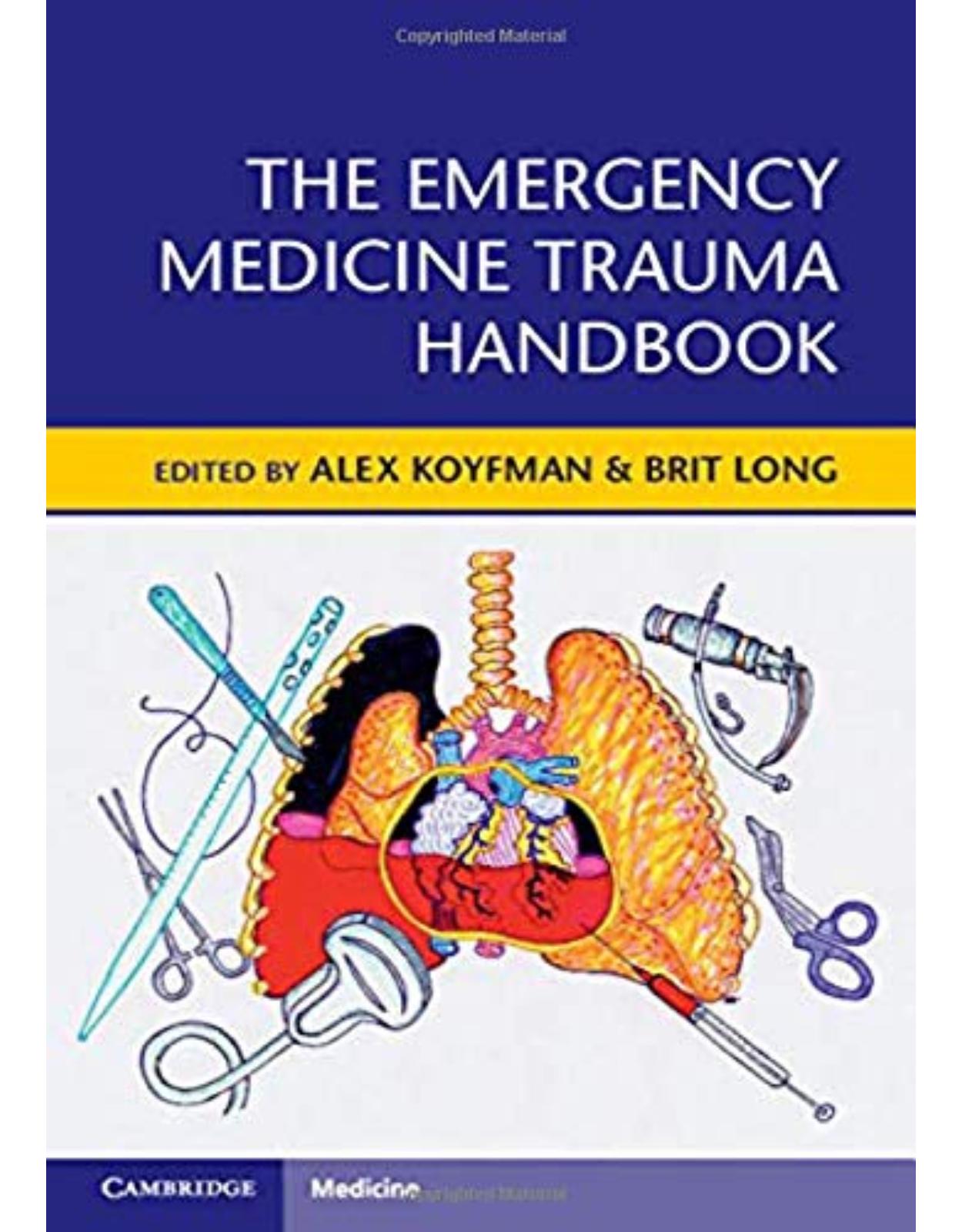
The Emergency Medicine Trauma Handbook
Livrare gratis la comenzi peste 500 RON. Pentru celelalte comenzi livrarea este 20 RON.
Disponibilitate: La comanda in aproximativ 4-6 saptamani
Autor: Alex Koyfman, Brit Long
Editura: Cambridge University Press
Limba: Engleza
Nr. pagini: 400
Coperta: Paperback
Dimensiuni: 157 x 234 x 17 mm
An aparitie: 17 Oct 2019
Description:
Trauma is a leading cause of death and disability around the world, and the leading cause of death in those aged under forty-five years. Conditions such as airway obstruction, hemorrhage, pneumothorax, tamponade, bowel rupture, vascular injury, and pelvic fracture can cause death if not appropriately diagnosed and managed. This essential book provides emergency physicians with an easy-to-use reference and source for traumatic injury evaluation and management in the emergency department. It covers approaches to common, life-threatening, and traumatic diseases in the emergency department, for use on shift and as a reference for further learning. Each chapter includes a succinct overview of common traumatic injuries, with evaluation and management pearls and pitfalls. Highly illustrated with images from one of the busiest trauma centers in the US, and featuring expert contributions from a diverse set of attending physicians, this is an essential text for all emergency medicine practitioners.
Table of Contents:
Chapter 1 General Approach to Traumatic Injuries
Introduction
The Trauma Team
Emergency Medical Services (EMS)
Primary Survey
A – Airway (Key Question: Do We Need to Take Control of the Airway Right Now?)
B – Breathing (Key Questions: Are Chest Tubes Required? Is There Bleeding in the Chest?)
C – Circulation (Key Question: Where Are They Bleeding and Do They Need Blood?)
Hypotension/Shock
D – Disability (Key Question: Is There a Major Neurologic Deficit?)
E – Expose (Key Question: What Injuries Haven't Been Found Yet?)
Secondary Survey
Imaging
Trauma Bay Imaging
CT Scans
Disposition and Ongoing Care
References
Chapter 2 Trauma Airway
Important Considerations for Airway Management in Trauma Patients
Consider Pre-existing Difficult Airway
Trauma Immobilization
Mechanical Distortion of the Airway and Contiguous Structures
Indications for Airway Intervention
Traumatic Injuries with Associated Difficult Airways
Closed Head Injury
Maxillofacial Trauma
Direct Airway Trauma
Cervical Spine Injury
Thoracic Trauma
Burns
Rapid-Sequence Intubation (RSI)
Rapid Sequence Intubation (RSI): The Technique
P – Plan B
P – Predict a Difficult Intubation
L – Look Externally
E – Evaluate Internally: The 3-3-2 Rule
M – Mallampati
O – Obstruction
N – Neck mobility
S – Saturation
P – Prepare
P – Preoxygenate
P – Position
P – Put to Sleep
P – Paralyze
P – Pass the Tube
Video Laryngoscopy
The GlideScope
The C-MAC
P – Prove Placement
P – Post-Intubation Management
Ventilator Settings
Post-Intubation Ventilator Settings
Post-Intubation Sedation/Analgesia
Post-Intubation Paralysis
P – Problem Solving
Difficult Intubation
Devices That Facilitate Intubation
Endotracheal Tube Introducer
Flexible Fiberoptic Bronchoscope (FFB)
Devices That Temporarily Substitute for Endotracheal Intubation
Supraglottic Airways
Laryngeal Mask Airway (LMA), the Intubating Laryngeal Mask Airway (ILMA), and the i-Gel
The Failed Airway
Surgical Airway
Conclusion
References
Chapter 3 Transfusion in Trauma
Introduction
Blood Products
Negatives of Crystalloid Resuscitation
Trauma Coagulopathy and the Lethal Triad
Damage Control Resuscitation
Permissive Hypotension
Minimal Volume Normotension
Balanced Resuscitation
Primary Literature for MTP
PROMMTT
PROPPR
When Should Massive Transfusion Be Activated?
How to Run MTP
Other Products
Tranexamic Acid (TXA)
Calcium
PCC
Factor VII
Product Guided Resuscitation
Resuscitation Goals
Anticoagulants
Transfusion Complications
Controversies – Whole Blood
References
Chapter 4 Trauma in Pregnancy
Epidemiology
Anatomical Changes in Pregnancy
Physiologic Changes in Pregnancy
Overview of Initial Evaluation and Management
Airway
Breathing
Circulation
Cardiopulmonary Resuscitation
Peri-Mortem Cesarean Section Procedure
Secondary Survey
Fetal Assessment
Ultrasound Examination
Diagnostic Evaluation
Therapeutic Considerations
Other Considerations
Pitfalls in ED Evaluation and Management
References
Chapter 5 Pediatric Trauma
Epidemiology
Anatomy and Physiology
Anatomical Differences
Airway
Developing musculoskeletal system
Physiological differences
Age-Specific Vitals (Table 5.1)
Breathing
Thermoregulation
Regions
Head
Anatomy
PECARN
Airway Management
Neck
Thorax
Abdomen
Extremities
Pitfalls in ED Evaluation and Management
References
Chapter 6 Geriatric Trauma
Importance
Triage of Elderly Trauma Patients
Prehospital
After Arrival at the Trauma Center
Pathophysiology Concerns in the Elderly
Supporting Evidence
Mechanisms and Injuries That are More Concerning in the Elderly
Falls
Head Injury
Chest Wall Injuries
Cervical Spine Injuries
Pedestrian Struck by Vehicle
Trauma Survey Principles in the Elderly
Elder Abuse
Anticoagulants
Pitfalls
References
Chapter 7 Head Trauma
Neurologic Injuries
Physiologic Goals
Secondary Injuries
ED Evaluation and Management
Airway Management
Pretreatment
Induction
Paralysis
Post Intubation
Blood Pressure Control
Hypotension
Neurogenic Shock
Vasopressors
Hypertension
Hyperosmolar Therapy
Mannitol
HTS
ICP Monitoring
Utility of US in ICP Evaluation
Surgical Indications
Coagulopathy Reversal in Traumatic Intracerebral Hemorrhage
Seizure Management
Tranexamic Acid in Head Trauma
Pitfalls in ED Evaluation and Management
References
Chapter 8 Facial Trauma
Introduction
The ABCs
Anatomy
Approach
History
Examination
Imaging
Management
Specific Injuries
Orbit
Ear
Nasal Injury
Zygomatic Bone Fractures
Tripod Fracture
Maxillary Fractures
TMJ
Mandible Fractures
Parotid Duct
Dental Injuries
Pitfalls in Facial Trauma
References
Chapter 9 Eye Trauma
Introduction and Epidemiology
Approach to the Patient With Eye Trauma
Treat Life-Threatening Conditions First
Involve the Ophthalmologist Early
Focused History
Immediately Identify Acute Visual Threats
Examination of the Injured Eye
Visual Acuity
Periocular Examination
Orbit and Orbital Rim
Lids and Lid Function
Eyelid and Periocular Sensation
Ocular Examination
Pupils
Extraocular Motility
Anterior Segment
Retina and Optic Nerve
Intraocular Pressure Measurement
Fluorescein Staining
Diagnostic Imaging
Vision-Threatening Conditions
Ocular Chemical Burns
Orbital Compartment Syndrome
Carotid-Cavernous Fistula
Open Globe Injury
Traumatic Hyphema
Vitreous Hemorrhage
Retinal Trauma
Optic Nerve Injury
Other Traumatic Eye Presentations
Traumatic Uveitis
Eyelid Lacerations
Corneal Abrasions and Corneal Foreign Bodies
Conjunctival Injury
Subconjunctival Hemorrhages
Conjunctival Lacerations
Conjunctival Foreign Bodies
Conjunctival Abrasions
Orbital Fractures
References
Chapter 10 Cervical Spine Trauma
Anatomy
History and Physical Examination
Immobilization
Decision to Image
Imaging
Types of Fractures/Patterns
General ED Management
Airway Management
Steroids
Complications
Clinical Clearance of Cervical Spine
Consultation Indications
Pitfalls in ED Management
References
Chapter 11 Thoracolumbar Trauma
Anatomy
Pre-Hospital Management
Examination of Suspected Thoracolumbar Trauma
Imaging Thoracolumbar Trauma
Classification
Injury Patterns (Table 11.4)
Compression Fractures
Burst Fractures
Chance Fractures
Translational/Fracture-Dislocation Injuries
Transverse Process, Spinous Process, Pars Interarticularis Fractures
Incomplete Spinal Cord Syndromes
Brown-Sequard Syndrome
Central Cord Syndrome
Anterior Cord Syndrome
Pitfalls
References
Chapter 12 Neck Trauma
Anatomy
Penetrating Injuries
Blunt Injuries
Strangulation and Hanging
Airway Management
Pitfalls
References
Chapter 13 Pulmonary Trauma
Airway Obstruction
Procedure – In-Line Stabilization
Tension Pneumothorax
Procedure – Needle Decompression
Procedure – Pleur-Evac Setup and Troubleshooting
Procedure – Chest Tube Removal
Open Pneumothorax
Massive Hemothorax
Procedure - Autologous Transfusion
Flail Chest
Pulmonary Contusion
Traumatic Diaphragmatic Injury
Tracheobronchial Disruption
Esophageal Disruption
Pitfalls in Thoracic Trauma
References
Chapter 14 Cardiac Trauma
Causes
Anatomy
Pathophysiology
Approach
History
Examination
Penetrating Cardiac Trauma
Cardiac Tamponade
Cardiac Missile
Iatrogenic Injury
Blunt Trauma
Cardiac Rupture
Coronary Vessel Injury/MI
Injury to the Valves, Papillary Muscles, Chordae Tendinae, and Septum
Pericardial Injury
Cardiac Dysfunction
Commotio Cordis
Multisystem Trauma
Great Vessel Injury
Evaluation
Electrocardiogram (ECG)
Laboratory Assessment with Cardiac Biomarkers
Imaging
Ultrasound
Transesophageal Echocardiography (TEE)
Chest X-ray
Computed Tomography (CT)
MRI
Management
Penetrating Trauma
Pericardiocentesis
ED Thoracotomy (EDT)
Blunt Trauma
Great Vessel Injury
Pitfalls in Cardiac Trauma
References
Chapter 15 Abdominal and Flank Trauma
Introduction
Causes
Anatomy
Pathophysiology
History
Examination
Differential Diagnosis
Hepatic Injury
Splenic Injury
Duodenal Injury
Pancreatic Injury
Diaphragmatic Injury
Renal Injury
Evaluation
Laboratory Testing
Imaging
Ultrasound (US)
Radiography
Computed Tomography (CT)
Diagnostic Peritoneal Lavage (DPL)
Management
Pitfalls in Abdominal and Flank Trauma
References
Chapter 16 Genitourinary Trauma
Overview
Signs and Symptoms
Diagnostic Studies
Kidney
Anatomy
Evaluation
Management
Complications
Ureter
Anatomy
Evaluation
Management
Complications
Bladder
Anatomy
Evaluation
Management
Complications
Urethra
Anatomy
Evaluation
Management
Complications
Scrotum and Testicle
Anatomy
Evaluation
Management
Complications
Penis
Anatomy
Evaluation
Management
Complications
Female GU
Anatomy
Evaluation
Management
Complications
ED Management
Surgical Indications
Pitfalls in ED Evaluation and Management
References
Chapter 17 Peripheral Vascular Injury
Vascular Injury Goals
Physiologic Goals
Secondary Injuries
ED Evaluation and Management
Low Risk Penetrating Extremity Injury Management and Disposition
Surgical Indications
Pitfalls in ED Evaluation and Management
References
Chapter 18 Pelvic Trauma
Pelvic Fractures
Anatomy
Diagnosis
Treatment
Pitfalls
Hip Fractures
Anatomy
Diagnosis
Treatment
Pitfalls in ED Management
Hip Dislocations
Diagnosis
Treatment
Techniques
Allis Technique for Reduction of Posterior Hip Dislocation (Figure 18.8)
Stimson's Technique for Reduction of Posterior Hip Dislocation (Figure 18.8)
Captain Morgan Technique for Reduction of Posterior Hip Dislocation
Whistler Technique for Reduction of Posterior Hip Dislocation
Pitfalls
References
Chapter 19 Upper Extremity Trauma
Introduction
Hand and Finger Injuries
Bone
Distal Phalanges
Proximal and Middle Phalanges
Metacarpal Head
Metacarpal Neck
Metacarpal Shaft
Thumb Metacarpal
Joint Dislocations
Nerve
Soft Tissue
Wrist and Distal Forearm Injuries
Bone
Distal Radius
Distal Ulna
Carpal Fractures
Subluxations and Dislocations
Soft Tissue
Elbow and Forearm
Bone
Distal Humerus Fractures
Ulnar Fractures11
Proximal Radius/Radial Head Fractures
Elbow Dislocations
Nerve Injury
Soft Tissue Injury
Biceps Tendon Rupture
Triceps Tendon Rupture
Shoulder and Upper Arm
Bone
Proximal Humerus Fractures
Humeral Shaft Fractures
Distal Humerus Fractures
Clavicular Fractures
Scapular Fractures
Dislocation
Vessel Injury
Pitfalls
References
Chapter 20 Lower Extremity Trauma
Femur
Mechanism of Injury
Physical Examination
Femoral Shaft Fractures
Distal Femur Fractures
Traction vs Splinting
Pediatric Femur Fractures
Knee
Tibial Plateau Fractures
Mechanism of Injury
Segond Fracture
Reverse Segond Fracture
Imaging Considerations
Complications and Associated Injuries
Management
Tibial Spine Fracture
Knee Dislocation
Mechanism of Injury
Examination
Associated Injuries (Figure 20.3)
Imaging
Initial Management
Definitive Management
Patella Dislocations
Patella Fractures
Examination - Superior Displacement of the Patella (''High Riding'')
Imaging - AP, Lateral, and ''Sunrise'' Views (Figure 20.5)
Initial Management
Definitive Management
Evaluation for an Open Knee Joint
Saline Load Test
CT
Proximal Third Tibial Fractures
Initial Management
Tibial Shaft Fractures
Anatomy
Imaging
Vascular Injuries – Compartment Syndrome
Proximal Fibula Fractures
Ankle
Imaging
Isolated Distal Fibular Fractures
Foot
Imaging
Hindfoot
Talus Fractures
Calcaneal Fractures
Midfoot
Cuboid, Navicular, Cuneiform Fractures
Lisfranc Fracture
Forefoot Fractures
Phalanx Fractures
Pitfalls
References
Chapter 21 Burns and Electrical Injuries
Burns
Pathophysiology
ED Evaluation and Management
Airway Management
Estimating Burn Size and Depth
Fluid Resuscitation
Escharotomies
Carbon Monoxide Poisoning
Cyanide Poisoning
Other Considerations
Transfer to a Burn Center
Wound Care
Electrical Injuries
Mechanism of Injury
Current
Voltage
Contact
ED Evaluation and Management
Clinical Features
Cardiopulmonary Injury
Nervous System Injury
Vascular and Muscle Injury
GI Injury
Cutaneous Injury
ENT Injury
Lip Burns in Children
Lightning Strikes
Disposition
Pitfalls in ED Evaluation and Management
References
Chapter 22 Procedural Sedation and Analgesia in Trauma
Background
Indications
Preparation
Medication Choices
Ketamine
Propofol
Ketofol (Ketamine and Propofol)
Etomidate, Midazolam, and Fentanyl
Discharge Considerations
Special Populations – Pediatrics
Special Populations – Pregnancy
Special Populations – Elderly
Pitfalls
References
Chapter 23 Commonly Missed Traumatic Injuries
Introduction
Scope of the Problem
Missed Injuries by Location
Head
Neck
Spine
Thoracic
Abdomen
Pelvis
Extremities
References
Index
| An aparitie | 17 Oct 2019 |
| Autor | Alex Koyfman, Brit Long |
| Dimensiuni | 157 x 234 x 17 mm |
| Editura | Cambridge University Press |
| Format | Paperback |
| ISBN | 9781108450287 |
| Limba | Engleza |
| Nr pag | 400 |



Clientii ebookshop.ro nu au adaugat inca opinii pentru acest produs. Fii primul care adauga o parere, folosind formularul de mai jos.