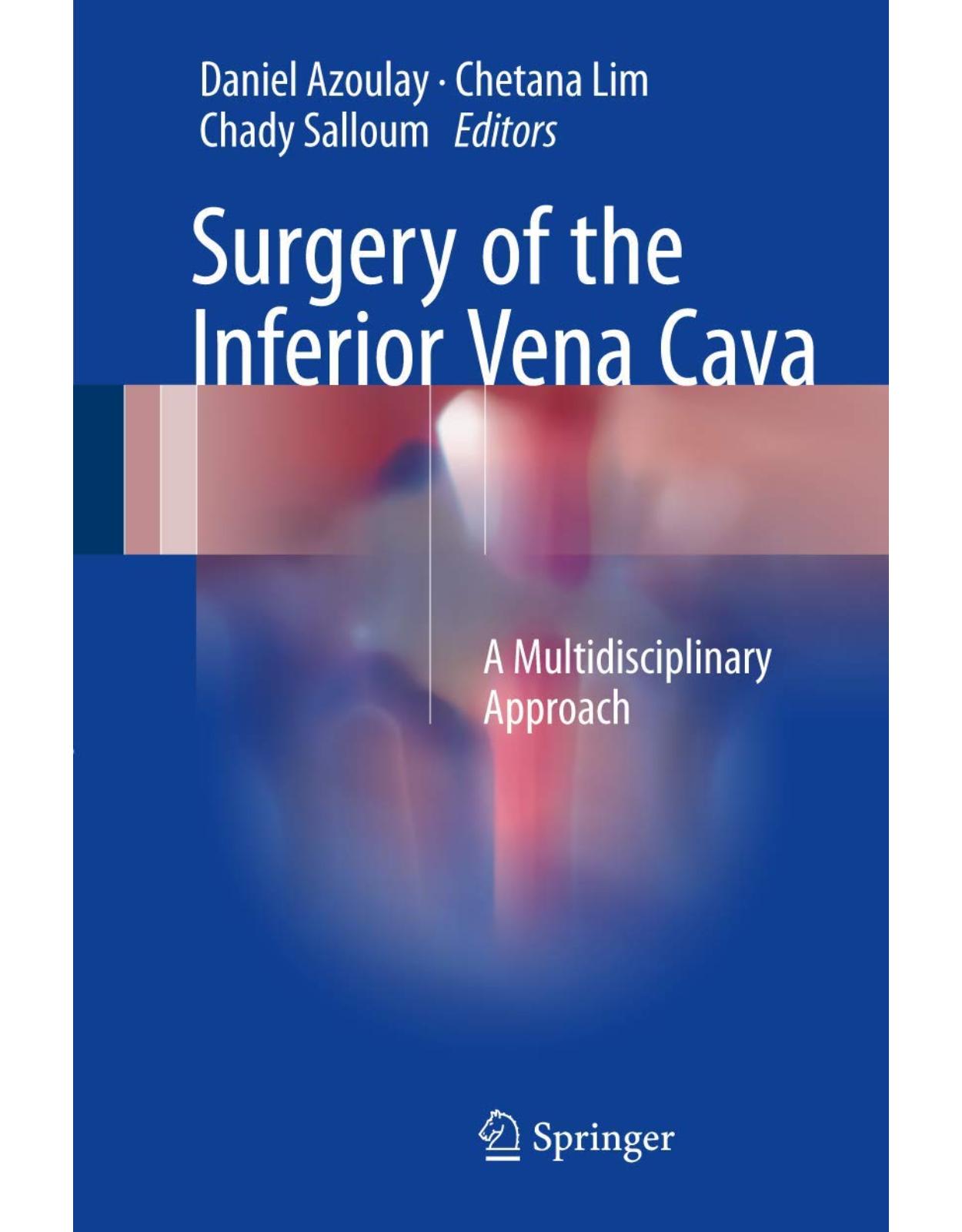
Surgery of the Inferior Vena Cava: A Multidisciplinary Approach
Livrare gratis la comenzi peste 500 RON. Pentru celelalte comenzi livrarea este 20 RON.
Disponibilitate: La comanda in aproximativ 4-6 saptamani
Editura: Springer
Limba: Engleza
Nr. pagini: 232
Coperta: Hardcover
Dimensiuni: 16.28 x 1.98 x 24.49 cm
An aparitie: 26 April 2017
Description:
This book addresses an urgent need, offering an updated, detailed and multidisciplinary review of surgery of the inferior vena cava (IVC). Over the past two decades, tremendous advances have been made in this field and related specialties. Many journals and/or textbooks on different surgical or medical specialties have reported on individual aspects, so it is high time to gather all of them in a single resource. To date, surgery of the IVC remains a major challenge, not only in terms of technical difficulties, but also in terms of perioperative management. The book sheds new light on cutting-edge developments, including: Innovations in imaging; Neoadjuvant and adjuvant treatments for tumors of or involving the IVC; Intraoperative anesthetic management; and Technical and technological advances in surgery. As such, it will be of a great interest to surgeons (cardiac, vascular and digestive) and urologists, as well as radiologists and anesthesiologists.
Table of Contents:
1: Imaging and Radiological Assessment of the Inferior Vena Cava
1.1 Introduction
1.2 IVC Imaging Techniques
1.2.1 Conventional Venography
1.2.2 Ultrasound
1.2.3 Multi-Detector Computed Tomography (MDCT)
1.2.4 Magnetic Resonance Imaging (MRI)
1.2.5 PET-CT
1.3 Main Imaging Diagnostic Feature
1.3.1 IVC Obstruction
1.3.1.1 Artifactual Filling Defect and Bland Thrombus
1.3.1.2 Benign Tumor Invasion Within the IVC
1.3.1.3 Malignant Tumor Invasion Within the IVC
1.3.1.4 IVC Obstruction Consequences and Therapeutic Implications
Budd-Chiari Syndrome
Collateral Pathways
1.3.2 IVC Anatomical Variants
1.3.2.1 Left IVC
1.3.2.2 Double IVC
1.3.2.3 Retrocaval Ureter
1.3.2.4 Retroaortic and Circumaortic Left Renal Vein
1.3.2.5 Interruption of the IVC and Azygos/Hemiazygos Continuation
1.3.2.6 Absence of the IVC
1.3.2.7 Portocaval Shunt
1.3.2.8 IVC Webs
1.3.3 IVC Trauma
1.3.4 Postoperative Imaging
1.3.4.1 Post-Liver Transplantation
1.3.4.2 Post-Portocaval Shunt
1.4 Interventional Imaging of the IVC
1.4.1 Inferior Vena Cava Filter
1.4.2 IVC Obstruction and Endovascular Management
References
2: Anesthetic and Hemodynamic Considerations of Inferior Vena Cava Surgery
2.1 Introduction
2.2 Anatomic and Physiologic Considerations on the IVC Circulation
2.2.1 Spontaneous Portocaval Shunts
2.2.1.1 Congenital Physiologic Bypasses
2.2.1.2 Acquired Shunts
2.2.2 Circulatory Models
2.2.2.1 Generalities
2.2.2.2 The “One-Compartment” Circulatory Model
2.2.2.3 The “Two-Compartment” Circulatory Model
2.3 The Different Venous Clampings
2.3.1 IVC Clamping Below the Renal Venous Confluence
2.3.2 IVC Clamping Above the Renal Venous Confluence (Below the Liver)
2.3.3 IVC Clamping Above the Hepatic Venous Confluence
2.3.4 Total Hepatic Vascular Exclusion
2.3.4.1 Hemodynamic Effects (Fig. 2.8)
2.3.4.2 Neurohormonal Effects
2.3.5 Inferior Vena Cava Clamping Combined with Supra-Celiac Aortic Clamping
2.4 The Role of the Anesthesiologist
2.4.1 Preoperative
2.4.2 Intraoperative
2.4.2.1 The Risk of Hemorrhage
2.4.2.2 The Risk of Gaseous Embolism
2.4.2.3 The Management of the Consequences of the Clamping
2.4.3 Postoperative
References
3: Primary Leiomyosarcoma of the Inferior Vena Cava
3.1 Introduction
3.2 Epidemiology, Etiology, and Predisposing Factors
3.3 Histopathology, Genetics, and Staging
3.4 Localization, Clinical Presentation, and Symptoms
3.5 Diagnostics
3.6 Treatment
3.6.1 Resection
3.6.2 A Resection Techniques
3.6.2.1 Limited Resection
3.6.2.2 Extended Resection (Single Organ Versus Multi-organ Resection)
3.6.3 Reconstruction Techniques
3.6.4 Morbidity and Mortality Rates
3.6.5 Neoadjuvant/Adjuvant Treatment
3.6.5.1 Chemotherapy
3.6.5.2 Radiotherapy
3.7 Oncological Outcomes and Prognostic Factors
References
4: Retroperitoneal Sarcoma Involving the Inferior Vena Cava
4.1 Introduction
4.2 Retroperitoneal Sarcoma
4.3 Technical Considerations on IVC Management
4.4 IVC Resection and Reconstruction
References
5: Renal Cell Carcinoma Involving the Inferior Vena Cava
5.1 Introduction
5.2 Clinical Presentation
5.3 Prognostic Factors
5.4 Imaging Diagnosis
5.5 Staging and Classification of Intracaval Extension
5.6 Patient Selection
5.7 Factors Determining the Choice of Surgical Technique
5.8 Preoperative Considerations
5.9 Surgical Steps and Technical Details
5.10 Adjunct Procedures: IVC Resection and Reconstruction
5.11 Postoperative Complications
5.12 Oncologic Outcomes After Radical Nephrectomy and Tumor Thrombectomy
References
6: Surgery of the Inferior Vena Cava Combined to Liver Resection
6.1 Introduction
6.2 General Principles
6.2.1 Caval Cross Clamping
6.2.2 Hemodynamic Changes of the Caval Cross Clamping
6.2.3 Hypothermic Perfusion Techniques
6.2.4 Venovenous Bypass
6.3 Perioperative Management
6.3.1 Anesthetic Management
6.3.2 Anticoagulation Protocol
6.4 Specific Technical Aspects
6.4.1 Total Vascular Exclusion of the Liver [4, 6, 7]
6.4.2 Hypothermic In Situ Liver Resection [10, 20, 25, 41]
6.4.3 Hypothermic Ante Situm Liver Resection [21, 24, 42–51]
6.4.4 Reperfusion
6.5 Surgical Indications and Outcomes
References
7: Ex Situ Resection of the Inferior Vena Cava with Hepatectomy
7.1 Introduction
7.2 Alternative Complex Techniques to Ex Situ Resection
7.2.1 In Situ Hypothermic Perfusion
7.2.2 The Ante Situm Procedure
7.3 Patient Assessment
7.3.1 Cardiorespiratory Assessment
7.3.2 Hepatic Reserve Assessment
7.3.3 Radiology Assessment
7.4 Preoperative Preparation
7.5 Anaesthesia
7.6 Operative Technique
7.6.1 Operability Assessment
7.6.2 Liver Mobilisation and Excision
7.6.3 Hepatic Perfusion and Preservation
7.6.4 Ex Situ Resection and Reconstruction
7.6.5 Hepatic Reimplantation and Reperfusion
7.7 Postoperative Care
7.8 Complications of Ex Situ Liver and IVC Resection
7.8.1 Vascular Thrombosis and Stenosis
7.8.2 Graft-Associated Sepsis
7.8.3 Biliary Strictures
7.9 Follow-Up
7.10 Long-Term Results
References
8: Surgical Treatment of Adrenocortical Carcinoma with Caval Invasion
8.1 Introduction
8.2 ACC and Inferior Vena Cava Extension
8.3 Preoperative Workup
8.4 Neoadjuvant Treatment
8.5 Surgical Management
8.5.1 Tumor Thrombus Located Below the Suprahepatic Veins
8.5.1.1 Thrombus Extension Limited to Infrahepatic IVC
8.5.1.2 Thrombus Extension to Retrohepatic IVC
8.5.2 Tumor Thrombus Extending Above the Suprahepatic Veins and Below the Cavo-Atrial Junction
8.5.3 Tumor Thrombus Is Located Above the Cavo-Atrial Junction
8.6 Results
References
9: Malignancy with Cavoatrial Extension
9.1 Introduction
9.2 Preoperative Staging
9.3 Surgical Technique
9.4 Postoperative Management
9.5 Discussion
References
10: Injuries of the Juxtahepatic Vena Cava
10.1 Introduction
10.2 Pattern of Injuries
10.3 Emergency Surgical Techniques in the Management of Retrohepatic Caval Injuries
10.3.1 Direct Suture
10.3.2 Vascular Exclusion of the Liver
10.3.3 Cavo-Caval Venous Bypass Procedures
10.3.4 Liver Resection to Obtain Access to the Retrohepatic Vena Cava
10.3.5 Perihepatic Packing (PHP)
10.3.6 Nonoperative Management
10.3.7 Liver Transplantation
10.4 Management Strategies
10.4.1 Hemodynamically Unstable Patient: Emergency Laparotomy Mandatory
10.4.2 Hemodynamically Stable Patient
References
11: Inferior Vena Cava Reconstruction in Liver Transplantation
11.1 Introduction
11.2 Conventional Vena Cava Technique
11.3 Venovenous Bypass
11.4 Piggyback Technique
11.5 Piggyback Variants: End-to-Side, Side-to-Side Cavocavostomy, and Reverse Piggyback
11.6 Piggyback Variant: Anterior Approach Hepatectomy
11.7 Temporary Portocaval Shunt
11.8 Reconstruction in Special Situations
11.8.1 Domino Transplantation
11.8.2 Budd-Chiari Syndrome
11.8.3 Outflow Obstruction
11.8.4 Split-Liver Transplantation
11.8.5 IVC as Source of Portal Inflow
References
12: Inferior Vena Cava Reconstruction in Living Donor Liver Transplantation
12.1 Introduction
12.2 Preservation and Preparation of Native IVC in LDLT
12.3 Outflow Reconstruction in LDLT
12.3.1 Right Liver
12.3.2 Left Liver
12.4 Orthotopic Position of the Partial Graft
12.5 Reconstruction of Short Hepatic Veins
12.6 Reconstruction of Middle Hepatic Vein Tributaries
12.7 Grafts Used to Reconstruct IVC, Autograft, Allograft, Cryopreserved Allograft, and Artificia
12.8 IVC Replacement in LDLT
12.9 IVC Reconstruction for Outflow Block
References
13: Vena Cava Filters: State of the Art
13.1 Introduction
13.2 History of the Vena Cava Filtration
13.3 Vena Cava Filter Classification
13.4 Indications for VCF Placement
13.4.1 Absolute Indications
13.4.2 Relative Indications
13.4.3 Prophylactic Indications
13.5 VCF Placement Procedure
13.6 VCF Retrieval Procedure
13.7 Efficiency
13.8 Long-Term Complications of VCF
13.9 Management of Patients with VCF and Strategies for Retrieval
References
| An aparitie | 26 April 2017 |
| Autor | Daniel Azoulay , Chetana Lim, Chady Salloum |
| Dimensiuni | 16.28 x 1.98 x 24.49 cm |
| Editura | Springer |
| Format | Hardcover |
| ISBN | 9783319255637 |
| Limba | Engleza |
| Nr pag | 232 |
-
1,96000 lei 1,74400 lei


Clientii ebookshop.ro nu au adaugat inca opinii pentru acest produs. Fii primul care adauga o parere, folosind formularul de mai jos.