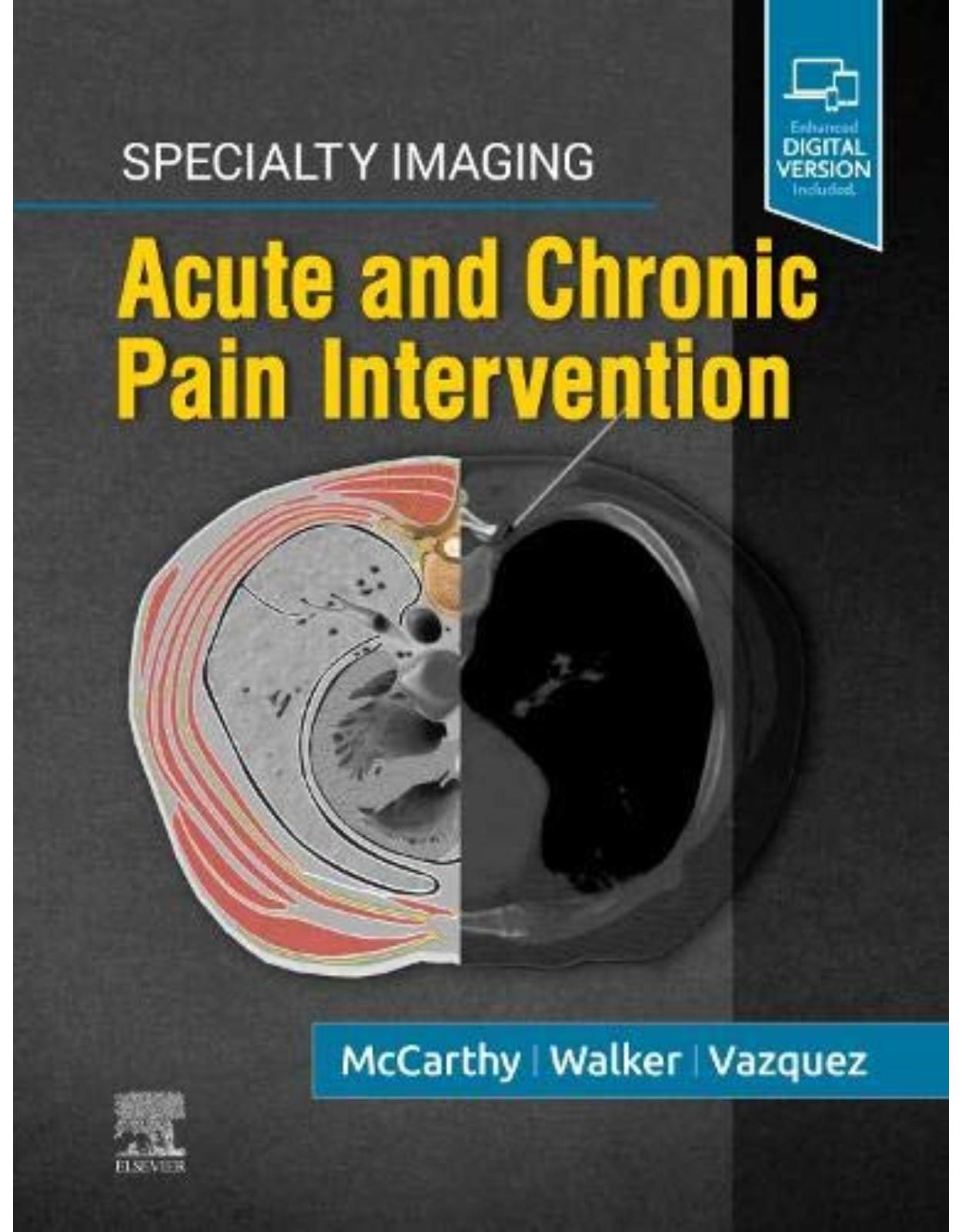
Specialty Imaging: Acute and Chronic Pain Intervention
Livrare gratis la comenzi peste 500 RON. Pentru celelalte comenzi livrarea este 20 RON.
Disponibilitate: La comanda in aproximativ 4-6 saptamani
Editura: Elsevier
Limba: Engleza
Nr. pagini: 416
Coperta: Hardcover
Dimensiuni: 22.23 x 2.54 x 28.58 cm
An aparitie: 4 Aug. 2020
Description:
Practical and clinically oriented, Specialty Imaging: Acute and Chronic Pain Intervention provides unique, authoritative guidance on the use of image-guided techniques for periprocedural analgesia and pain management procedures. Ideal for practicing and trainee interventional radiologists, pain physicians, and anesthesiologists, this one-stop resource is tailored to your decision support needs, with coverage of everything from neuroanatomy and specific pain conditions to interventional procedures for acute and chronic pain.
Provides up-to-date content informed by best practices and the perspectives of both interventional radiology and anesthesiology
Discusses key topics such as multimodal opioid sparing techniques as adjuncts and alternatives to the use of opioids for acute pain management, as well as shared decision making in interventional radiology pain management
Demonstrates the new fascial pain blocks as well as sympathetic nerve blocks for periprocedural analgesia during interventional procedures
Covers adult and pediatric acute and chronic pain conditions
Integrates neuroanatomy and the "why" of clinical procedures for a better understanding of the pathways and various options for therapeutic intervention
Presents information consistently, using a highly templated format with bulleted text for quick, easy reference
Begins each section with a discussion of neuroanatomy, followed by succinct chapters that provide "how-to" information on a clinically useful, imaging-guided interventional procedure for treating a specific acute or chronic pain condition
Features procedural videos and clear, high-quality drawings for visual reinforcement, e.g., sequential illustrations that show where nerves are located through successive peeling of anatomic layers
Table of Contents:
SECTIONS
SECTION 1: INTRODUCTION
Chapter 1: Treatment of Acute and Periprocedural Pain
Key Facts
Key Images
Main Text
Tables
Chapter 2: Pain Neuroanatomy
Key Facts
Key Images
Main Text
Tables
Chapter 3: Visceral Pain
Key Facts
Key Images
Main Text
Image Gallery
Chapter 4: Opioid Reduction Strategies
Key Facts
Key Images
Main Text
Tables
Chapter 5: Acute on Chronic Cancer Pain
Key Facts
Key Images
Main Text
Tables
Chapter 6: Pediatric Pain
Key Facts
Key Images
Main Text
Image Gallery
Chapter 7: Shared Decision-Making in Analgesia
Key Facts
Key Images
Main Text
Tables
SECTION 2: ACUTE PAIN TREATMENT MODALITIES
Chapter 8: Peripheral Nerve Blocks
Key Facts
Key Images
Main Text
Tables
SECTION 3: CHRONIC PAIN TREATMENT MODALITIES
Chapter 9: Blocks for Chronic Pain: Facet and Nerve
Key Facts
Key Images
Main Text
Image Gallery
Chapter 10: Thermal Ablation for Malignancy-Associated Pain
Key Facts
Key Images
Main Text
Image Gallery
Chapter 11: Spinal Cord Stimulator
Key Facts
Key Images
Main Text
Image Gallery
SECTION 4: IMAGING MODALITIES
Chapter 12: Radiography
Key Facts
Key Images
Main Text
Image Gallery
Chapter 13: Ultrasound
Key Facts
Key Images
Main Text
Image Gallery
Chapter 14: Computed Tomography
Key Facts
Key Images
Main Text
Image Gallery
Chapter 15: Magnetic Resonance Imaging
Key Facts
Key Images
Main Text
Chapter 16: Radiation Safety
Key Facts
Key Images
Main Text
Tables
Image Gallery
Chapter 17: Contrast Agents
Key Facts
Key Images
Main Text
Image Gallery
SECTION 5: HEAD, NECK, AND UPPER EXTREMITY
Chapter 18: Functional Anatomy of Head and Neck
Main Text
Image Gallery
ACUTE PAIN PROCEDURES
Chapter 19: Cervical Epidural
Key Facts
Key Images
Main Text
Image Gallery
Chapter 20: Brachial Plexus Nerve Block
Key Facts
Key Images
Main Text
Image Gallery
Video Gallery
Chapter 21: Upper Extremity Blocks
Key Facts
Key Images
Main Text
Image Gallery
CHRONIC PAIN PROCEDURES
Chapter 22: Scalp Block for Migraines and Facial Pain
Key Facts
Key Images
Main Text
Chapter 23: Temporomandibular Joint Injection
Key Facts
Key Images
Main Text
Image Gallery
Chapter 24: Trigger Points
Key Facts
Key Images
Main Text
Chapter 25: Stellate Ganglion Blocks
Key Facts
Key Images
Main Text
Video Gallery
SECTION 6: THORAX AND CHEST WALL
Chapter 26: Functional Anatomy of Thorax and Chest Wall
Main Text
Image Gallery
ACUTE PAIN PROCEDURES
Chapter 27: Erector Spinae Plane Block
Key Facts
Key Images
Main Text
Image Gallery
Video Gallery
Chapter 28: Intercostal Nerve Block
Key Facts
Key Images
Main Text
Image Gallery
Chapter 29: Serratus Anterior Plane Block
Key Facts
Key Images
Main Text
Image Gallery
Chapter 30: Intermediate Cervical Plexus Block
Key Facts
Key Images
Main Text
Image Gallery
Chapter 31: PECS 1 and PECS 2 Blocks
Key Facts
Key Images
Main Text
Image Gallery
Video Gallery
CHRONIC PAIN PROCEDURES
Chapter 32: Facet Joint Injection
Key Facts
Key Images
Main Text
Image Gallery
Chapter 33: Scapulothoracic Bursa Injection
Key Facts
Key Images
Main Text
Image Gallery
SECTION 7: ABDOMEN AND ABDOMINAL WALL
Chapter 34: Functional Anatomy of Abdominal Wall
Main Text
Image Gallery
ACUTE PAIN PROCEDURES
Chapter 35: Paravertebral Block
Key Facts
Key Images
Main Text
Image Gallery
Video Gallery
Chapter 36: TAP Block
Key Facts
Key Images
Main Text
Image Gallery
Video Gallery
Chapter 37: Quadratus Lumborum Block
Key Facts
Key Images
Main Text
Image Gallery
Video Gallery
Chapter 38: Ilioinguinal-Iliohypogastric Nerve Block
Key Facts
Key Images
Main Text
Image Gallery
Video Gallery
CHRONIC PAIN PROCEDURES
Chapter 39: Celiac Neurolysis
Key Facts
Key Images
Main Text
Image Gallery
Chapter 40: Genitofemoral Nerve Block
Key Facts
Key Images
Main Text
SECTION 8: PELVIS AND PERINEUM
Chapter 41: Functional Anatomy of Pelvis and Perineum
Main Text
Image Gallery
ACUTE PAIN PROCEDURES
Chapter 42: Sacroiliac Joint Injection
Key Facts
Key Images
Main Text
Image Gallery
Chapter 43: Pubic Symphysis Injection
Key Facts
Key Images
Main Text
Image Gallery
Chapter 44: Piriformis Injection
Key Facts
Key Images
Main Text
Image Gallery
Chapter 45: Sacral Nerve Root Block
Key Facts
Key Images
Main Text
Image Gallery
Chapter 46: Pudendal Nerve Block
Key Facts
Key Images
Main Text
Image Gallery
Chapter 47: Penile Nerve Block
Key Facts
Key Images
Main Text
Chapter 48: Superior and Inferior Hypogastric Blocks
Key Facts
Key Images
Main Text
CHRONIC PAIN PROCEDURES
Chapter 49: Ganglion Impar Block
Key Facts
Key Images
Main Text
SECTION 9: SPINE
Chapter 50: Functional Anatomy of Spine
Main Text
Image Gallery
ACUTE PAIN PROCEDURES
Chapter 51: Epidural Steroid Injection
Key Facts
Key Images
Main Text
Image Gallery
Chapter 52: Facet Blocks
Key Facts
Key Images
Main Text
Image Gallery
Chapter 53: Medial Branch Blocks
Key Facts
Key Images
Main Text
Image Gallery
CHRONIC PAIN PROCEDURES
Chapter 54: RFA Facet Joint
Key Facts
Key Images
Main Text
Image Gallery
Chapter 55: Vertebroplasty and Kyphoplasty
Key Facts
Key Images
Main Text
Image Gallery
SECTION 10: LOWER EXTREMITY
Chapter 56: Functional Anatomy of Lower Extremity
Main Text
Image Gallery
ACUTE PAIN PROCEDURES
Chapter 57: Femoral Nerve Block
Key Facts
Key Images
Main Text
Image Gallery
Chapter 58: Adductor Canal Nerve Block (ACNB)
Key Facts
Key Images
Main Text
Image Gallery
Video Gallery
Chapter 59: Lateral Femoral Cutaneous Nerve Block
Key Facts
Key Images
Main Text
Image Gallery
Chapter 60: Fascia Iliaca Compartment Block
Key Facts
Key Images
Main Text
Image Gallery
Video Gallery
Chapter 61: Sciatic Block
Key Facts
Key Images
Main Text
Image Gallery
Video Gallery
Chapter 62: Genicular Nerve Block
Key Facts
Key Images
Main Text
Chapter 63: Peroneal Nerve Block
Key Facts
Key Images
Main Text
Chapter 64: Tibial Nerve Block
Key Facts
Key Images
Main Text
Image Gallery
Chapter 65: Saphenous Nerve Block
Key Facts
Key Images
Main Text
Chapter 66: Ankle Block
Key Facts
Key Images
Main Text
Chapter 67: Obturator Nerve Block
Key Facts
Key Images
Main Text
Image Gallery
CHRONIC PAIN PROCEDURES
Chapter 68: Lumbar Sympathetic Blockade and Neurolysis
Key Facts
Key Images
Main Text
Chapter 69: Infiltration Between Popliteal Artery and Capsule of Posterior Knee (iPACK) Block
Key Facts
Key Images
Main Text
Image Gallery
Video Gallery
SECTION 11: PEDIATRIC PAIN
ACUTE PAIN PROCEDURES
Chapter 70: Pediatric Acute and Postoperative Pain
Key Facts
Key Images
Main Text
Image Gallery
CHRONIC PAIN PROCEDURES
Chapter 71: Pediatric Recurrent and Chronic Pain
Key Facts
Key Images
Main Text
INDEX
| An aparitie | 4 Aug. 2020 |
| Autor | Colin J. McCarthy MB BCh BAO MRCSI FFR (RCSI) , T. Gregory Walker MD FSIR |
| Dimensiuni | 22.23 x 2.54 x 28.58 cm |
| Editura | Elsevier |
| Format | Hardcover |
| ISBN | 9780323680363 |
| Limba | Engleza |
| Nr pag | 416 |


Clientii ebookshop.ro nu au adaugat inca opinii pentru acest produs. Fii primul care adauga o parere, folosind formularul de mai jos.