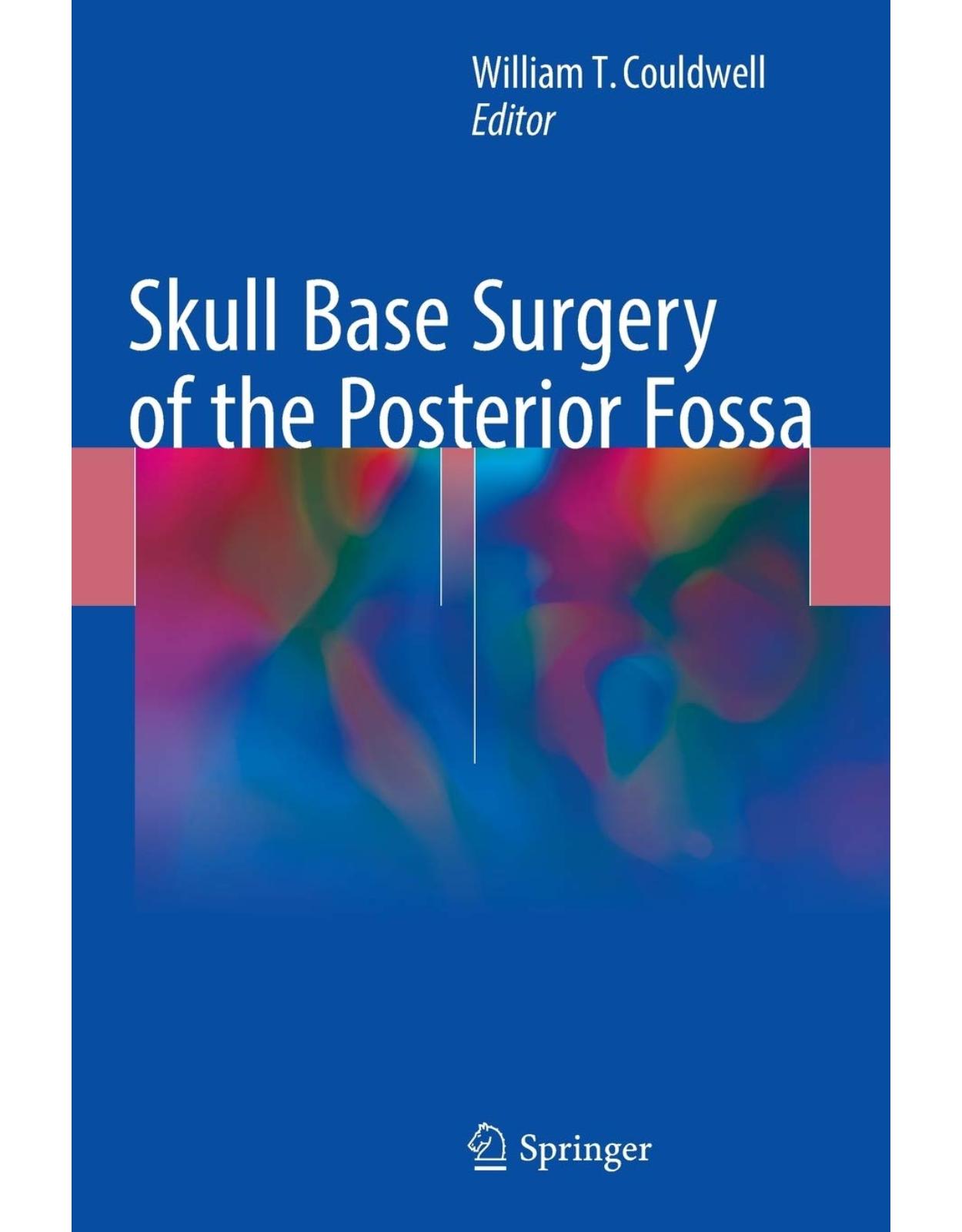
Skull Base Surgery of the Posterior Fossa
Livrare gratis la comenzi peste 500 RON. Pentru celelalte comenzi livrarea este 20 RON.
Description:
This text provides a comprehensive and contemporary overview of surgical approaches to lesions of the posterior fossa. It will serve as a resource for neurosurgeons and otologists who treat patients with tumors and vascular diseases of the posterior fossa. It provides a concise review of surgical strategies that address the most important pathologies affecting the posterior fossa. It is richly illustrated with photographs and illustrations of the surgical strategies covered. All chapters are written by experts with world-wide recognition for their contributions in their respective subspecialty. Skull Base Surgery of the Posterior Fossa will be of great utility to Neurosurgeons, Otolaryngologists, and Radiation Therapists with an interest in diseases that affect the posterior fossa, as well as Senior Residents in Neurosurgery and Otolaryngology, and Fellows of Skull Base Surgery and Otology.
Table of Contents:
Part I: Introduction
1: Surgical Anatomy of the Posterior Fossa
Introduction
Enclosure of the Posterior Fossa
Skull Base
Neural Foramina
Tentorium
Venous Sinuses
Obstacles to Surgery
Contents of the Posterior Fossa
Arteries and Rule of Three
Veins
Special Regions
Cavernous Sinus
Clivus and Petroclival Region
Internal Auditory Meatus and Otological Structures
Jugular Foramen
Surgical Corridors and Related Anatomy
Posterior
Occipital Transtentorial
Supracerebellar Infratentorial
Suboccipital, Telovelar
Posterolateral: Retrosigmoid
Far-Lateral Approach
Posterolateral: Presigmoid
Mastoidectomy
Transcrusal and Translabyrinthine Approaches
Transcochlear and Transotic Approaches
Combined Supra-/Infratentorial Petrosal Approaches
Exposure of Jugular Foramen
Lateral
Anterolateral
Anterior
Credits
References
Part II: Approaches
2: Retrosigmoid Craniotomy and Its Variants
Background
Indications
Anatomy
Cranial and Extradural Anatomy
Intradural Anatomy
Surgical Technique
Positioning
Craniotomy Technique
Reconstruction and Closure
Complications
Conclusions
References
3: Middle Fossa and Translabyrinthine Approaches
Introduction
Preoperative Evaluation
Indications for Surgery
Patient Counseling
Surgery
General Preoperative Preparation
Surgical Technique: Middle Fossa Approach
Surgical Technique: Translabyrinthine Approach
Postoperative Care
Outcomes
Hearing Preservation
Facial Nerve Function
Complications
Cerebrospinal Fluid Leak and Meningitis
Hemorrhage
Sigmoid Sinus Thrombosis
Venous Thromboembolism
Major Neurological Complications
Summary
References
4: Posterior and Combined Petrosal Approaches
Introduction
Preoperative Planning
Role of Intraoperative Monitoring
Petrosal Approach (or Posterior Petrosal Approach)
Patient Positioning
Skin Incision and Craniotomy
Combined Petrosal Approach
Patient Positioning
Skin Incision and Craniotomy
Tumor Resection
Closure
Tricks and Pitfalls
Conclusion
References
5: Far Lateral Approach and Its Variants
Introduction
Anesthetic Technique and Positioning
Incision and Muscle Dissection
Extradural Exposure
Transcondylar Variants
Supracondylar Variants
Paracondylar Variants
Intradural Exposure
Conclusions
References
6: Endoscopic Endonasal Approach for Posterior Fossa Tumors
Introduction
Endoscopic Endonasal Transclival Approach
Extradural Posterior Fossa Tumors
Chondrosarcomas and Chordomas
Preoperative Radiological Assessment
Approach Selection
The Role of EEA
Intradural Posterior Fossa Tumors
Meningiomas
Preoperative Radiological Assessment
Pathological Anatomy
Petroclival Meningiomas
Clival Meningiomas
Foramen Magnum Meningiomas
EEA Indications in Posterior Fossa Meningiomas
Advantages and Limitations
Skull Base Reconstruction
References
Part III: Specific Diseases
7: Petroclival Meningiomas
Introduction
Natural History and Clinical Presentation
Preoperative Assessment and Planning
Radiological Imaging
Preoperative Hearing Status
Preparation for Cerebral Revascularization
The Role of Intraoperative Electrophysiological Neuromonitoring and Safe Anesthetic Techniques
Decision-Making and Treatment Strategies
Surgical Approaches
Transpetrosal Approaches
Anterior Transpetrosal (Kawase’s) Approach
Indications and Limitations
Surgical Technique and Nuances
Complications and Their Avoidance
Posterior Transpetrosal Approach
Indications and Limitations
Surgical Technique and Nuances
Complications and Their Avoidance
Combined Transpetrosal Approach
Indications and Limitations
Surgical Technique and Nuances
Complications and Their Avoidance
Treatment Outcomes in the Multimodality Management Era
Conclusions
References
8: Meningiomas of the Cerebellopontine Angle
Anatomic Classification
Surgical Approaches
APFM
MPFM
PPFM
Case Examples
Case 1: APFM
Case 2: MPFM
Case 3: PPFM: Small-Sized Tumor
Case 4: PPFM: Large-Sized Tumor
Discussion
References
9: Tentorial Meningiomas
Introduction
Incisural Type
Surgical Planning
Illustrative Case
Case 1: Retrosigmoid Approach (Fig. 9.2)
Case 2: Retrosigmoid Approach with Drilling of the Petrous Apex (Fig. 9.3)
Case 3: Anterior Transpetrosal Approach (Fig. 9.4)
Case 4: Combined Transpetrosal Approach (Fig. 9.5)
Falcotentorial Type
Surgical Planning
Illustrative Case
Case 5: Posterior Interhemispheric Transtentorial Approach (Fig. 9.7)
Lateral Type
Surgical Planning
Illustrative Case
Case 6: Retrosigmoid Approach (Fig. 9.8)
Case 7: Combined Presigmoid and Retrosigmoid Approach with Resection of Tumor Invading the Sigm
Posterior Type
Surgical Planning
Illustrative Case
Case 8: Suboccipital Approach with Resection of Invasion into the Transverse Sinus (Fig. 9.10)
Case 9: Aggressive Resection with Reconstruction of the Superior Sagittal Sinus (Fig. 9.11)
Conclusions
References
10: Foramen Magnum Meningiomas
Introduction
Clinical Presentation
Evolution of Surgical Approach to FMM
Classification
Preoperative Assessment
Surgical Considerations
Monitoring
Specific Microsurgical Considerations
Outcomes and Complications
Conclusions
References
11: Vestibular Schwannomas
Introduction
Retrosigmoid Approach
Indications
Surgical Technique
Surgical Risks and Complications
Surgical Outcome
Middle Fossa Approach
Indications
Relevant Surgical Anatomy
IAC Localization Techniques in the Petrous Bone
Surgical Technique
Bony Exposure
Dural Opening and Tumor Dissection Techniques
Limitations of the MFA
Surgical Risk and Complications
Surgical Outcome for MFA
Translabyrinthine Approach for Vestibular Schwannoma
Indications
Tumor Removal Techniques
Surgical Risks and Complications
Facial Nerve Preservation with the Translabyrinthine Approach
Limitations of Translabyrinthine Approach
Specific Perioperative Considerations
Conclusion
References
12: Epidermoid Cyst
Introduction
Clinical Presentation
Imaging and Differential Diagnosis
Management
Surgical Approach
Complication Avoidance
Surgical Outcome
Management of Recurrence and Malignant Transformation
Conclusion
References
13: Metastasis to the Posterior Fossa
Introduction
Metastasis to the Cerebellum
Metastasis to the Base of Skull
Patient Presentation
Diagnosis and Selection for Surgical Intervention
Perioperative Care and Surgical Techniques for Metastasis to the Posterior Fossa
Preoperative Care
Surgical Approaches to the Posterior Fossa
Midline Approach for Vermial or Medial Hemispheric Lesions
Paramedian and Retrosigmoid Approaches for Lateral and Far Anterolateral Lesions
Superior Cerebellar Approach
Excision of Metastatic Lesions
Postoperative Care and Complications
Stereotactic Radiosurgery for Treatment of Posterior Fossa Metastasis
Conclusions
References
14: Microsurgical Management of Posterior Fossa Vascular Lesions
Introduction
Surgical Vascular Anatomy of the Posterior Circulation
Posterior Circulation Aneurysms
Incidence of Aneurysms of the Posterior Circulation
Presentation of Aneurysms of the Posterior Circulation
Natural History of Aneurysms of the Posterior Circulation
Indications for Interventions for Aneurysms of the Posterior Circulation
Posterior Fossa Arteriovenous Malformations
Incidence of Arteriovenous Malformations of the Posterior Fossa
Presentation of Arteriovenous Malformations of the Posterior Fossa
Natural History of Arteriovenous Malformations of the Posterior Fossa
Indications for Interventions for Arteriovenous Malformations of the Posterior Fossa
Brainstem and Cerebellar Cavernous Malformations
Incidence of Cavernous Malformations of the Brainstem and Cerebellum
Presentation of Cavernous Malformations of the Brainstem and Cerebellum
Natural History of Cavernous Malformations of the Brainstem and Cerebellum
Indications for Intervention for Cavernous Malformations of the Brainstem and Cerebellum
Preoperative Evaluation
Patient History and General Considerations
Diagnostic Imaging
Operative Adjuncts
Neuromonitoring and Mapping
Intraoperative Evaluation of Blood Flow
Cerebral Protection and Hypothermia
Pharmacological Cardiac Arrest
Approaches and Approach Selection to Posterior Fossa Vascular Lesions
Approaches to the Ventral Midbrain/Posterior Fossa
Approaches to the Lateral Midbrain
Approaches to the Dorsal Midbrain
Approaches to the Ventral Pons
Approaches to the Lateral Pons
Approaches to the Dorsal Pons
Approaches to the Ventral Medulla
Approaches to the Lateral Medulla
Approaches to the Dorsal Medulla and Cervicomedullary Junction
Outcomes of Microsurgery
Posterior Circulation Aneurysms
Basilar and Vertebral Artery Aneurysms
Posterior Inferior Cerebellar Artery Aneurysms
Posterior Cerebral Artery, Superior Cerebral Artery, and Anterior Inferior Cerebellar Artery Aneur
Posterior Fossa Arteriovenous Malformations
Brainstem and Cerebellar Cavernous Malformations
Conclusions
References
Index
| An aparitie | 2018 |
| Autor | Couldwell |
| Dimensiuni | 17.78 x 1.42 x 25.4 cm |
| Editura | Springer |
| Format | Hardcover |
| ISBN | 9783319670379 |
| Limba | Engleza |
| Nr pag | 237 |






Clientii ebookshop.ro nu au adaugat inca opinii pentru acest produs. Fii primul care adauga o parere, folosind formularul de mai jos.