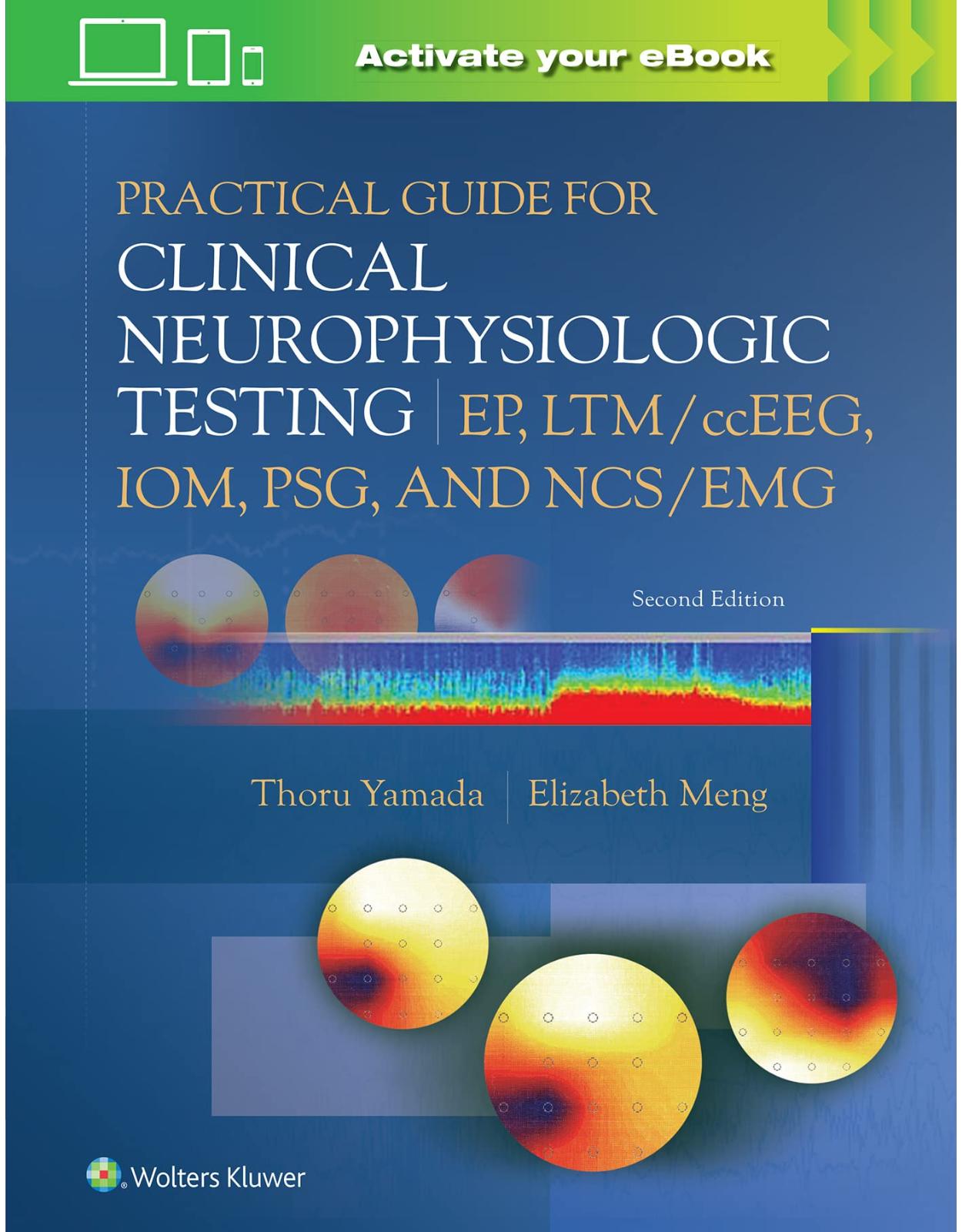
Practical Guide for Clinical Neurophysiologic Testing
Livrare gratis la comenzi peste 500 RON. Pentru celelalte comenzi livrarea este 20 RON.
Disponibilitate: La comanda in aproximativ 4 saptamani
Autor: Thoru Yamada, Elizabeth Meng
Editura: LWW
Limba: Engleza
Nr. pagini: 400
Coperta: Paperback
Dimensiuni: 213 x 276 mm
An aparitie: 08 Nov 2022
Description:
Focusing on the technical aspects of clinical neurophysiologic testing, Practical Guide for Clinical Neurophysiologic Testing: EP, LTM/ccEEG, IOM, PSG, and NCS/EMG 2nd Edition, offers comprehensive guidance on neurophysiologic testing that picks up where the companion Practical Guide for Clinical Neurophysiologic Testing: EEG ends. Dr. Thoru Yamada and Elizabeth Meng provide advanced content on evoked potentials, intraoperative monitoring, long-term EEG monitoring, epilepsy monitoring, sleep studies, and nerve conduction studies. All chapters have been updated to incorporate recent advancements and new studies and articles.
Table of Contents:
Online Videos
Section 1: Evoked Potentials
1 Principles of Evoked Potentials
Introduction
Principles of the Averaging Method
Digital Process
Vertical (Voltage) Resolution and Range
Horizontal (Time) Resolution
Amplifier
Filter Setting of Amplifier
Automatic Artifact Rejection
Data Measurement and Offline Analyses
Polarity Convention
Wave Form Nomenclature
Determination of Normality and Abnormality
Technical Problems in Recording EPs
No Appreciable Response
“Noisy” Responses
Large Stimulus Artifacts
Excessive Sample Rejection (With Auto-Rejection Mode)
Appendix
2 Visual Evoked Potentials
Introduction
Anatomy of the Visual System
Technical Parameters
Stimulus Devices
Visual Angle
Check Size
Luminance
Contrast
Stimulus Repetition Rate
Replication
Dealing with “Noisy” Responses
Physiological Factors
Full-Field Versus Half-Field and Monocular Versus Binocular Stimulation
Age
Visual Acuity
Gender
Body Temperature
Pupillary Size
Amplifier and Signal Averager Settings
Recording Electrodes
Normal VEP in Full-Field Monocular Stimulation
Normal Waveforms and Distribution
Normal Variations
Half-Field Stimulation
Method
Normal VEP to Half-Field Stimulation: “Paradoxical Lateralization”
Abnormal VEP
Latency Abnormalities
Amplitude Abnormalities
Topographic Abnormalities
Waveform Abnormalities
VEP and Clinical Correlates
Optic Neuritis
Multiple Sclerosis
Compressive or Axonal Optic Nerve Lesions
Spinocerebellar Degeneration
Charcot–Marie–Tooth Disease
Parkinson’s Disease
Transverse Myelitis
Chiasmal Lesion
Postchiasmal Lesion
Albinism
Huntington’s Chorea
Cortical Blindness
Functional Blindness, Hysteria, or Malingering
Electroretinogram
Steady-State VEP
VEP by Other Stimulation
Flash and Led Goggle VEPs
Other Stimulus Devices for VEP
3 Brainstem Auditory Evoked Potentials and Auditory Evoked Potentials
Anatomy of Auditory Pathway in Brainstem
Brainstem Auditory Evoked Potentials
Technical Parameters
Stimulation
Filter Setting
Analysis Time
Recording Electrodes
Normal BAEP
Wave I
Wave II
Wave III
Wave IV/V Complex
Electrocochleogram
Cochlear Microphonic Potentials
Summating Potential
Action Potential
Factors that Affect BAEP
Physiological Factors
Nonphysiological Features
Technical Modification to Improve Waveform Identification
Evaluation of the BAEP
Absence of One or More Waves
Latency Abnormalities
Clinical Application of BAEP
Multiple Sclerosis
Cerebellopontine Angle Tumors
Coma and Brain Death
Brainstem Tumor
Vascular Lesions
BAEP in Infants and Young Children
Other Diseases that May Show Abnormalities in BAEP
Middle- and Long-Latency Auditory Evoked Potential
Middle-Latency Auditory Evoked Potential
Long-Latency Auditory Evoked Potential
Auditory P300 ERP
Mismatch Negativity
4 Somatosensory Evoked Potentials
Anatomy of the Sensory System
Somatosensory Evoked Potentials
Methodology/Recording Technique
Stimulation
Frequency Filter Setting
Amplification and Summation
Generation Mechanism of Far-Field Potential
Physiological Factors that Affect SSEP
Age
Body Size
Temperature
Sleep
Short-Latency Upper Extremity SSEP
P9 and N9 Potentials
P11 and N11 Potentials
P13/P14 Potentials
N18 Potential
N20, P20, P22 Potentials
Cervical Responses
Recording Montages for Short-Latency SSEP of Upper Extremity Nerves
Medium- and Long-Latency SSEPs from Upper Extremity Stimulation
Lower Extremity SSEP
Spinal Response
Scalp Potential
Paradoxical Lateralization
Recording Montages for Lower Extremity Nerve
Abnormal Criteria
Obligate Potentials
Anatomical Correlates of SSEP Abnormalities
Peripheral Nerve Lesions
Cervical Cord/Spinal Cord Lesions
High Cervical Cord and Brainstem Lesions
Thalamic Lesions
Cortical/Hemispheric Lesions
SSEP in Specific Neurologic Conditions
Surgical Monitoring
Multiple Sclerosis
Friedreich’s Ataxia
Motor Neuron Disease
Spinocerebellar Degeneration
Huntington’s Chorea
Coma and Brain Death
Myoclonic Epilepsy
Appendix Figures 4A-1 through 4A-12
Appendix
Section 2: Intraoperative Neurophysiologic Monitoring
5 The Technologist’s Role in Neurophysiologic Intraoperative Monitoring
Introduction
IONM Technologist Responsibilities and Skills
Preoperative Preparation
Intraoperative Phase
Postoperative Phase
Equipment and Supplies
Somatosensory Evoked Potentials
Alarm Criteria
Transcranial Motor Evoked Potentials
Alarm Criteria
Contraindication
Pedicle Screw Monitoring
Choosing Proper Modalities
Brainstem Auditory Evoked Potentials
Alarm Criteria
Electroencephalography
Electrocorticography and Intracranial Recording
Electrocorticography
Cortical Somatosensory Evoked Potential
Troubleshooting in the Operating Room
The Effect of Anesthesia
Inhalation Anesthetics
Total Intravenous Anesthesia
Muscle Relaxant
Patient Safety
6 Brain Function Monitoring for Carotid Endarterectomy and Aortic Arch Surgery
Introduction
Anesthesia
EEG Changes during Induction
EEG Patterns at Sub-Mac Concentration
EEG Patterns during Recovery from Anesthesia
Other Factors That Affect EEG
EEG Monitoring
Recording Technique
Preclamp Focal EEG Abnormalities in Relationship to Anesthesia
EEG Changes after Cross-Clamping the Carotid Artery
Predictability of EEG Change
Regional Anesthesia for Awake CEA and EEG Correlates
The Incidence of Stroke after CEA
The Use of SSEP (Somatosensory Evoked Potential) for CEA
The Use of tcMEP (Transcranial Motor Evoked Potential)
Other Monitoring Methods
Aortic Arch Surgery
7 Spinal Cord Monitoring
Introduction
Monitoring Modalities
Somatosensory Evoked Potentials
Upper Extremity SSEP Monitoring
Lower Extremity SSEP Monitoring
Transcranial Motor Evoked Potentials
Other Spinal Cord Monitoring Methods
Recording MEPs From the Epidural or Subdural Space After Transcranial Electric Stimulations
Spinal Cord Stimulation With Recording From Spinal Cord, Scalp, Muscle, or Peripheral Nerve
Peripheral Nerve Stimulation With Spinal Cord Recording
Anesthesia During MEP and SSEP Monitoring
EMG Monitoring for Pedicle Screw Fixation
Intraoperative Monitoring During Selective Dorsal Rhizotomy (SDR)
Pediatric Tethered Cord Repair
Summary and Conclusion
8 Intraoperative Monitoring of the Brainstem and Cranial Nerve Function
Anatomy
Indications for Intraoperative Monitoring of Brainstem Function
Modalities
Brainstem Auditory Evoked Potentials
Somatosensory Evoked Potentials
Motor Evoked Potentials
Visual Evoked Potentials
EMG and Monitoring and Mapping of Cranial Nerve and Nuclei
Anesthesia
Application and Interpretation
Conclusion
Section 3: Long-Term EEG Monitoring
9 Diagnostic Video-EEG Monitoring for Epilepsy and Spells: Indications, Application, and Interpretation
Introduction
Appropriate Selection of Patients for Monitoring
Indications for Video-EEG Monitoring
Seizure/Spell Classification
Seizure Localization
Seizure Frequency
Therapeutic Assessment and Tailoring
Application
Outpatient Monitoring
Inpatient Epilepsy Monitoring
Recording Equipment and Technical Aspects
Digital Reformatting
Seizure and Spike Detection Software
Care of the Monitored Patient
Counseling the Patient Prior to Admission
Admission Orders
Seizure Precautions
Seizure First Aid and Nursing Care
Peri-ictal Patient Assessment
Seizure Provocation
Emergencies in Epilepsy Monitoring Unit Practice
Interpretation
Interictal Recording
Ictal Recording
Spell Classification: Nonepileptic Spells
Nonepileptic Behavioral Events
Physiologic
Seizure Classification
Generalized Seizures
Focal Seizures
Focal to Bilateral Tonic–Clonic
Seizures in Infant and Pediatric Video-EEG Monitoring
Synthesizing the Epilepsy Syndrome: Integrating Video-EEG Data into the Clinical Picture and Other Appropriate Investigations
Pitfalls in Video-EEG Monitoring
Conclusions
Acknowledgements
10 Invasive Video EEG Monitoring in Epilepsy Surgery Candidates: Indications, Technique, and Interpretation
Introduction
Definitions
Indications for Invasive Video-EEG
The Noninvasive Evaluation
The Role of the Epilepsy Surgery Case Conference
The Epilepsy Monitoring Unit: Considerations for Performing Invasive Video-EEG
Personnel
Space Considerations
Safety
The Technique of Invasive Video-EEG
Recording
Stereo-electroencephalography
Data Storage
Review
Outcome Measurements
Concluding Remarks
11 Long-Term Bedside EEG Monitoring for Acutely Ill Patients (LTM/ccEEG)
Introduction
Indication ccEEG
Incidence of NCS or NCSE in the ICU
Recording Technology of ccEEG
Evaluation of ccEEG for NCS or NCSE
The Assessment of Progress for Acute Cerebral Dysfunction
Seizure and Spike Detection Using Computer Software
The use of Quantitative EEG (qEEG) Analyses for ccEEG
Monitoring in the Neonatal ICU
Standardized Critical Care EEG Terminology
EEG Background Descriptions
Rhythmic and Periodic Patterns (RPPs)
Sporadic Epileptiform Discharges
Brief Potentially Ictal Rhythmic Discharges
Ictal–Interictal Continuum
Section 4: Sleep Studies
12 Technology of Polysomnography
Introduction
The Technologists’ Role in the Sleep Lab
Preparing for the Patient
Review of the Physician’s Order
Review of the Medical Record
Preparing for Hookup
Selecting a Montage
Calibration
After Patient Arrives
Orientation
Questionnaires
Interview
Consent Forms
Electrode and Sensor Application
Placement and Application
Additional Recording Parameters
The Recording
Physiologic Calibrations
Observation and Documentation
Ending the Study
Sleep Stage Scoring in the Normal Adult Population
Sleep Cycles
Scoring
Scoring of Periodic Limb Movements in Sleep
Respiratory Event Scoring
Apneas
Hypopneas
Respiratory Effort-Related Arousal
Hypoventilation
Cheyne–Stokes Breathing
Report
Summary
13 Sleep Physiology and Pathology
Introduction
Basic CNS Anatomy and Physiology of Sleep
History
Wakefulness Mechanisms
Sleep Onset Mechanisms
Sleep Pathology
REM Sleep Mechanisms Appreciated Through Narcolepsy with Cataplexy
Obstructive Sleep Apnea and Stroke
Summary
Acknowledgments
14 Sleep Apnea and Related Conditions
Introduction
Obstructive Sleep Apnea
Epidemiology
Pathophysiology
Risk Factors for OSA and Associated Conditions
Other Medical Conditions Associated with Increased OSA Prevalence
Clinical Manifestations of OSA
Physical Examination
OSA Screening
Diagnostic Evaluation of OSA
Management and Treatments
Positive Airway Pressure
Oral Appliance
Hypoglossal Nerve Stimulator
Surgical Approaches
Positional Therapy
Weight Loss
Exercise
Others
Central Apnea
Epidemiology
Pathophysiology
Risk Factors and Associated Conditions
Clinical Manifestations
Physical Examination
Diagnostic Evaluation
Management and Treatment
Treatment of Hyperventilation-Related CSA
CPAP
Supplemental Oxygen during Sleep
Phrenic Nerve Stimulation
Treatment of Hypoventilation-Related CSA
Sleep-Related Hypoventilation
Obesity Hypoventilation Syndrome
Congenital Central Alveolar Hypoventilation
Late-Onset Central Hypoventilation with Hypothalamic Dysfunction or Rapid-Onset Obesity with Hypothalamic Dysfunction, Hypoventilation, and Autonomic Dysregulation (ROHHAD)
Sleep-Related Hypoventilation due to a Medication or Substance
Sleep-Related Hypoventilation Due to a Medical Disorder
Idiopathic Central Alveolar Hypoventilation
Sleep-Related Hypoxemia
15 Evaluating Narcolepsy and Related Conditions
Introduction
Clinical Aspects
Narcolepsy
Other Hypersomnias of Central Origin
Multiple Sleep Latency Test
Pretest Preparation
Performing the MSLT
Concluding the Test
Scoring and Interpreting the MSLT
Interpretation Pitfalls
Maintenance of Wakefulness Test
Procedure
Interpretation
Final Thoughts
16 Parasomnias
Introduction
Polysomnography
Specific Parasomnias
Types of Parasomnias
The Differential Diagnosis
Sleep-Related Movement Disorders
Sleep-Related Epilepsy
Summary
17 Electrophysiological Measurement and Rules for Sleep-Related Movement Disorders
Introduction
Normal Physiologic Movements of Sleep
Phasic Twitches during REM Sleep
Major Body Movements
Isolated Sleep-Related Movements and Normal Variants
Excessive Fragmentary Myoclonus
Alternating Leg Muscle Activation
Hypnagogic Foot Tremor
Sleep Starts (Hypnic Jerks)
Sleep-Related Movement Disorders
Periodic Limb Movements in Sleep
Sleep-related Leg cramps
Sleep-related Bruxism
Sleep-related Rhythmic Movement Disorder
Benign Sleep Myoclonus of Infancy
Propriospinal Myoclonus at Sleep Onset
Restless Legs Syndrome
Dopamine Agonists
α∂ Ligands
Opioids
Benzodiazepines
Section 5: Nerve Conduction Studies
18 Nerve Conduction and Electromyography Studies
Introduction and Recording Technology
NCS Techniques
Motor Study
Sensory Study
F Waves
H Reflex
The Facial Nerve (VII)
The Blink Reflex (Trigeminal V, Facial VII)
Waveform Analysis
Uncommon NCS Techniques
Artifacts
Electrical
Physiological
Understanding Electromyography for the Technologist
Introduction to Electromyography
Waveform Patterns of the Resting Phase
Denervation Potentials
Myotonic Discharges
Complex Repetitive Discharges
Fasciculation Potentials and Myokymic Discharges
Waveform Patterns of the Activation Phase
Motor Unit Potentials
Firing Pattern
Waveform Analysis
Contraindications to EMG
Summary
Index
| An aparitie | 08 Nov 2022 |
| Autor | Thoru Yamada, Elizabeth Meng |
| Dimensiuni | 213 x 276 mm |
| Editura | LWW |
| Format | Paperback |
| ISBN | 9781975193577 |
| Limba | Engleza |
| Nr pag | 400 |
| Versiune digitala | DA |













Clientii ebookshop.ro nu au adaugat inca opinii pentru acest produs. Fii primul care adauga o parere, folosind formularul de mai jos.