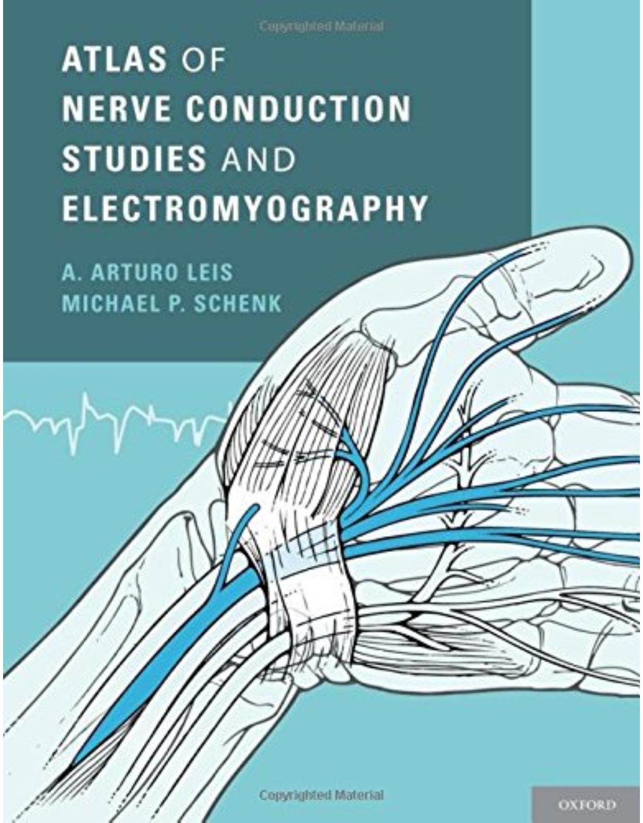
Atlas of Nerve Conduction Studies and Electromyography
Livrare gratis la comenzi peste 500 RON. Pentru celelalte comenzi livrarea este 20 RON.
Disponibilitate: La comanda in aproximativ 4-6 saptamani
Editura: Oxford University Press
Limba: Engleza
Nr. pagini: 336
Coperta: Hardcover
Dimensiuni: 222 x 280 x 22 mm
An aparitie: 21 Mar 2013
Description:
Beautifully and lavishly illustrated, Atlas of Nerve Conduction Studies and Electromyography demystifies the major conditions affecting peripheral nerves and provides electrodiagnostic strategies for confirming suspected lesions of the peripheral nervous system.Building on the success of the landmark Atlas of Electromyography, this new text is divided into sections based on the major peripheral nerves. It contains detailed illustrations of each nerve along with a discussion of its anatomy, followed by a thorough outline of the clinical conditions and entrapment syndromes that affect the nerve, including a list of the etiologies, clinical features, and electrodiagnostic strategies used for each syndrome. Routine and special motor and sensory nerve conduction studies are shown in an anatomical illustration. In addition, each muscle supplied by the peripheral nerve is illustrated showing the root, plexus, and peripheral nerve supply to the muscle and is accompanied by a corresponding human photograph. Written text provides information about the nerve conduction studies, muscle origin, tendon insertion, voluntary activation maneuver, and the site of optimum needle insertion, which is identified in the figures by a black dot or a needle electrode.Atlas of Nerve Conduction Studies and Electromyography is the perfect anatomical guide for neurologists, specialists in physical medicine and rehabilitation, and electrodiagnostic medicine consultants, while also providing support for individuals in residency training programs, critical care medicine, neurological surgery, and family practice.
Table of Contents:
1. Overview of Nerve Conduction Studies
How the Peripheral Nervous System Conveys Information
Stimulating and Recording Electrodes
Electrode Amplifiers and Ground Electrode
Reducing Artifacts and Interference
Electrical Safety
Temperature Effect
Effect of Aging
Motor Nerve Conduction
Sensory Nerve Conduction
Role of Dorsal Root Ganglia (DRG) in Localizing Lesions
Late Responses: F-wave and H-reflex
Types of Nerve Injury
Neurapraxia
Axonal Loss Injury
Ion channel Disorders (Channelopathies)
2. Overview of Electromyography (EMG)
The Motor Unit
Needle Electrodes
Muscle Selection for Needle EMG
The Needle EMG Examination
Assessment of Insertional Activity
Assessment of Spontaneous Activity
Assessment of Motor Unit Potentials (MUPs)
Assessment of Firing Pattern and Recruitment
Complications Related to Needle Electromyography
3. Brachial Plexus
Upper Trunk Lesion
Middle Trunk Lesion
Lower Trunk Lesion
Plexus Cord Lesions
4. Median Nerve
Carpal Tunnel Syndrome
Anterior Interosseous Nerve Syndrome
Pronator Teres Syndrome
Ligament of Struthers' Syndrome
Median Nerve Conduction Studies
Median Motor Nerve Conduction Study
Short Segment Stimulation across the Palm ("inching technique”)
Median F-waves
Martin-Gruber Anastomosis
Median Sensory Nerve Conduction Study
Median and Ulnar Palmar Comparative Study for the Diagnosis of CTS
Median and Ulnar Digit 4 (ring finger) Comparative Study
Median and Superficial Radial Digit 1 (thumb) Comparative Study
Combined Sensory Index (CSI)
Standards for Severity of Carpal Tunnel Syndrome
Digital Branch Injury
Needle Electromyography
Abductor Pollicis Brevis
Opponens Pollicis
Flexor Pollicis Brevis
1st, 2nd Lumbricals
Pronator Quadratus
Flexor Pollicis Longus
Flexor Digitorum Profundus, Digits 2 and
Flexor Digitorum Superficialis (sublimis)
Palmaris Longus
Flexor Carpi Radialis
Pronator Teres
5. Ulnar Nerve
Ulnar Neuropathy at the Elbow (Retrocondylar Groove)
Ulnar Neuropathy at the Elbow (Cubital Tunnel Syndrome)
Ulnar Neuropathy at the Wrist (Guyon's Canal)
Ulnar Nerve Conduction Studies
Ulnar Motor Nerve Conduction Study from Abductor Digiti Minimi (ADM)
Ulnar Motor Nerve Conduction Study from First Dorsal Interosseous (FDI)
Short Segment Stimulation across the Elbow ("inching technique”)
Ulnar F-waves
Martin-Gruber Anastomosis
Riches-Cannieu Anastomosis (RCA)
Ulnar Sensory Nerve Conduction Study
Dorsal Ulnar Cutaneous (DUC) Nerve Conduction Study
Anomalous Superficial Radial Innervation to Ulnar Dorsum of Hand
Needle Electromyography
Adductor Pollicis
Flexor Pollicis Brevis
First Dorsal Interosseous
2nd, 3rd, 4th Dorsal Interossei
Palmar Interossei
3rd and 4th Lumbricals
Abductor Digiti Minimi
Opponens Digiti Minimi
Flexor Digiti Minimi
Flexor Digitorum Profundus, Digits 4 and 5
Flexor Carpi Ulnaris
6. Radial Nerve
Radial Nerve Lesion in the Arm
Radial Nerve Lesion in the Axilla
Posterior Interosseous Nerve Syndrome
Superficial Radial Nerve Lesion
Radial Nerve Conduction Studies
Radial Motor Nerve Conduction Study
Superficial Radial Sensory Nerve Conduction Study
Needle Electromyography
Extensor Indicis
Extensor Pollicis Brevis
Extensor Pollicis Longus
Abductor Pollicis Longus
Extensor Digitorum Communis and Extensor Digiti Minimi
Extensor Carpi Ulnaris
Supinator
Extensor Carpi Radialis, Longus and Brevis
Brachioradialis
Anconeus
Triceps, Lateral Head
Triceps, Long Head
Triceps, Medial Head
7. Axillary Nerve
Axillary Nerve Lesion
Axillary Motor Nerve Conduction Study
Needle Electromyography
Deltoid, Anterior Fibers
Deltoid, Middle Fibers
Deltoid, Posterior Fibers
Teres Minor
8. Musculocutaneous Nerve
Musculocutaneous Nerve Lesion
Musculocutaneous Nerve Conduction Studies
Musculocutaneous Motor Nerve Conduction Study
Lateral Cutaneous Nerve of the Forearm (Lateral Antebrachial Cutaneous) Conduction Study
Needle Electromyography
Brachialis
Biceps Brachii
Coracobrachialis
9. Medial Cutaneous Nerve of the Forearm
(Medial Antebrachial Cutaneous Nerve)
Lesion of the Medial Cutaneous Nerve of the Forearm
Medial Cutaneous Nerve of the Forearm Conduction Study
10. Suprascapular Nerve
Suprascapular Nerve Lesion
Needle Electromyography
Infraspinatus
Supraspinatus
11. Dorsal Scapular Nerve
Dorsal Scapular Nerve Lesion
Rhomboideus Major and Minor
Levator Scapulae
12. Long Thoracic Nerve
Long Thoracic Nerve Lesion
Needle Electromyography
Serratus Anterior
13. Subscapular Nerves and the Thoracodorsal Nerve
Needle Electromyography
Teres Major
Latissimus Dorsi
14. Medial and Lateral Pectoral Nerves
Needle Electromyography
Pectoralis Major
Pectoralis Minor
15. Cervical Plexus
16. Phrenic Nerve
Phrenic Nerve Lesion
Phrenic Nerve Conduction Study
Needle Electromyography
Diaphragm
17. Sacral Plexus
Sacral Plexus Lesion
18. Sciatic Nerve
Sciatic Nerve Lesion
Needle Electromyography
Semitendinosus
Semimembranosus
Biceps Femoris (Long Head)
Biceps Femoris (Short Head)
19. Tibial Nerve
Tarsal Tunnel Syndrome
Tibial Nerve Conduction Studies
Tibial Motor Nerve Conduction Studies
Tibial F-waves
Sural Sensory Nerve Conduction Study
H-reflex
Medial and Lateral Plantar Nerve Conduction Studies
Needle Electromyography
Gastrocnemius, Medial Head
Gastrocnemius, Lateral Head
Soleus
Tibialis Posterior
Flexor Digitorum Longus
Flexor Hallucis Longus
Popliteus
Abductor Hallucis
Flexor Digitorum Brevis
Flexor Hallucis Brevis
Abductor Digiti Minimi (Quinti)
Adductor Hallucis
20. Common Peroneal Nerve
Common Peroneal Mononeuropathy at the Knee
Common Peroneal Nerve Conduction Studies
Common Peroneal Motor Nerve Conduction Study from Extensor Digitorum Brevis
Peroneal F-waves
Accessory Deep Peroneal Nerve
Common Peroneal Motor Nerve Conduction Study from Tibialis Anterior
Short Segment Stimulation across the Fibular Head ("Inching Technique”)
Superficial Peroneal Sensory Nerve Conduction Study
Needle Electromyography
Tibialis Anterior
Extensor Digitorum Longus
Extensor Hallucis Longus
Peroneus Tertius
Extensor Digitorum Brevis
Peroneus Longus
Peroneus Brevis
21. Superior Gluteal Nerve
Needle Electromyography
Gluteus Medius
Gluteus Minimus
Tensor Fasciae Latae
22. Inferior Gluteal Nerve
Needle Electromyography
Gluteus Maximus
23. Pudendal Nerve
Pudendal Nerve Lesion
Needle Electromyography
Sphincter Ani Externus (External Anal Sphincter)
Levator Ani
24. Lumbar Plexus
Lesion of the Lateral Femoral Cutaneous Nerve (Meralgia Paresthetica)
Lateral Femoral Cutaneous Nerve Conduction Study
Needle Electromyography
External Oblique, Internal Oblique, and Transversus Abdominis
25. Femoral Nerve
Femoral Nerve Lesion
Femoral Nerve Conduction Studies
Femoral Motor Nerve Conduction Study
Saphenous Sensory Nerve Conduction Study
Needle Electromyography
Iliacus (Iliopsoas)
Pectineus
Sartorius
Rectus Femoris
Vastus Lateralis
Vastus Intermedius
Vastus Medialis
26. Obturator Nerve
Obturator Nerve Lesion
Needle Electromyography
Adductor Longus
Adductor Brevis
Adductor Magnus
Gracilis
27. Paraspinal Muscles
Needle Electromyography
Cervical Paraspinal
Thoracic Paraspinal
Lumbosacral Paraspinal
28. Cranial Nerves and Muscles
Spinal Accessory Nerve Conduction Study
Facial Motor Nerve Conduction Study
Blink Reflex Study
Needle Electromyography
Sternocleidomastoid
Trapezius
Frontalis
Orbicularis Oculi
Orbicularis Oris
Masseter
Tongue
29. Dermatomes and Peripheral Nerve Cutaneous Distributions
Index
| An aparitie | 21 Mar 2013 |
| Autor | A. Arturo Leis, Michael P. Schenk |
| Dimensiuni | 222 x 280 x 22 mm |
| Editura | Oxford University Press |
| Format | Hardcover |
| ISBN | 9780199754632 |
| Limba | Engleza |
| Nr pag | 336 |
-
88800 lei 84600 lei
-
77200 lei 69300 lei







Clientii ebookshop.ro nu au adaugat inca opinii pentru acest produs. Fii primul care adauga o parere, folosind formularul de mai jos.