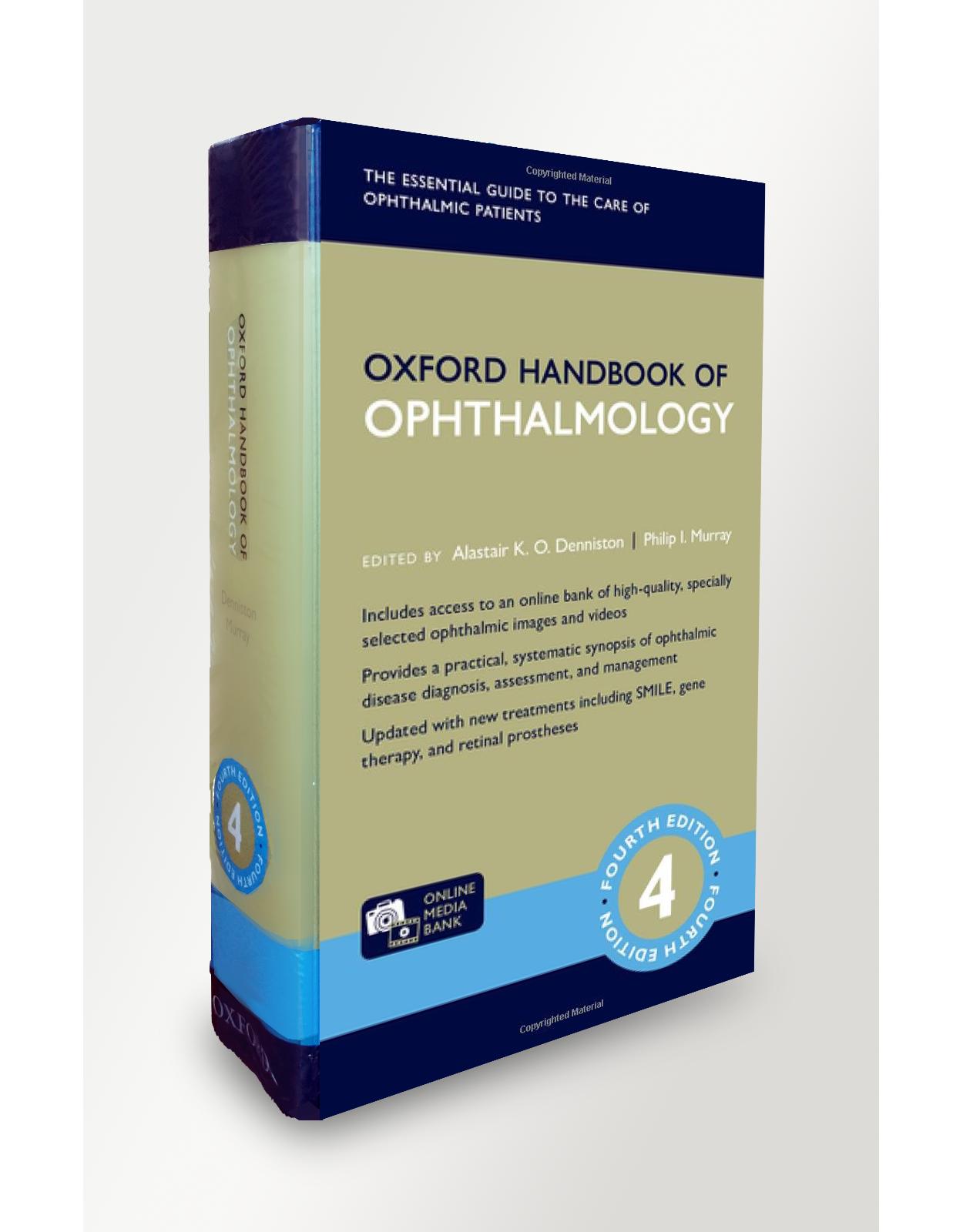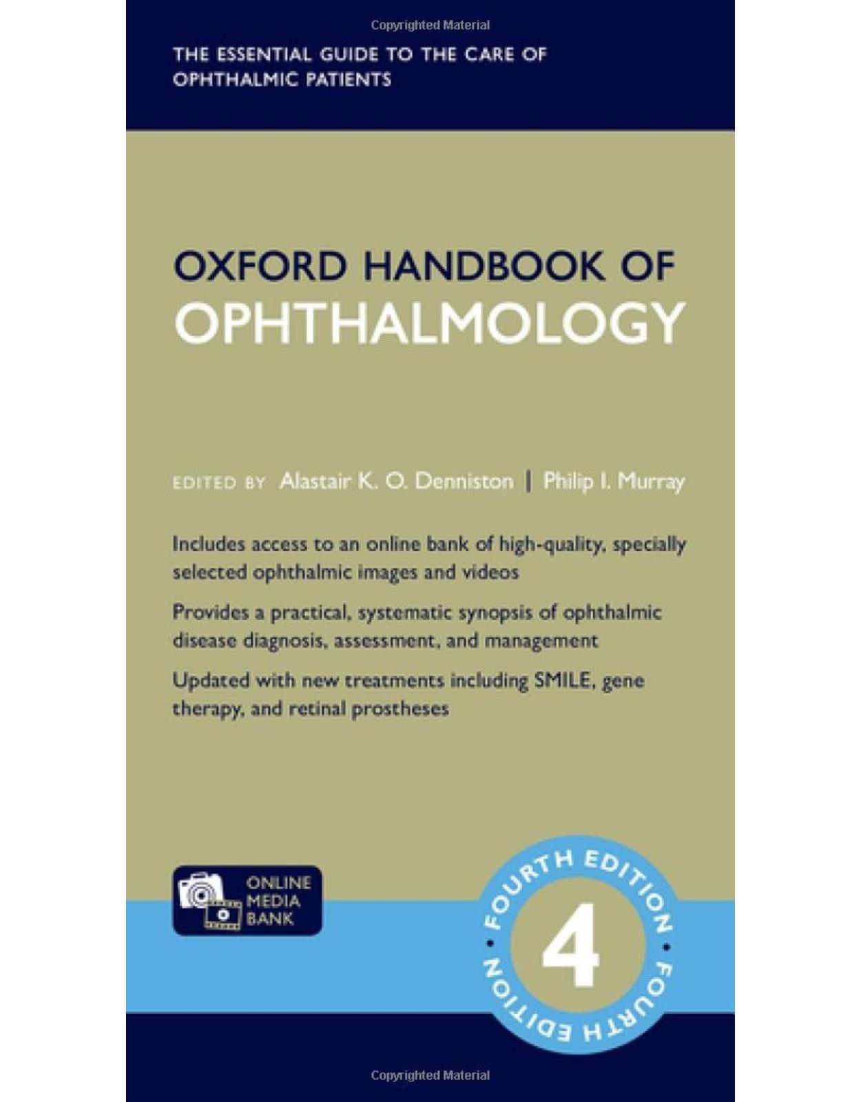
Oxford Handbook of Ophthalmology
Livrare gratis la comenzi peste 500 RON. Pentru celelalte comenzi livrarea este 20 RON.
Disponibilitate: La comanda in aproximativ 4-6 saptamani
Editura: Oxford
Limba: Engleza
Nr. pagini: 1248
Coperta: Flexicover
Dimensiuni: 18.5 x 4.6 x 10.9 cm
An aparitie: 2018
Description:
Fully revised and updated throughout, the fourth edition of the Oxford Handbook of Ophthalmology now includes free access to the ophthalmic online media bank, a selection of high-quality clinical images and videos for a wide breadth of key ophthalmic diseases. Clear, concise, and practical, this handbook provides immediate access to the detailed clinical information you need, in casualty, clinic, theatre, and on the wards. The core of the book comprises a systematic synopsis of ophthalmic disease directed towards diagnosis, interim assessment, and ongoing management. Assessment boxes for common clinical conditions and algorithms for important clinical presentations illustrate this practical approach. The information is easily accessible, presented in a clear format with areas of importance highlighted. Key sections for the trainee include: Clinical Skills, Aids to Diagnosis, Investigations and their Interpretation, Perioperative Care, Theatre Notes and Therapeutics. The wider practise of eye-care is supported by expanded chapters on Refractive Ophthalmology, Vision in Context, Evidence Based Ophthalmology and Resources for Ophthalmologists. Now including newer treatments across a range of specialities such as SMILE, gene-therapy and retinal prostheses, as well as greater emphasis on the evidence underlying current clinical practice and guidelines, this handbook has never been more essential for all those working in eye-care. Whether you want to learn about patient-reported outcomes, identify a surgical instrument, interpret a statistical test, or diagnose and treat ophthalmic emergencies, you will find it here. Whatever your role in caring for patients with eye disease: ophthalmologist, optometrist, orthoptist, ophthalmic nurse, or other health profession - discover for yourself why this handbook has become the 'go-to' resource for tens of thousands of eye-care professionals around the world.
Table of Contents:
Orthoptic abbreviations
1. Clinical skills
Taking an ophthalmic history
Assessment of vision: acuity
Assessment of vision: clinical tests in children and tests of binocular status
Assessment of vision: contrast and colour
Biomicroscopy: slit-lamp overview
Biomicroscopy: use of the slit-lamp
Anterior segment examination (1)
Techniques for anterior segment examination
Anterior segment examination (2)
Gonioscopy
Posterior segment examination
Pupil examination
Ocular motility examination
Visual field (VF) examination
Lids/ptosis examination
Orbital examination
Nasolacrimal system examination
Refraction: outline (1)
Refraction: outline (2)
Focimetry
2. Investigations and their interpretation
Visual field testing: general
Static automated perimetry: performance and interpretation
Automated perimetry: protocols
Glaucoma progression analysis
Goldmann perimetry
Goldmann perimetry (2)
Anterior segment imaging (1)
Anterior segment imaging (2)
Posterior segment imaging
Fundus fluorescein angiography (FFA)
Indocyanine green (ICG) angiography and other vascular assessments
Imaging the retinal nerve fibre layer
Adaptive optics
Optical coherence tomography
OCT angiography (OCTA)
Ophthalmic ultrasonography (1)
Ophthalmic ultrasonography (2)
Electrodiagnostic tests (1)
Electrodiagnostic tests (2)
Electrodiagnostic tests (3)
Ophthalmic radiology: X-ray, dacryocystography (DCG), and dacryoscintigraphy (DSG)
Ophthalmic radiology: CT and CT angiography (CTA)
Ophthalmic radiology: MRI and MR angiography (MRA)/MR venography (MRV)
3. Ocular trauma
Ocular trauma: assessment
Tetanus status and prophylaxis
Chemical injury: assessment
Chemical injury: treatment
Thermal injury/burns: assessment
Thermal injury/burns: management
Orbital fractures: assessment
Orbital fractures: treatment
Lid lacerations
Blunt trauma: assessment
Blunt trauma: treatment
Penetrating trauma/intraocular foreign bodies: assessment
Penetrating trauma/intraocular foreign bodies: treatment
Corneal foreign bodies and abrasions
Hyphaema
Laser trauma
4. Lids
Anatomy and physiology (1)
Anatomy and physiology (2)
Eyelash disorders
Blepharitis and meibomian gland dysfunction (MGD) (1)
Blepharitis and meibomian gland dysfunction (2)
Lid lumps: cysts and abscesses
Lid lumps: benign and premalignant tumours
Lid lumps: malignant tumours (1)
Lid lumps: malignant tumours (2)
Repair of eyelid defects
Ectropion
Entropion
Ptosis: acquired
Ptosis: congenital, p. 180).
Miscellaneous lid disorders
5. Lacrimal
Anatomy and physiology
The watery eye: assessment
The watery eye: treatment
Dacryocystorhinostomy
Lacrimal system infections
6. Conjunctiva
Anatomy and physiology, pp. 196–7.
Conjunctival signs
Conjunctival diagrams
Bacterial conjunctivitis (1)
Bacterial conjunctivitis (2)
Viral conjunctivitis
Chlamydial conjunctivitxis
Allergic conjunctivitis (1)
Allergic conjunctivitis (2)
Cicatricial conjunctivitis (1)
Cicatricial conjunctivitis (2)
Cicatricial conjunctivitis (3)
Cicatricial conjunctivitis (4)
Dry eyes: clinical features (1)
Dry eyes: clinical features (2)
Dry eyes: clinical features (3)
Dry eyes: treatment (1)
Dry eyes: treatment (2)
Ocular neuropathic pain
Miscellaneous conjunctivitis and conjunctival degenerations
Pigmented conjunctival lesions
Non-pigmented conjunctival lesions (1)
Non-pigmented conjunctival lesions (2)
7. Cornea
Anatomy and physiology
Corneal signs
Corneal diagrams
Microbial keratitis: assessment
Microbial keratitis: treatment (1)
Microbial keratitis: treatment (2)
Microbial keratitis: treatment of corneal perforation
Acanthamoeba keratitis
Fungal keratitis: treatment
Fungal keratitis: treatment
Herpes simplex keratitis (1)
Herpes simplex keratitis (2)
Herpes zoster ophthalmicus
Thygeson’s superficial punctate keratopathy
Recurrent corneal erosion syndrome (RCES)
Persistent epithelial defects, p. 272).
Limbal epithelial stem cell deficiency
Corneal degenerative disease (1)
Corneal degenerative disease (2)
Corneal dystrophies: anterior
Corneal dystrophies: stromal (1)
Corneal dystrophies: stromal (2)
Corneal dystrophies: posterior
Keratoconus
Other corneal ectasias
Peripheral ulcerative keratitis (PUK)
Other peripheral corneal diseases
Neurotrophic keratopathy
Exposure keratopathy
Deposition keratopathies
Keratoplasty: penetrating keratoplasty
Keratoplasty: lamellar and endothelial keratoplasty
Keratoplasty: complications
Corneal collagen cross-linking
Amniotic membrane transplantation
Donor eye retrieval and eye banks
8. Sclera
Anatomy and physiology
Episcleritis
Anterior scleritis: outline
Anterior scleritis: non-necrotizing
Anterior scleritis: necrotizing
Posterior scleritis
9. Lens
Anatomy and physiology
Cataract: introduction
Cataract: types
Cataract surgery: assessment
Cataract surgery: consent and planning
Cataract surgery: perioperative, p. 346).
Phacoemulsification (1)
Phacoemulsification (2)
Phacoemulsification (3)
Femtosecond laser (FSL) cataract surgery
Extracapsular, manual small incision, and intracapsular cataract extraction
Intraocular lenses (1)
Intraocular lenses (2)
Intraocular lenses (3)
Cataract surgery: post-operative
Cataract surgery and concurrent eye disease
Cataract surgery: complications
Post-operative endophthalmitis
Toxic anterior segment syndrome
Post-operative cystoid macular oedema
Refractive surprise
Abnormalities of lens size, shape, and position
10. Glaucoma
Anatomy and physiology
Glaucoma: assessment
Ocular hypertension
Primary open-angle glaucoma
Normal-tension glaucoma
Primary angle-closure glaucoma
Acute primary angle closure
Pseudoexfoliation syndrome
Pigment dispersion syndrome
Neovascular glaucoma
Inflammatory glaucoma: general
Inflammatory glaucoma: syndromes
Lens-related glaucoma
Other secondary open-angle glaucoma
Other secondary closed-angle glaucoma
Iatrogenic glaucoma
Pharmacology of IOP-lowering agents
Laser procedures in glaucoma
Surgery for glaucoma
Filtration surgery: trabeculectomy
Filtration surgery: antifibrotics
Filtration surgery: complications of penetrating procedures (1)
Filtration surgery: complications of penetrating procedures (2)
Non-penetrating glaucoma surgery
Micro-invasive glaucoma surgery
11. Uveitis
Anatomy and physiology
Classification of uveitis (1)
Classification of uveitis (2)
Uveitis: assessment
Uveitis: systemic review
Uveitis: investigations (1)
Uveitis: investigations (2)
Uveitis: complications and treatment
Acute anterior uveitis
Uveitis with seronegative spondyloarthropathies
Anterior uveitis syndromes (1), p. 466)
Anterior uveitis syndromes (2)
Uveitis with juvenile idiopathic arthritis
Intermediate uveitis
Retinal vasculitis
Sarcoidosis (1)
Sarcoidosis (2)
Multiple sclerosis
Behçet’s disease
Vogt–Koyanagi–Harada disease
Sympathetic ophthalmia
Viral uveitis (1)
Viral uveitis (2)
Viral uveitis (3)
HIV-associated disease: anterior segment
HIV-associated disease: posterior segment
Mycobacterial disease (1)
Mycobacterial disease (2)
Spirochaetal and other bacterial uveitis
Protozoan uveitis
Nematodal uveitis
Fungal uveitis
White dot syndromes (1)
White dot syndromes (2)
White dot syndromes (3)
12. Vitreoretinal
Anatomy and physiology
Retinal detachment: assessment
Peripheral retinal degenerations
Retinal breaks
Posterior vitreous detachment
Rhegmatogenous retinal detachment (1)
Rhegmatogenous retinal detachment (2)
Tractional retinal detachment
Exudative retinal detachment
Retinoschisis
Hereditary vitreoretinal degenerations
Choroidal detachments and uveal effusion syndrome
Epiretinal membranes
Vitreomacular interface
Submacular (subfoveal) haemorrhage: Treatment, p. 554).
Laser retinopexy and cryopexy for retinal tears
Pneumatic retinopexy
Scleral buckling procedures
Vitrectomy: outline
Vitrectomy: heavy liquids and tamponade agents
Gene therapy for inherited retinal diseases
Retinal prosthesis
13. Medical retina
Anatomy and physiology (1), pp. 570–1.
Anatomy and physiology (2)
Age-related macular degeneration (1)
Age-related macular degeneration (2)
Age-related macular degeneration (3)
Age-related macular degeneration (4)
Age-related macular degeneration (5)
Anti-vascular endothelial growth factor therapy: outline
Anti-vascular endothelial growth factor therapy: in practice
Photodynamic therapy
Diabetic eye disease: general
Diabetic eye disease: assessment
Diabetic eye disease: management
Diabetic eye disease: screening
Central serous chorioretinopathy
Cystoid macular oedema
Degenerative myopia
Angioid streaks
Choroidal folds
Retinal vein occlusion: CRVO (1)
Retinal vein occlusion: CRVO (2)
Retinal vein occlusion: BRVO, HRVO
Retinal artery occlusion (1)
Retinal artery occlusion (2)
Ocular ischaemic syndrome
Hypertensive retinopathy
Haematological disease
Retinal conditions associated with renal disease
Retinal telangiectasias
Other retinal vascular anomalies
Radiation retinopathy
Retinitis pigmentosa (1)
Retinitis pigmentosa (2)
Congenital stationary night blindness
Inherited disorders of cone function
Macular dystrophies (1)
Macular dystrophies (2)
Chorioretinal dystrophies
Albinism
Toxic retinopathies (1)
Toxic retinopathies (2)
Toxic retinopathies (3)
Miscellaneous disorders
14. Orbit
Anatomy and physiology
Orbital and preseptal cellulitis
Mucormycosis (phycomycosis)
Thyroid eye disease: general
Thyroid eye disease: assessment
Thyroid eye disease: management
Other orbital inflammations (1)
Other orbital inflammations (2)
Cystic lesions
Orbital tumours: lacrimal and neural
Orbital tumours: vascular
Orbital tumours: lymphoproliferative
Orbital tumours: other
Vascular lesions
Disorders of the anophthalmic socket
15. Intraocular tumours
Iris tumours
Ciliary body tumours
Choroidal melanoma
Choroidal naevus
Choroidal haemangiomas
Other choroidal tumours
Retinoblastoma (1)
Retinoblastoma (2)
Retinoblastoma (3)
Retinal vascular tumours (1)
Retinal vascular tumours (2)
Other retinal tumours
Retinal pigment epithelium tumours
Lymphoma
16. Neuro-ophthalmology
Anatomy and physiology (1)
Anatomy and physiology (2)
Anatomy and physiology (3)
Optic neuropathy: assessment
Typical optic neuritis
Multiple sclerosis
Neuromyelitis optica spectrum disorders (NMOSD)
Atypical optic neuritis
Anterior ischaemic optic neuropathy, p. 743);
Arteritic AION and giant cell arteritis
Temporal artery biopsy
Non-arteritic AION
Posterior ischaemic optic neuropathy (PION)
Other optic neuropathies/atrophies
Papilloedema
Idiopathic intracranial hypertension
Pseudopapilloedema
Congenital optic disc anomalies
Chiasmal disorders
Retrochiasmal disorders
Migraine
Supranuclear eye movement disorders
Supranuclear eye movement disorders (2)
Third nerve disorders
Fourth nerve disorders
Sixth nerve disorders
Seventh nerve disorders
Anisocoria, pp. 794–5.
Anisocoria: sympathetic chain, pp. 796–7.
Anisocoria: parasympathetic chain
Argyll Robertson pupils
Nystagmus (1)
Nystagmus (2)
Nystagmus (3)
Saccadic oscillations and intrusions
Myasthenia gravis
Other disorders of the neuromuscular junction
Myopathies, pp. 812–13,
Blepharospasm and other dystonias
Functional visual loss
17. Strabismus
Anatomy and physiology (1)
Anatomy and physiology (2)
Binocular single vision
Strabismus: assessment
Strabismus diagrams
Hess and Lees charts
Strabismus: outline, p. 834.
Concomitant strabismus: esotropia (1)
Concomitant strabismus: esotropia (2)
Concomitant strabismus: exotropia
Incomitant strabismus
Restriction syndromes
Alphabet patterns
Strabismus surgery: general
Strabismus surgery: horizontal
18. Paediatric ophthalmology
Embryology (1)
Embryology (2)
Genetics
Ophthalmic assessment in a child (1)
Ophthalmic assessment in a child (2)
The child who does not see
➲Amblyopia
p
Common clinical presentations: red eye, watering, and photophobia
Common clinical presentations: proptosis and globe size
Common clinical presentations: cloudy cornea and leucocoria
Intrauterine infections (1)
Intrauterine infections (2)
Ophthalmia neonatorum, pp. 882–3.
➲Orbital and preseptal cellulitis (paediatric)
➲ Congenital cataract: assessment
➲ Congenital cataract: surgery
➲ Congenital cataract: complications
Uveitis in children
➲Glaucoma in children: assessment
➲Glaucoma in children: treatment
➲Retinopathy of prematurity (1)
Retinopathy of prematurity (2)
Other retinal disorders
Developmental abnormalities: craniofacial and globe
Developmental abnormalities: anterior segment
Developmental abnormalities: posterior segment
Chromosomal syndromes
Metabolic and storage diseases (1)
Metabolic and storage diseases (2)
Phakomatoses
Child abuse
19. Refractive ophthalmology
Refractive error: introduction
Myopia, hypermetropia, and astigmatism
Spectacles: types
Spectacles: materials
Spectacles: prescribing
Contact lenses: outline
Contact lenses: hard and RGP lenses
Contact lenses: hydrogel lenses
Contact lenses: fitting
Contact lenses: complications
Introduction to refractive surgery
Biophysics of refractive lasers (1)
Biophysics of refractive lasers (2)
Excimer laser refractive surgery: preoperative evaluation
Excimer laser refractive surgery: contraindications
Excimer laser refractive surgery: surface treatments
Excimer laser refractive surgery: LASIK
Femtosecond laser refractive surgery: SMILE
Complications of refractive laser: immediate and early
Complications of refractive laser: late
Incisional refractive surgery
Intracorneal ring segments
Collagen shrinkage procedures
➲ Lens-based techniques
20. Aids to diagnosis
Acute red eye
Sudden/recent loss of vision
Gradual loss of vision
The watery eye
Flashes and floaters
Headache
Diplopia
Anisocoria
Nystagmus
Ophthalmic signs: external
Ophthalmic signs: anterior segment (1)
Ophthalmic signs: anterior segment (2)
Ophthalmic signs: anterior segment (3)
Ophthalmic signs: posterior segment (1)
Ophthalmic signs: posterior segment (2)
Ophthalmic signs: visual fields
21. Vision in context
Low vision: taking a history
Low vision: assessing visual function
Low vision: doing something useful (1)
Low vision: doing something useful (2)
Visual impairment registration (1)
Visual impairment registration (2)
Driving standards (1)
(2), pp. 1024–
Pilot standards
NHS (UK), p. 1030).
22. Surgery: anaesthetics and perioperative care
Preoperative assessment
Preoperative preparation
Preoperative management: patients with diabetes
Preoperative management: other special patient groups
Ocular anaesthesia: topical and local
Ocular anaesthesia: sub-Tenon’s block
Ocular anaesthesia: peribulbar block
Ocular anaesthesia: general anaesthesia, p. 1046).
Treatment of anaphylaxis
Hypoglycaemia
Needle-stick injuries
Management of severe local anaesthetic toxicity
Basic and advanced life support
23. Surgery: theatre notes
Sterilization services
Hand hygiene
Suture materials and needle types
Surgical instruments (1)
Surgical instruments (2)
Surgical instruments (3)
24. Laser
Outline
Laser reactions
Clinical applications
Laser safety in the clinic
Laser procedures in retina
Laser procedures in glaucoma (1)
Laser procedures in glaucoma (2), p. 1086).
Laser procedures in lens/cataract
25. Therapeutics
Principles and delivery of ocular drugs
Intracameral injections
Sub-Tenon’s and peribulbar injections
Intravitreal injections
Topical antimicrobials
Topical anti-inflammatory agents
Topical glaucoma medications
Topical mydriatics
Topical anaesthetics
Topical tear replacement
Systemic medication: antimicrobials
Systemic medication: glaucoma
➲Systemic corticosteroids: general
➲Systemic corticosteroids: prophylaxis
➲Antimetabolites, calcineurin inhibitors, and cytotoxics
➲Biologics
26. Evidence-based ophthalmology
Evidence-based medicine
Study design (1)
Study design (2)
Critical appraisal
Clinical guidelines
NICE (National Institute for Health and Care Excellence) guidelines
Healthcare economics
Patient-reported outcomes (PROs)
Statistical terms
Statistical tests
Risks, odds, and number needed to treat (NNT)
Statistical issues relating to two eyes
Investigations, p. 1154).
Bayesian vs frequentist approaches
27. Resources
Eponymous syndromes
Web resources for ophthalmologists (1)
Web resources for ophthalmologists (2)
Web resources for ophthalmologists (3)
Web resources for patients
Reference intervals
Image Bank Introduction
Chapter 1 Clinical skills Image Bank
Chapter 2 Investigations and their interpretation Image Bank
Chapter 3 Ocular trauma Image Bank
Chapter 4 Lids Image Bank
Chapter 5 Lacrimal Image Bank
Chapter 6 Conjunctiva Image Bank
Chapter 7 Cornea Image Bank
Chapter 8 Sclera Image Bank
Chapter 9 Lens Image Bank
Chapter 10 Glaucoma Image Bank
Chapter 11 Uveitis Image Bank
Chapter 12 Vitreoretinal Image Bank
Chapter 13 Medical retina Image Bank
Chapter 14 Orbit Image Bank
Chapter 15 Intraocular tumours Image Bank
Chapter 16 Neuro-ophthalmology Image Bank
Chapter 17 Strabismus Image Bank
Chapter 18 Paediatric ophthalmology Image Bank
Chapter 19 Refractive ophthalmology Image Bank
Chapter 23 Surgery: theatre notes Image Bank
Index
| An aparitie | 2018 |
| Autor | Alastair K. O. Denniston, Philip I. Murray |
| Dimensiuni | 18.5 x 4.6 x 10.9 cm |
| Editura | Oxford |
| Format | Flexicover |
| ISBN | 9780198804550 |
| Limba | Engleza |
| Nr pag | 1248 |


