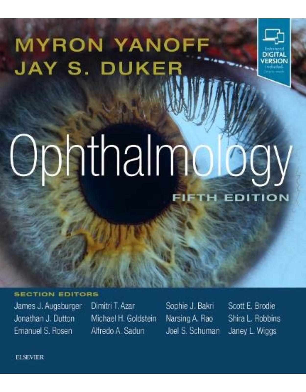
Ophthalmology, 5th Edition
Livrare gratis la comenzi peste 500 RON. Pentru celelalte comenzi livrarea este 20 RON.
Disponibilitate: La comanda in aproximativ 4 saptamani
Autor: Myron Yanoff, Jay S. Duker
Editura: Elsevier
Limba: Engleza
Nr. pagini: 1440
Coperta: Hardback
Dimensiuni:
An aparitie: End of Oct. 2018
Description:
Long considered one of ophthalmology’s premier texts, this award-winning title by Drs. Myron Yanoff and Jay S. Duker remains your go-to reference for virtually any topic in this fast-changing field. It offers detailed, superbly illustrated guidance on nearly every ophthalmic condition and procedure you may encounter, making it a must-have resource no matter what your level of experience. Extensive updates throughout keep you current with all that’s new in every subspecialty area of the field.
Table of Contents:
Part 1 Genetics
1.1 Fundamentals of Human Genetics
Key Features
DNA and the Central Dogma of Human Genetics
Human Genome
Basic Mendelian Principles
Mutations
Genes and Phenotypes
Patterns of Human Inheritance
Molecular Mechanisms of Disease
Gene Therapy
Key References
References
1.2 Molecular Genetics of Selected Ocular Disorders
Key Features
Introduction
Dominant Corneal Dystrophies
Aniridia, Peter's Anomaly, Autosomal Dominant Keratitis
Rieger's Syndrome
Juvenile Glaucoma
Congenital Glaucoma
Nonsyndromic Congenital Cataract
Retinitis Pigmentosa
Stargardt Disease
X-Linked Juvenile Retinoschisis
Norrie's Disease
Sorsby's Macular Dystrophy
Gyrate Atrophy
Color Vision
Retinoblastoma
Albinism
Leber's Optic Neuropathy
Congenital Fibrosis Syndromes and Disorders of Axon Guidance
Autosomal Dominant Optic Atrophy
Complex Traits
Key References
References
1.3 Genetic Testing and Genetic Counseling
Key Features
Genetic Testing
Genetic Counseling
Key References
References
Part 2 Optics and Refraction
2.1 Light
Key Features
Introduction
Geometrical Optics
Basic Stigmatic Optics
Astigmatic Optics
Wave Properties of Light
Key References
References
2.2 Optics of the Human Eye
Key Features
Introduction
Cornea
Pupil
Lens
Accommodation
Scattering
Aberrations
Retina
Resolution and Focal Length
Refractive Errors
Key References
References
2.3 Clinical Refraction
Key Features
Introduction
History
Visual Acuity
Spherical Equivalent
Detecting Astigmatism
Refracting at Near
Additional Subjective Techniques
Retinoscopy
Key References
References
2.4 Correction of Refractive Errors
Key Features
Introduction
Spectacle Correction
Contact Lenses
Intraocular Lenses
Keratorefractive Surgery
Corneal Inlays
Key References
References
2.5 Ophthalmic Instruments
Key Features
Introduction
Direct Ophthalmoscope
Binocular Indirect Ophthalmoscope
Fundus Camera
Optical Coherence Tomography
Slit-Lamp Biomicroscope
Slit-Lamp Fundus Lenses
Goldmann Applanation Tonometer
Operating Microscope
Keratometer and Corneal Topographer
Lensmeter
Automated Refractor
Magnifying Devices
Key References
References
2.6 Wavefront Optics and Aberrations of the Eye
Key Features
Introduction
Ray Aberrations
The Wavefront Approach to Aberrations
Spherical Aberration
Coma
Distortion
Field Curvature (FC)
Oblique Astigmatism (OA)
Higher-Order Aberrations (HOA)
Chromatic Aberration
Measurement of Ocular Aberrations
An Overall Perspective on Aberration Theory
Key References
References
Part 3 Refractive Surgery
3.1 Current Concepts, Classification, and History of Refractive Surgery
Key Features
Associated Features
Introduction
Excimer Laser and Ablation Profiles
Concepts in Development
Classification of Refractive Procedures
Summary
Key References
References
3.2 Preoperative Evaluation for Refractive Surgery
Key Features
Introduction
Ophthalmic Examination
Ancillary Testing
Key References
References
3.3 Excimer Laser Surface Ablation
Key Features
Introduction
Ablation Profiles
Indications
Preoperative Evaluation
PRK Surgical Technique
Results
Complications
Conclusions
Key References
References
3.4 LASIK
Key Features
Associated Features
Historical Review
LASIK
LASIK Enhancements
LASIK in Complex Cases
Summary
Key References
References
3.5 Small Incision Lenticule Extraction (SMILE)
Key Features
Introduction
Femtosecond Laser System
Treatment Range for SMILE
Patient Evaluation
Surgical Procedure
Complications
Higher-Order Aberrations
Biomechanical Stability
Refractive and Visual Outcome
Retreatments
Conclusions
Key References
References
3.6 Wavefront-Based Excimer Laser Refractive Surgery
Key Features
Introduction
Wavefront Optics
Higher-Order Aberrations
Ideal Corneal Shape
Measurements of Wavefront Aberrations
Quality of Vision and Measures of Optical Quality
Wavefront-Measuring Devices
Wavefront-Based Surgery
Results
Wavefront Platforms (Table 3.6.1)
Conclusions
Key References
References
3.7 Phakic Intraocular Lenses
Key Features
Associated Features
Introduction
History of Phakic Lenses
Indications of Phakic Lenses
Advantages and Disadvantages of Phakic Iols
Intraocular Lens Power Calculation
Visual Outcomes
Anterior Chamber Angle-Supported Phakic Intraocular Lenses
Iris-Fixated Phakic Intraocular Lenses
Posterior Chamber Phakic Intraocular Lenses
Bioptics
Conclusion
Key References
References
3.8 Astigmatic Keratotomy
Key Features
Historical Review
Surgical Techniques for Astigmatic and Radial Keratotomy
Outcome Comparison for Various Astigmatism Correction Methods
Complications and Management of Astigmatism Correction Methods
Conclusions
Key References
References
3.9 Intrastromal Corneal Ring Segments and Corneal Cross-Linking
Key Features
Introduction
Intracorneal Ring Segments
Surgical Procedure: ICRS
Surgical Procedure: CXL
CXL Clinical Outcomes
Combining ICRS With CXL
Conclusions
Key References
References
3.10 Surgical Correction of Presbyopia
Key Features
Introduction
Presbyopia Correction at the Corneal Level
Key References
References
Part 4 Cornea and Ocular Surface Diseases
Section 1 Basic Principles
4.1 Corneal Anatomy, Physiology, and Wound Healing
Key Features
Introduction
Corneal Wound Healing
Key References
References
4.2 Anterior Segment Imaging Modalities
Key Features
Introduction
Anterior Segment Optical Coherence Tomography
Specular Microscopy
Ultrasound Biomicroscopy
Meibography
In vivo Confocal Microscopy
Topography and Tomography
Wavefront Analysis
Summary
Key References
References
Section 2 Congenital Abnormalities
4.3 Congenital Corneal Anomalies
Key Features
Introduction
Size and Shape Anomalies
Anomalies of Corneal Clarity
Key References
References
Section 3 External Diseases
4.4 Blepharitis
Key Features
Associated Features
Introduction
Epidemiology
Pathogenesis
Ocular Manifestations
Diagnosis and Ancillary Testing
Treatment
Key References
References
4.5 Herpes Zoster Ophthalmicus
Key Features
Associated Features
Epidemiology and Pathogenesis
Clinical Manifestations
Herpes Zoster Ophthalmicus in Acquired Immune Deficiency Syndrome
Diagnosis
Management
Prevention
Key References
References
Section 4 Conjunctival Diseases
4.6 Conjunctivitis
Key Features
Infectious Conjunctivitis
Noninfectious Conjunctivitis
Key References
References
4.7 Allergic Conjunctivitis
Key Features
Acute Allergic Conjunctivitis: Seasonal/Perennial
Chronic Atopic Keratoconjunctivitis
Vernal Conjunctivitis
Treatment of Allergic/Atopic Keratoconjunctivitis
Allergic Dermatoconjunctivitis
Microbial Allergic Conjunctivitis
Giant Papillary Conjunctivitis
Key References
References
4.8 Tumors of the Conjunctiva
Key Features
Conjunctival Malignant Neoplasms
Benign Conjunctival Neoplasms and Neoplasias
Nonneoplastic Lesions and Disorders Simulating Malignant Conjunctival Neoplasms and Neoplasias
Management of Conjunctival Tumors Suspected of Being Malignant Neoplasms or Neoplasias
Key References
References
4.9 Pterygium and Conjunctival Degenerations
Key Features
Associated Features
Introduction
Pinguecula
Pterygium
Senile Scleral Plaques
Conjunctival Amyloid
Conjunctival Melanosis
Key References
References
4.10 Ocular Cicatricial Pemphigoid/Mucous Membrane Pemphigoid
Key Features
Associated Features
Introduction
Pathogenesis
Clinical Findings
Diagnosis
Treatment
Conclusions
Key References
References
Section 5 Scleral and Episcleral Diseases
4.11 Episcleritis and Scleritis
Key Features
Associated Feature
Key Features
Associated Features
Introduction
Inflammatory Diseases
Key References
References
Section 6 Corneal Diseases
4.12 Bacterial Keratitis
Key Feature
Associated Features
Introduction
Epidemiology and Pathogenesis
Clinical Features
Diagnosis
Differential Diagnosis
Systemic Associations
Pathology
Treatment
Outcome
Key References
References
4.13 Fungal Keratitis
Key Feature
Associated Features
Introduction
Epidemiology and Pathogenesis
Clinical Features
Diagnosis
Differential Diagnosis
Systemic Associations
Pathology
Treatment
Outcome
Key References
References
4.14 Parasitic Keratitis
Key Feature
Associated Features
Introduction
Acanthamoeba Keratitis
Microsporidiosis
Onchocerciasis
Key References
References
4.15 Herpes Simplex Keratitis
Key Features
Associated Features
Epidemiology
Herpes Simplex Virus
Primary HSV Infection
Recurrent HSV Infections
Diagnosis
Herpetic Eye Disease Study
Treatment
Future Directions
Key References
References
4.16 Peripheral Ulcerative Keratitis
Key Features
Associated Features
Introduction
Anatomy and Pathogenesis
Ocular Manifestations
Systemic Associations
Differential Diagnosis
Diagnostic and Ancillary Testing
Treatment
Course and Outcome
Key References
References
4.17 Noninfectious Keratitis
Key Feature
Associated Feature
Introduction
Thygeson's Superficial Punctate Keratitis
Superior Limbic Keratoconjunctivitis of Theodore
Mooren's Ulcer
Neurotrophic Keratitis
Terrien's Marginal Degeneration
Lax Eyelid Condition, Lax Eyelid Syndrome, and Floppy Eyelid Syndrome
Treatment
Key References
References
4.18 Keratoconus and Other Ectasias
Key Features
Associated Features
Keratoconus
Pellucid Corneal Degeneration
Keratoglobus
Posterior Keratoconus
Post–Refractive Surgery Corneal Ectasia
Key References
References
4.19 Anterior Corneal Dystrophies
Key Features
Associated Features
Introduction
Anterior Basement Membrane Dystrophy
Meesmann's Epithelial Dystrophy
Reis–Bückler Dystrophy
Thiel–Behnke Dystrophy
Key References
References
4.20 Stromal Corneal Dystrophies
Key Features
Associated Features
Introduction
Gelatinous Drop-Like Dystrophy
Lattice Dystrophy Type I
Systemic Amyloidosis With Corneal Lattice
Granular Corneal Dystrophy Type I
Granular Corneal Dystrophy Type 2
Macular Corneal Dystrophy
Pathology
Schnyder's Corneal Dystrophy
Central Cloudy Dystrophy
Fleck Dystrophy
Posterior Amorphous Corneal Dystrophy
Congenital Stromal Corneal Dystrophy
Key References
References
4.21 Diseases of the Corneal Endothelium
Key Features
Associated Features
Introduction
Fuchs' Dystrophy
Congenital Hereditary Endothelial Dystrophy
Posterior Polymorphous Corneal Dystrophy
Key References
References
4.22 Corneal Degenerations
Key Features
Associated Features
Introduction
Corneal Arcus (Arcus Senilis)
Lipid Keratopathy
Vogt's White Limbal Girdle
Senile Corneal Furrow Degeneration
Terrien's Marginal Corneal Degeneration
Peripheral Corneal Guttae
Calcific Band Keratopathy
Spheroidal Degeneration
Iron Deposition
Crocodile Shagreen
Cornea Farinata
Salzmann's Corneal Degeneration
Corneal Keloids
Corneal Amyloid Degeneration
Key References
References
4.23 Dry Eye Disease
Key Features
Associated Features
Introduction
Epidemiology
Pathogenesis
Ocular Manifestations
Diagnosis and Ancillary Testing
Treatment
Key References
References
Section 7 Miscellaneous Conditions
4.24 Complications of Contact Lens Wear
Key Features
Introduction
Toxic, Allergic and Mechanical Reactions
Conditions Reflecting Metabolic Challenge
Corneal Inflammatory Events and Microbial Keratitis
Unsupervised Lens Wear
FDA and CDC Recommendations
Conclusions
Key References
References
4.25 Corneal and External Eye Manifestations of Systemic Disease
Key Feature
Associated Feature
Introduction
Congenital Disorders
Chromosomal Disorders
Inherited Connective Tissue Disorders
Metabolic Disorders
Other Oculosystemic Disorders
Conclusions
Key References
References
Section 8 Trauma
4.26 Acid and Alkali Burns
Key Features
Associated Feature
Introduction
Alkali Injuries
Acid Injuries
Pathophysiology
Clinical Course
Therapy
Key References
References
Section 9 Surgery
4.27 Corneal Surgery
Key Features
Associated Feature
Keratoplasty
Superficial Corneal Procedures
Phototherapeutic Keratectomy
Key References
References
4.28 Conjunctival Surgery
Key Features
Associated Feature
Historical Review
Anesthesia
Specific Techniques
Key References
References
4.29 Endothelial Keratoplasty
Key Feature
Associated Features
Introduction
Evolution of EKP Techniques
Indications
Surgical Technique
Combined Procedures
Outcomes
Outlook
Key References
References
4.30 Surgical Ocular Surface Reconstruction
Key Features
Associated Features
Introduction
Historical Perspectives
Preoperative Considerations
Operative Procedures
Special Considerations in Ocular Surface Reconstruction
Conclusions
Key References
References
4.31 Management of Corneal Thinning, Melting, and Perforation
Key Features
Associated Features
Introduction
Corneal Thinning From Noninflammatory Disorders
Corneal Thinning and Melting From Inflammatory Disorders
Surgical Treatment of Corneal Perforations
Conclusions
Key References
References
Part 5 The Lens
5.1 Basic Science of the Lens
Key Features
Introduction
Anatomy of the Lens
Physiology of the Lens
Biophysics
Biochemistry
Lens Crystallins
Age Changes
Secondary Cataract
Key References
References
5.2 Evolution of Intraocular Lens Implantation
Key features
Introduction
Lens Design and Fixation
Recent Advances
Key References
References
5.3 Epidemiology, Pathophysiology, Causes, Morphology, and Visual Effects of Cataract
Morphology and Visual Effects of Cataract
Key References
References
5.4 Patient Workup for Cataract Surgery
Assessment of Lens Opacities,
Investigations for Further Surgical Refinement
Good Clinical Practice (Social and Legal Aspects)
Key References
References
5.5 Intraocular Lens Power Calculations
Key Features
Introduction
Ocular Biometry
IOL Power Formulas
IOL Calculations in Special Eyes
Toric IOL Calculation
Intraoperative Wavefront Aberrometry
Postoperative IOL Adjustment
Conclusion
Key References
References
5.6 Indications for Lens Surgery/Indications for Application of Different Lens Surgery Techniques
Key Features
Introduction
Medical Indications for Lens Surgery
Refractive Indications for Lens Surgery
Indications for Different Lens Surgery Techniques
Key References
References
5.7 The Pharmacotherapy of Cataract Surgery
Key Features
Introduction
Preoperative Medications
Anesthetics
Intraoperative Medications
Ophthalmic Viscosurgical Devices
Postoperative Medications
Late Postoperative Medications
Key References
References
5.8 Anesthesia for Cataract Surgery
Key Features
Introduction
Medical Aspects of Anesthesia for Cataract Surgery
Local Anesthesia
Sedative Agents
General Anesthesia
Postoperative Care
Key References
References
5.9 Phacoemulsification
Key Features
Introduction
Handpieces and Tips
Power Modulation
Pumps and Fluidics
Postocclusion Surge
Key References
References
5.10 Refractive Aspects of Cataract Surgery
Key Features
Introduction
Value of Corneal Topography
Intraoperative Management of Preoperative Corneal Astigmatism to Prevent Induction of Corneal Astigmatism
To Treat Preoperative Corneal Astigmatism
Toric Intraocular Lens Implantation
Postoperative Management of Residual or Induced Corneal Astigmatism
Post–Cataract Surgery Piggyback IOLs
Light-Adjustable Intraocular Lens Implant
Key References
References
5.11 Small Incision and Femtosecond Laser-Assisted Cataract Surgery
Key Features
Associated Feature
Introduction
Incision Construction and Architecture
Continuous Curvilinear Capsulorrhexis
Hydrodissection and Hydrodelineation
Nucleofractis Techniques
Power Modulations
Biaxial Microincision Cataract Surgery
B-MICS Vertical Chop Technique
Femtosecond Laser-Assisted Cataract Surgery
Conclusions
Key References
References
5.12 Manual Cataract Extraction
Key Features
Introduction
Manual (Large-Incision) Cataract Surgery
Intracapsular Cataract Extraction
Extracapsular Cataract Extraction
Complications
Discussion
Key References
References
5.13 Combined Procedures
Key Features
Introduction
Combined Glaucoma Surgery
Lens Surgery Combined With Keratoplasty
Combined Phacovitrectomy
Key References
References
5.14 Cataract Surgery in Complex Eyes
Key Features
Associated Features
Introduction
Zonular Instability
Uveitis
Compromised Endothelium
Key References
References
5.15 Pediatric Cataract Surgery
Key Features
Introduction
Historical Review
Preoperative Evaluation and Diagnostic Approach
Alternatives to Surgery
Anesthesia
General Techniques
Specific Techniques
Choices for Correction of Aphakia in Children
Complications
Outcome
Key References
References
5.16 Complications of Cataract Surgery
Key Features
Associated Feature
Introduction
Intraoperative Complications
Postoperative Complications
Key References
References
5.17 Secondary Cataract
Key Features
Introduction
Pathogenesis
Intraocular Lenses Maintaining the Capsular Bag Open or Expanded
Key References
References
5.18 Outcomes of Cataract Surgery
Key Features
Introduction
Evaluation of Outcomes
Five Parameters That Describe Visual Function
Objective Findings of Cataract Surgery Outcomes
Subjective Findings of Cataract Surgery Outcomes
Cataract Surgery of One or Both Eyes
Cataract Surgery in Eyes With Ocular Comorbidity
Summary
Key References
References
Part 6 Retina and Vitreous
Section 1 Anatomy
6.1 Structure of the Neural Retina
Key Feature
Introduction
Center of the Macula: Umbo
Foveola
Fovea
Parafovea
Perifovea
Macula, or Central Area
Extra-Areal Periphery
Layers of the Neural Retina
Key References
References
6.2 Retinal Pigment Epithelium
Key Features
Introduction
Structure and Metabolism
Membrane Properties and Fluid Transport
Photoreceptor–Retinal Pigment Epithelium Interactions
Repair, Regeneration, and Therapy
References
6.3 Retinal and Choroidal Circulation
Key Features
Introduction
Posterior Segment Vascular Anatomy
Blood–Retinal Barrier
Retinal and Choroidal Blood Flow
Retina and Choroidal Circulation Evaluated by OCTA
Regulation of Retinal and Choroidal Blood Flows
Key References
References
6.4 Vitreous Anatomy and Pathology
Key Features
Associated Features
Introduction
Molecular Morphology
Vitreous Anatomy
Age-Related Changes
Key References
References
Section 2 Ancillary Tests
6.5 Contact B-Scan Ultrasonography
Key Features
Associated Features
Introduction
Devices
Technique of Examination
Concepts of B-Scan Interpretation
Display Presentation and Documentation
Digital Contact Ultrasonography
What's New?
Summary
Key References
References
6.6 Camera-Based Ancillary Retinal Testing
Key Features
Key Features
Key Features
Fluorescein Angiography
Indocyanine Green Angiography
Fundus Autofluorescence
Acknowledgments
Key References
References
6.7 Optical Coherence Tomography in Retinal Imaging
Key Features
Associated Features
Introduction
OCT Technology Platforms
Anatomical Results
Image Optimization
OCT Image Interpretation
Key References
References
6.8 Optical Coherence Tomography Angiography
Key Features
Introduction
Biological Basis of OCTA
OCTA Versus Dye-Based Angiography Methods
Key Applications of SD-OCTA
Conclusions
Key References
References
6.9 Retinal Electrophysiology
Key Features
Introduction
Full-Field ERG
Multifocal ERG
Electro-oculography
Key References
References
Section 3 Basic Principles of Retinal Surgery
6.10 Light and Laser Injury
Key Features
Associated Features
Introduction
Light Interaction With the Retina
Photic Retinopathy
Light Exposure and Age-Related Macular Degeneration
Laser Injury
Laser Pointers
Complications of Therapeutic Retinal Laser Photocogulation
Key References
References
6.11 Scleral Buckling Surgery
Key Features
Associated Features
Introduction
Historical Review
Preoperative Evaluation and Diagnostic Approach
Differential Diagnosis
Alternatives to Scleral Buckling
Anesthesia
General Techniques
Complications
Outcome
Key References
References
6.12 Vitrectomy
Key Features
Introduction
Historical Review
Preoperative Evaluation and Diagnostic Approach
Indications and Alternatives to Surgery
Anesthesia
General Techniques
Specific Techniques
Complications
Outcomes
Key References
References
6.13 Intravitreal Injections and Medication Implants
Key Features
Associated Features
Introduction
Preinjection Preparation
Injection
Postinjection
Complications
Other Considerations
Implants
Conclusions
Key References
References
Section 4 Dystrophies
6.14 Progressive and “Stationary” Inherited Retinal Degenerations
Key Features
Associated Features
Key Features
Associated Features
Progressive Diffuse/Panretinal Degenerations
“Stationary” Retinal Disorders
Female Carriers of X-Linked Retinal Degenerations
Treatment of Retinal Degenerations
Key References
References
6.15 Macular Dystrophies
Stargardt Disease and Fundus Flavimaculatus
Vitelliform Macular Dystrophy (Best Disease)
Adult Vitelliform Macular Dystrophy/ Adult-Onset Foveomacular Dystrophy (Pattern Dystrophy)
Dominant Drusen (Doyne's Drusen, Malattia Leventinese)
Pattern Dystrophy
Dominant Cystoid Macular Edema
Sorsby's Macular Dystrophy
North Carolina Macular Dystrophy
Atrophia Areata
Cone Dystrophy
Central Areolar Choroidal Dystrophy
Key References
References
6.16 Choroidal Dystrophies
Choroideremia
Gyrate Atrophy
Key References
References
6.17 Hereditary Vitreoretinopathies
Stickler's Syndrome
X-Linked Juvenile Retinoschisis
Autosomal Dominant Vitreoretinochoroidopathy
Familial Exudative Vitreoretinopathy
Norrie's Disease
Key References
References
Section 5 Vascular Disorders
6.18 Hypertensive Retinopathy
Chronic Hypertensive Retinopathy
Malignant Acute Hypertensive Retinopathy
Key References
References
6.19 Retinal Arterial Obstruction
Central Retinal Artery Obstruction
Branch Retinal Artery Obstruction
Central Retinal Artery Obstruction
Branch Retinal Artery Obstruction
Ophthalmic Artery Obstruction
Cilioretinal Artery Obstruction
Combined Artery and Vein Obstructions
Key References
References
6.20 Venous Occlusive Disease of the Retina
Central Retinal Vein Occlusion
Branch Retinal Vein Occlusion
Central Retinal Vein Occlusion
Branch Retinal Vein Occlusion
Key References
References
6.21 Retinopathy of Prematurity
Key Features
Associated Features
Introduction
Pathogenesis
Clinical Features and Classification
Diagnosis and Screening
Role of Telemedicine in ROP Screening
Differential Diagnosis
Pathology
Treatment
Ablation of Peripheral Avascular Retina
Role of Anti-VEGF Therapy in ROP Treatment
Surgery in ROP Treatment
Late Complications of ROP
Key References
References
6.22 Diabetic Retinopathy
Key Features
Associated Features
Introduction
Epidemiology
Pathogenesis
Ocular Manifestations
Diagnosis and Ancillary Testing
Differential Diagnosis
Pathology
Treatment
Conclusions
Key References
References
6.23 Ocular Ischemic Syndrome
Key Features
Associated Features
Introduction
Epidemiology and Pathogenesis
Ocular Manifestations
Diagnosis and Ancillary Testing
Differential Diagnosis
Systemic Associations
Pathology
Treatment, Course, and Outcome
Key References
References
6.24 Hemoglobinopathies
Key Features
Associated Features
Introduction
Epidemiology and Pathogenesis
Ocular Manifestations
Diagnosis
Differential Diagnosis
Systemic Associations
Treatment
Course and Outcome
Key References
References
6.25 Coats' Disease and Retinal Telangiectasia
Key Features
Associated Features
Introduction
Epidemiology
Ocular Manifestations
Diagnosis
Ancillary Testing
Differential Diagnosis
Systemic Associations
Treatment
Complications of Treatment
Course and Outcome
Key References
References
6.26 Radiation Retinopathy and Papillopathy
Key Features
Associated Features
Radiation Retinopathy
Radiation Papillopathy
Summary
Key References
References
6.27 Proliferative Retinopathies
Key Features
Associated Features
Introduction
Retinal Angiogenesis
Overview of Diagnosing Neovascularization
Overview of Treating Neovascularization
Entities Associated With Retinal Neovascularization
Key References
References
6.28 Retinal Arterial Macroaneurysms
Key Features
Associated Features
Introduction
Epidemiology and Pathogenesis
Ocular Manifestations
Diagnosis and Ancillary Testing
Differential Diagnosis
Systemic Associations
Pathology
Treatment, Course, and Outcome
Key References
References
Section 6 Macular Disorders
6.29 Age-Related Macular Degeneration
Key Features
Associated Features
Introduction
Epidemiology
Pathogenesis
Ocular Manifestations
Diagnosis and Ancillary Testing
Differential Diagnosis
Pathology
Natural History and Prognosis
Treatment and Prevention
Conclusions
Key References
References
6.30 Secondary Causes of Choroidal Neovascularization
Key Features
Associated Feature
Traumatic Ruptures of Bruch's Membrane
Angioid Streaks
Pathological Myopia
Inflammatory Disorders
Key References
References
6.31 Central Serous Chorioretinopathy
Key Features
Introduction
Epidemiology and Pathogenesis
Ocular Manifestations
Diagnosis
Differential Diagnosis
Systemic Associations
Pathology
Treatment
Course and Outcome
Key References
References
6.32 Macular Hole
Key Features
Associated Features
Introduction
Epidemiology and Pathogenesis
Ocular Manifestations
Diagnosis and Ancillary Testing
Differential Diagnosis
Pathology
Treatment
Course and Outcome
Key References
References
6.33 Epiretinal Membrane
Key Features
Associated Features
Introduction
Epidemiology and Pathogenesis
Ocular Manifestations
Diagnosis and Ancillary Testing
Differential Diagnosis
Pathology
Treatment
Course and Outcomes
Key References
References
6.34 Vitreomacular Traction
Key Features
Associated Features
Introduction
Natural History of Vitreomacular Traction
Diagnosis and Ancillary Testing
Differential Diagnosis
Pathology
Treatment
Conclusions
Key References
References
6.35 Cystoid Macular Edema
Key Features
Associated Features
Introduction
Pathogenesis and Etiology
Diagnosis and Ancillary Testing
Treatment
Conclusions
Key References
References
6.36 Coexistent Optic Nerve and Macular Abnormalities
Key Features
Associated Features
Introduction
Congenital Anomalies of the Optic Disc
Other Optic Nerve Abnormalities Associated With Macular Pathology
Key References
References
Section 7 Retinal Detachment
6.37 Peripheral Retinal Lesions
Key Features
Associated Features
Introduction and Anatomy
Ocular Manifestations and Diagnosis of Peripheral Retinal Lesions
Key References
References
6.38 Retinal Breaks
Key Features
Associated Features
Introduction
Epidemiology and Pathogenesis
Ocular Manifestations
Diagnosis and Ancillary Testing
Differential Diagnosis
Systemic Associations
Treatment of Retinal Breaks
Course and Outcome
Key References
References
6.39 Rhegmatogenous Retinal Detachment
Key Features
Associated Features
Introduction
Epidemiology and Pathogenesis
Ocular Manifestations
Diagnosis
Differential Diagnosis
Pathology
Treatment
Course and Outcome
Key References
References
6.40 Serous Detachments of the Neural Retina
Key Features
Associated Features
Introduction
Pathophysiology
Alterations in Choroidal Flow
Poor Scleral Outflow
Breakdown of the RPE and Retina
Miscellaneous
Diagnostic and Ancillary Testing
Differential Diagnosis
Treatment
Course and Outcome
Key References
References
6.41 Choroidal Hemorrhage
Key Features
Associated Features
Introduction
Epidemiology and Pathogenesis
Ocular Manifestations
Diagnosis and Ancillary Testing
Differential Diagnosis
Treatment
Course and Outcome
Key References
References
6.42 Proliferative Vitreoretinopathy
Key Features
Associated Features
Introduction
Epidemiology and Pathogenesis
Ocular Manifestations
Diagnosis
Differential Diagnosis
Systemic Associations
Pathology
Treatment
Course and Outcome
Key References
References
Section 8 Trauma
6.43 Posterior Segment Ocular Trauma
Key Features
Associated Features
Introduction
Ocular Manifestations and Clinical Examination
Nonpenetrating Trauma
Penetrating Trauma
Intraocular Foreign Bodies
Course and Outcome
Key References
References
6.44 Distant Trauma With Posterior Segment Effects
Terson Syndrome
Purtscher Retinopathy
Shaken Baby Syndrome
Terson Syndrome
Purtscher Retinopathy
Shaken Baby Syndrome
Miscellaneous Conditions
Key References
References
6.45 Retinal Toxicity of Systemically Administered Drugs
Key Features
Associated Features
Introduction
Chloroquine and Hydroxychloroquine
Sildenafil
Thioridazine
Niacin
Canthaxanthine
Tamoxifen
Fingolimod
Paclitaxel
Deferoxamine
Didanosine
Clofazimine
Thiazolidinediones
Imatinib
Mitogen-Activated Protein Kinase Inhibitors and Immune Checkpoint Inhibitors
Key References
References
Part 7 Uveitis and Other Intraocular Inflammations
Section 1 Basic Principles
7.1 Anatomy of the Uvea
Key Feature
Associated Features
Introduction
Iris
Ciliary Body
Choroid
Key References
References
7.2 Mechanisms of Uveitis
Key Feature
Associated Features
Introduction
Innate and Adaptive Immunities
Cells of the Immune System
Molecules of the Immune System Involved in Uveitis
Tolerance and Autoimmunity
Mechanisms That Trigger and Promote Uveitogenic Processes
Mechanisms of Inflammation
Immunopathogenic Processes of Uveitis in Humans
Mechanisms That Inhibit Inflammation in the Eye
Key References
References
7.3 General Approach to the Uveitis Patient and Treatment Strategies
Key Feature
Associated Features
Introduction
Classification
Epidemiology
Ocular Manifestations
Diagnosis and Ancillary Testing
Differential Diagnosis
Treatment
Course and Outcome
Key References
References
Section 2 Infectious Causes of Uveitis—Viral
7.4 Herpetic Viral Uveitis
Herpes Simplex and Varicella Zoster
Herpes Simples Virus and Varicella-Zoster Virus
Cytomegalovirus
Epstein–Barr Virus
Key References
References
7.5 Nonherpetic Viral Infections
West Nile Virus
Chikungunya
Zika
Ebola
Human T-Cell Lymphotropic Virus Type I
Measles Virus
Rubella Virus
Key References
References
Section 3 Infectious Causes of Uveitis—Bacterial
7.6 Syphilitic and Other Spirochetal Uveitis
Syphilitic Uveitis
Lyme Disease
Leptospirosis
Key References
References
7.7 Tuberculosis, Leprosy, and Brucellosis
Tuberculosis
Leprosy
Brucellosis
Acknowledgements
Key References
References
7.8 Bartonella-Related Infectious Uveitis (Cat Scratch Disease) and Whipple's Disease
Cat Scratch Disease: Bartonella Henselae–Associated Uveitis
Whipple's Disease: Tropheryma Whipplei–Associated Uveitis
Key References
References
7.9 Infectious Endophthalmitis
Key Features
Associated Features
Introduction
Epidemiology and Risk Factors
Pathology and Pathogenesis
Clinical Presentation and Evaluation
Microbiological Testing
Differential Diagnosis
Treatment
Outcomes
Conclusions
Key References
References
Section 4 Infectious Causes of Uveitis—Fungal
7.10 Histoplasmosis
Key Features
Associated Features
Introduction
Epidemiology and Pathogenesis
Ocular Manifestations
Diagnosis
Differential Diagnosis
Pathology
Treatment
Course and Outcome
Key References
References
7.11 Fungal Endophthalmitis
Candida
Aspergillus
Fusarium
Coccidioides Immitis—Ocular Coccidioidomycosis
Cryptococcal Endophthalmitis
Histoplasma Endophthalmitis
Key References
References
Section 5 Infectious Causes of Uveitis—Protozoal and Parasitic
7.12 Ocular Toxoplasmosis
Key Features
Associated Features
Introduction
Organism and Life Cycle
Epidemiology
Pathology and Pathogenesis
Clinical Manifestations
Diagnosis
Differential Diagnosis
Therapy
Course and Prognosis
Key References
References
7.13 Posterior Parasitic Uveitis
Ocular Cysticercosis
Ocular Toxocariasis
Onchocerciasis
Gnathostomiasis
Diffuse Unilateral Subacute Neuroretinitis
Amebiasis
Giardiasis
Malaria
Leishmaniasis
Pediatric Presumed Trematode Infection
Ophthalmomyiasis
Acknowledgement
Key References
References
Section 6 Uveitis Associated with Systemic Disease
7.14 Uveitis Related to HLA-B27 and Juvenile Idiopathic Arthritis–Associated Uveitis
Uveitis Related to HLA-B27
Juvenile Idiopathic Arthritis-Associated UveiTIS
Uveitis Related to HLA-B27
Juvenile Idiopathic Arthritis
Key References
References
7.15 Sarcoidosis
Key Features
Associated Features
Introduction
Epidemiology and Pathogenesis
Ocular Manifestations
Diagnosis
Differential Diagnosis
Systemic Associations
Pathology
Treatment
Course and Outcome
Key References
References
7.16 Behçet's Disease
Key Features
Associated Features
Introduction
Epidemiology and Pathogenesis
Ocular Manifestations
Diagnosis
Differential Diagnosis
Systemic Associations
Pathology
Treatment
Course and Outcome
Key References
References
7.17 Vogt–Koyanagi–Harada Disease
Key Features
Associated Features
Introduction
Epidemiology and Pathogenesis
Ocular Manifestations
Diagnosis and Ancillary Testing
Differential Diagnosis
Systemic Associations
Pathology
Treatment
Course and Outcome
Key References
References
Section 7 Traumatic Uveitis
7.18 Phacogenic Uveitis
Key Feature
Associated Features
Introduction
Epidemiology and Pathogenesis
Ocular Manifestations
Diagnosis
Differential Diagnosis
Pathology
Treatment
Course and Outcome
Key References
References
7.19 Sympathetic Uveitis
Key Features
Associated Features
Introduction
Epidemiology and Pathogenesis
Ocular Manifestations
Diagnosis
Differential Diagnosis
Systemic Associations
Pathology
Prevention
Treatment
Course and Outcome
Key References
References
Section 8 Uveitis of Unknown Causes
7.20 Idiopathic and Other Anterior Uveitis Syndromes
Key Features
Associated Features
Introduction
Epidemiology and Pathogenesis
Ocular Manifestations
Diagnosis and Ancillary Testing
Differential Diagnosis
Pathology
Treatment
Fuchs' Heterochromic Iridocyclitis
Posner–Schlossman Syndrome
Drug-Induced Anterior Uveitis
Schwartz–Matsuo Syndrome
Ellingson Syndrome
Key References
References
7.21 Pars Planitis and Other Intermediate Uveitis
Key Features
Associated Features
Introduction
Epidemiology and Pathogenesis
Ocular Findings and Complications
Diagnosis
Management
Prognosis
Key References
References
7.22 Posterior Uveitis of Unknown Cause—White Spot Syndromes
Key Feature
Associated Features
Introduction
Birdshot Chorioretinopathy
Acute Posterior Multifocal Placoid Pigment Epitheliopathy
Serpiginous Choroiditis
Relentless Placoid Chorioretinitis
Persistent Placoid Maculopathy
Multifocal Choroiditis/Punctate Inner Choroidopathy
Multiple Evanescent White Dot Syndrome
Acute Zonal Occult Outer Retinopathy
Acute Macular Neuoretinopathy
Acknowledgment
Key References
References
Section 9 Masquerade Syndromes
7.23 Masquerade Syndromes
Key Features
Associated Features
Introduction
Primary Neoplasms
Secondary Neoplasms and Metastases
Conclusions
Key References
References
Part 8 Intraocular Tumors
8.1 Malignant Intraocular Neoplasms
Primary Malignant Intraocular Neoplasms
Primary Nonophthalmic Malignant Neoplasms Metastatic to the Eye
Secondary Malignant Intraocular Neoplasms
Hematological Neoplasias Involving the Eyes
Key References
References
8.2 Benign Intraocular Neoplasms, Hamartomas, and Choristomas
Benign Intraocular Neoplasms
Retinal Astrocytoma
Retinal Capillary Hemangioma
Adenomas of Intraocular Neuroectodermal Epithelial Tissues
Intraocular Hamartomas
Intraocular Choristomas
Benign Cellular Tumors of Uncertain Category
Key References
References
8.3 Non-Neoplastic Intraocular Lesions and Disorders Simulating Malignant Intraocular Neoplasms
Non-Neoplastic Lesions and Disorders Simulating Malignant Intraocular Neoplasms of the Anterior Ocular Segment
Non-Neoplastic Lesions and Disorders Simulating Malignant Intraocular Neoplasms of the Posterior Ocular Segment Other Than Retinoblastoma
Non-Neoplastic Intraocular Lesions and Disorders Simulating Intraocular Retinoblastoma
Non-Neoplastic Lesions and Disorders Simulating Intraretinal Retinoblastoma
Lesions and Disorders Simulating Exophytic Retinoblastoma
Lesions and Disorders Simulating Endophytic Retinoblastoma
Endogenous Endophthalmitis Simulating Endophytic Retinoblastoma
Key References
References
8.4 Phakomatoses
Neurofibromatosis
Tuberous Sclerosis
Von Hippel–Lindau Syndrome
Sturge–Weber Syndrome
Wyburn–Mason Syndrome
Key References
References
Part 9 Neuro-Ophthalmology
Section 1 Imaging in Neuro-Ophthalmology
9.1 Principles of Imaging in Neuro-Ophthalmology
Key Features
Introduction
Computed Tomography
Magnetic Resonance Imaging
Angiography
Functional Imaging
Imaging Strategies in Neuro-Ophthalmology
Key References
References
9.2 Optical Coherence Tomography in Neuro-Ophthalmology
Neurodegenerative Diseases
Hereditary Optic Neuropathies
Key References
References
Section 2 The Afferent Visual System
9.3 Anatomy and Physiology
Key Features
Historical Review
General Anatomy
Constituent Elements
Four Portions of the Optic Nerve
Circulation of the Optic Nerve
Key References
References
9.4 Differentiation of Optic Nerve From Macular Retinal Disease
Key Features
Diagnostic Features
Introduction
Ocular Features
Diagnosis and Ancillary Testing
Key References
References
9.5 Congenital Optic Disc Anomalies
Key Feature
Associated Feature
Introduction
Optic Nerve Hypoplasia
Morning Glory Disc Anomaly
Optic Disc Coloboma
Optic Pit
Megalopapilla
Congenital Tilted Disc Syndrome
Congenital Optic Disc Pigmentation
Aicardi Syndrome
Key References
References
9.6 Papilledema and Raised Intracranial Pressure
Key Features
Associated Features
Introduction
Epidemiology and Pathogenesis
Ocular Manifestations
Diagnosis and Testing
Differential Diagnosis
Systemic Associations
Pathology
Treatment
Course and Outcome
Key References
Reference
9.7 Inflammatory Optic Neuropathies and Neuroretinitis
Key Features
Associated Feature
Introduction
Epidemiology and Pathogenesis
Ocular Manifestations
Diagnosis and Ancillary Testing
Differential Diagnosis
Pathology
Treatment
Course and Outcome
Key References
References
9.8 Ischemic Optic Neuropathy
Ischemic Optic Neuropathy
Diabetic Papillopathy
Key References
References
9.9 Hereditary, Nutritional, and Toxic Optic Atrophies
Key Features
Associated Features
Introduction
Epidemiology and Pathogenesis
Ocular Manifestations
Diagnosis and Ancillary Testing
Differential Diagnosis
Systemic Associations
Pathology
Treatment
Course and Outcome
Key References
References
9.10 Prechiasmal Pathways—Compression by Optic Nerve and Sheath Tumors
Optic Nerve Compression by Optic Nerve and Sheath Tumors
Optic Nerve Sheath Meningiomas
Key References
References
9.11 Traumatic Optic Neuropathies
Key Features
Associated Features
Introduction
Epidemiology and Pathogenesis
Ocular Manifestations
Diagnosis and Ancillary Testing
Differential Diagnosis
Pathology
Treatment
Key References
References
9.12 Lesions of the Optic Chiasm, Parasellar Region, and Pituitary Fossa
Key Feature
Associated Features
Introduction
Epidemiology and Pathogenesis
Ocular Manifestations
Diagnosis
Differential Diagnosis
Systemic Associations
Pathology
Treatment, Course, and Outcome
Optic Gliomas
Key References
References
9.13 Lesions of Retrochiasmal Pathways, Higher Cortical Function, and Nonorganic Visual Loss
Retrochiasmal Pathways and Higher Cortical Function
Cortical Representation of Vision
Key References
References
Section 3 The Efferent Visual System
9.14 Disorders of Supranuclear Control of Ocular Motility
Key Features
Associated Features
Introduction
Anatomy of Eye Movement
Diagnostic Testing
Disorders of Supranuclear Ocular Motility
Ocular Motility Disorders and the Cerebellum
Ocular Motility Disorders and the Vestibular System
Vergence Disorders
Development of the Ocular Motor System
Key References
References
9.15 Nuclear and Fascicular Disorders of Eye Movement
Key Features
Associated Features
Introduction
Epidemiology and Pathogenesis
Ocular Manifestations
Diagnosis
Treatment, Course, and Outcome
Key References
References
9.16 Paresis of Isolated and Multiple Cranial Nerves and Painful Ophthalmoplegia
Key Features
Associated Features
Introduction
Anatomy
Ocular Manifestations
Diagnosis
Differential Diagnosis
Treatment
Key References
References
9.17 Disorders of the Neuromuscular Junction
Key Feature
Associated Features
Myasthenia Gravis
Botulism
Lambert–Eaton Myasthenic Syndrome
Key References
References
9.18 Ocular Myopathies
Key Features
Associated Features
Introduction
Mitochondrial Myopathies
Dystrophic Myopathies
Inflammatory and Infiltrative Myopathies
Key References
References
9.19 Nystagmus, Saccadic Intrusions, and Oscillations
Key Features
Associated Features
Introduction
Epidemiology and Pathogenesis
Ocular Manifestations
Diagnosis
Differential Diagnosis
Treatment
Key References
References
9.20 Pupillary Signs of Neuro-Ophthalmic Disease
Key Features
Associated Features
Introduction
Relative Afferent Pupillary Defects
Efferent Pupillary Defects
Poor Pupil Dilation
Retinal Origin of the Pupil Light Reflex—The Melanopsin-Containing Retinal Ganglion Cell
Key References
References
9.21 Presbyopia and Loss of Accommodation
Key Feature
Associated Features
Introduction
Epidemiology and Pathogenesis
Ocular Manifestations
Diagnosis
Differential Diagnosis
Treatment
Key References
References
Section 4 The Brain
9.22 Headache and Facial Pain
Key Features
Associated Feature
Introduction
Differential Diagnosis of Headache Syndromes
Differential Diagnosis of Facial Pain
Key References
References
9.23 Tumors, Infections, Inflammations, and Neurodegenerations
Key Features
Associated Features
Tumors
Infections
Inflammations
Neurodegenerations
Key References
References
Section 5 Neuro-Ophthalmological Emergencies
9.24 Urgent Neuro-Ophthalmic Disorders
Key Features
Introduction
Epidemiology and Pathogenesis
Ocular Manifestations
Diagnosis and Ancillary Testing
Differential Diagnosis
Pathology
Treatment
Course and Outcomes
Key References
References
9.25 Trauma, Drugs, and Toxins
Key Features
Associated Features
Trauma and the Brain
Drugs, Toxins, and the Brain
Key References
References
9.26 Vascular Disorders
Key Feature
Associated Features
Introduction
Aneurysms
Carotid–Cavernous Sinus Fistulas and Dural Shunts
Transient Visual Loss
Transient Isolated Bilateral Loss of Vision
Strokes
Key References
References
Section 6 Neuro-Ophthalmic Electrophysiology
9.27 Electrophysiology
Key Features
Introduction
NonOrganic Vision Loss
Optic Nerve Disease
Key References
References
Part 10 Glaucoma
Section 1 Epidemiology and Mechanisms of Glaucoma
10.1 Epidemiology of Glaucoma
Key Features
Introduction
Prevalence and Rates of Associated Blindness
Primary Open-Angle Glaucoma
Primary Angle-Closure Glaucoma
Secondary Glaucomas
Ocular Hypertension
Glaucoma Suspects
Key References
References
10.2 Screening for Glaucoma
Key Features
Introduction
Historical Review
Purpose of the Test
Utility of the Test and Interpretation of Results
Procedure
Complications
Alternative Tests
Future Direction of Glaucoma Screening
Key References
References
10.3 Mechanisms of Glaucoma
Key Features
Introduction
Physiology of Aqueous Production
The Aqueous Humor Outflow Pathway
Pathophysiology of Glaucomatous Optic Neuropathy
Conclusions and Future Directions
Key References
References
Section 2 Evaluation and Diagnosis
10.4 Clinical Examination of Glaucoma
Key Features
Introduction
Obtaining Clinically Relevant Information
Examination Techniques
Testing for Glaucoma
Key References
References
10.5 Visual Field Testing in Glaucoma
Key Features
Associated Features
Introduction
Standard Automated Perimetry
Key References
References
10.6 Advanced Psychophysical Tests for Glaucoma
Key Features
Introduction
Test Strategies
Test Procedures
Analysis Methods
Conclusion
Key References
References
10.7 Optic Nerve Analysis
Key Features
Associated Features
Diagnostic Technologies
Introduction
Normal Anatomy
Clinical Examination: Glaucomatous Features
Imaging
Optic Disc Photography
Confocal Scanning Laser Ophthalmoscopy
Optical Coherence Tomography
Conclusions
Key References
References
10.8 Optic Nerve Blood Flow Measurement
Key Features
Associated Features
Introduction
Applied Anatomy
Physiology
Experimental Investigations
Clinical Studies
Optical Coherence Tomography Angiography
Retinal Oximetry
Pharmacology
Statement of Disclosure
Key References
References
10.9 Ocular Hypertension
Key Features
Introduction
Epidemiology and Pathogenesis
Diagnosis
Differential Diagnosis
Treatment
Course and Outcome
Key References
References
Section 3 Specific Types of Glaucoma
10.10 Primary Open-Angle Glaucoma
Key Features
Associated Features
Definition and Classification
Intraocular Pressure, Risk Factors, and Aspects of Molecular Pathogenesis
Diagnosis
Nature of Progressive Visual Loss
Treatment and Monitoring
Key References
References
10.11 Normal-Tension Glaucoma
Key Features
Associated Features
Introduction
Epidemiology and Pathogenesis
Ocular Manifestations
Diagnosis
Differential Diagnosis
Systemic Associations
Treatment
Course and Outcome
Key References
References
10.12 Angle-Closure Glaucoma
Key Features
Introduction
Epidemiology and Pathogenesis
Diagnosis
Differential Diagnosis
Management of Acute Angle Closure
Management of Chronic Angle-Closure Glaucoma
Management of Angle-Closure Glaucoma
Prognosis
Acknowledgments
Key References
References
10.13 Glaucoma Associated With (Pseudo)-Exfoliation Syndrome
Key Features
Associated Features
Introduction
Epidemiology and Genetics
Clinical Presentation and Ocular Manifestations
Glaucoma in Exfoliation Syndrome
Systemic Manifestations
Differential Diagnosis
Key References
References
10.14 Pigmentary Glaucoma
Key Features
Associated Features
Introduction
Epidemiology and Pathogenesis
Ocular Manifestations
Differential Diagnosis
Treatment
Course and Outcome
Key References
References
10.15 Neovascular Glaucoma
Key Features
Associated Features
Introduction
Epidemiology and Pathogenesis
Ocular Manifestations
Diagnosis
Differential Diagnosis
Treatment
Course and Outcome
Emerging Treatments
Key References
References
10.16 Inflammatory and Corticosteroid-Induced Glaucoma
Key Features
Associated Features
Introduction
Pathophysiology
Mechanisms of Elevated IOP
Principles of Management
Uveitis
Glaucoma
Specific Entities
Key References
References
10.17 Glaucoma Associated With Ocular Trauma
Key Features
Associated Features
Introduction
Immediate or Early-Onset Glaucoma After Ocular Trauma
Late-Onset Glaucoma After Ocular Trauma
Key References
References
10.18 Glaucoma With Raised Episcleral Venous Pressure
Key Feature
Associated Features
Introduction
Epidemiology and Pathogenesis
Ocular Manifestations
Diagnosis
Differential Diagnosis
Systemic Associations
Treatment
Course and Outcome
Key References
References
10.19 Malignant Glaucoma
Key Features
Associated Features
Introduction
Epidemiology and Pathogenesis
Ocular Manifestations
Diagnosis
Differential Diagnosis
Treatment
Key References
References
10.20 Glaucomas Secondary to Abnormalities of the Cornea, Iris, Retina, and Intraocular Tumors
Ghost Cell Hemolytic Glaucoma
Schwartz's Syndrome
Iridocorneal Endothelial Syndrome
Axenfeld–Rieger Syndrome
Epithelial Downgrowth and Fibrous Ingrowth (Proliferation)
Aniridia
Tumors and Glaucoma
Penetrating Keratoplasty
Alkali Chemical Trauma
Key References
References
10.21 Congenital Glaucoma
Key Features
Associated Features
Introduction
Epidemiology and Pathogenesis
Ocular Manifestations
Diagnosis and Ancillary Testing
Differential Diagnosis
Classification Schemes
Pathology
Treatment
Course and Outcome
Key References
References
Section 4 Therapy
10.22 When to Treat Glaucoma
Key Features
Introduction
Analysis of Risk Factors
Risk Factors
Principles of Initiation of Therapy
Initiation of Therapy in the Glaucoma Patient
Initiation of Therapy in the Glaucoma Suspect
Conclusions
Key References
References
10.23 Which Therapy to Use in Glaucoma
Key Feature
Introduction
Historical Review
Treatment Modalities
Treatment Algorithms
Conclusions
Key References
References
10.24 Current Medical Management of Glaucoma
Key Features
Introduction
Drugs That Decrease Aqueous Production
Drugs That Increase Aqueous Outflow
Fixed Combination Medications
Drug Development Pipeline
The Medical Armamentarium for Glaucoma Treatment
Key References
References
10.25 Laser Trabeculoplasty and Laser Peripheral Iridectomy
Key Features
Associated Features: Laser Trabeculoplasty
Associated Features: Laser Peripheral Iridectomy
Laser Trabeculoplasty
Laser Peripheral Iridectomy
Laser Iridoplasty
Micropulse Laser Trabeculoplasty
Key References
References
10.26 Cyclodestructive Procedures in Glaucoma
Key Features
Associated Features
Introduction
Historical Review
Preoperative Evaluation and Diagnostic Approach
Mechanism of Action
Alternatives to a Cyclodestructive Procedure
Anesthesia
Specific Techniques
Endoscopic Laser Cyclophotocoagulation
Complications
Outcome
Key References
References
10.27 Goniotomy and Trabeculotomy
Key Features
Associated Feature
Introduction
Indications
Instruments
Preoperative Care
Examination Under Anesthesia
Procedures
Postoperative Care
Results
Complications
Key References
References
10.28 Minimally Invasive and Microincisional Glaucoma Surgeries
Key Features
Introduction
Considerations for Patient Selection
Angle Surgeries
Subconjunctival Microshunts
Suprachoroidal Drainage Devices
Conclusions
Key References
References
10.29 Trabeculectomy
Key Features
Associated Features
Introduction
Indications
Surgical Planning
Preoperative Factors to Consider
Patient Counseling
Surgical Techniques
Postoperative Care
Conclusions
Key References
References
10.30 Antifibrotic Agents in Glaucoma Surgery
Key Features
Associated Features
Introduction
Types of Antifibrotic Agents
Indications for Antimetabolite Use
Application Techniques
Position of Drainage Area Under Eyelid
Closure of Scleral Flap and Associated Surgical Techniques
Postoperative Injections
Complications
Future Strategies to Prevent Fibrosis
Key References
References
10.31 Drainage Implants
Key Feature
Associated Features
Historical Perspective
Basic Concept
Indications
Preoperative Considerations
Implant Selection
Surgical Technique
Postoperative Management
Complications
Evidence From Randomized Clinical Trials
Conclusions
Key References
References
10.32 Complications of Glaucoma Surgery and Their Management
Key Features
Associated Features
Introduction
Trabeculectomy
Glaucoma Drainage Implants
Key References
References
10.33 Genes Associated With Human Glaucoma
Key Features
Associated Feature
Introduction
Congenital Glaucoma
Developmental Glaucoma
Primary Open-Angle Glaucoma: Juvenile Onset
Pigment Dispersion Syndrome and Glaucoma
Adult-Onset Primary Open-Angle Glaucoma
Normal-Tension Glaucoma
Exfoliation Syndrome and Glaucoma
Angle-Closure Glaucoma
Key References
References
10.34 Evidence-Based Medicine in Glaucoma
Key Features
Introduction
Tools of Evidence-Based Medicine
Evaluation of Diagnostic Testing in Glaucoma
Examples of Evidence-Based Medicine in Glaucoma Therapy
Barriers to the Practice of Evidence-Based Medicine
Future Directions
Conclusions
Key References
References
Part 11 Pediatric and Adult Strabismus
Tribute to Gary R. Diamond, MD (1949–2016)
Section 1 Basic Science
11.1 Anatomy and Physiology of the Extraocular Muscles and Surrounding Tissues
Key Features
Embryology
General Structure of Extraocular Muscles
Gross Anatomy of the Extraocular Muscles
The Orbital Infrastructure and Anatomy
Clinical Correlates
Extraocular Muscle Physiology
Key References
References
Section 2 Evaluation and Diagnosis
11.2 Evaluating Vision in Preverbal and Preliterate Infants and Children
Key Features
Introduction
Historical and Observational Techniques
Fixation Targets
Optokinetic Nystagmus
Visual Evoked Potentials
Forced-Choice Preferential Looking
Graded Optotypes
Maturation of Visual Acuity
Key References
References
11.3 Examination of Ocular Alignment and Eye Movements
Key Features
Evaluation of Ocular Alignment
Eye Movement Examinations
Key References
References
11.4 Sensory Adaptations in Strabismus
Key Features
Visual Confusion and Diplopia
Suppression and Anomalous Retinal Correspondence
Monofixation Syndrome
Key References
References
Section 3 Ocular Manifestations
11.5 Sensory Status in Strabismus
Key Features
Introduction
Sensory Fusion
Depth Perception and Stereopsis
Clinical Testing
Key References
References
11.6 Esotropia
Key Features of Infantile Esotropia
Associated Features (Often Appear After the First Year of Life)
Key Features of Accommodative Esotropia
Associated Feature
Key Features of Duane's Syndrome
Associated Features of Duane's Syndrome
Introduction
Infantile Esotropia
Accommodative Esotropia
Cyclic Esotropia
Moebius' Sequence
Duane's Syndrome
Strabismus Fixus
Esotropia in the Neurologically Impaired
Esotropia Associated With Visual Deficit (Sensory Esotropia)
The “Heavy Eye Syndrome” and the “Sagging Eye Syndrome”
Key References
References
11.7 Exotropia
Key Feature
Associated Features
Introduction
Epidemiology and Pathogenesis of Intermittent Exotropia
Ocular Manifestations of Intermittent Exotropia
Diagnosis and Ancillary Testing for Exotropia
Differential Diagnosis
Treatment
Course and Outcome
Key References
References
11.8 Torsional Strabismus
Key Features
Introduction
Clinical Observations
Etiology
Management
Summary
Key References
References
11.9 Paralytic Strabismus
Key Features
Associated Features
Introduction
Third Nerve Palsy (Oculomotor)
Fourth Nerve Palsy (Trochlear)
Sixth Nerve Palsy
Summary
Key References
References
11.10 Other Vertical Strabismus Forms
Key Features
Introduction
Dissociated Vertical Divergence
Primary Inferior Oblique Muscle Overaction
Monocular Elevation Deficiency (Previously Termed “Double Elevator Palsy”)
Brown's Syndrome
Congenital Fibrosis
Fractures of the Orbital Floor
Graves' Ophthalmopathy (Dysthyroid Orbitopathy)
Heavy Eye Syndrome
Key References
References
11.11 Amblyopia
Key Features
Associated Features
Experimental Findings
Introduction
Classification of Amblyopia (the Three Ds)
Pathophysiology
Diagnosis
Treatment
Course and Outcome
Considerations
Key References
References
Section 4 Treatment
11.12 Forms of Nonsurgical Strabismus Management
Key Feature
Orthoptics
Prisms
Botulinum Toxin
Bupivicaine
Occlusion
Key References
References
11.13 Techniques of Strabismus Surgery
Key Feature
Introduction
Historical Review
Preoperative Evaluation and Diagnostic Approach
Anesthesia
General Techniques
Specific Techniques
Complications
Muscle Rupture or Pulled in Two Syndrome (Pits)
Outcomes
Key References
References
Part 12 Orbit and Oculoplastics
Section 1 Orbital Anatomy and Imaging
12.1 Clinical Anatomy of the Eyelids
Key Features
Introduction
Anatomy of the Eyelids
Key References
References
12.2 Clinical Anatomy of the Orbit
Key Features
Introduction
General Organization
Osteology of the Orbit
Connective Tissue System
Muscles of Ocular Motility
Motor Nerves of the Orbit
Sensory Nerves of the Orbit
Arterial Supply to the Orbit
Venous Drainage From the Orbit
Key References
References
12.3 Orbital Imaging
Key Features
Introduction
Normal Orbital Anatomy in the Axial Plane
Normal Orbital Anatomy in the Coronal Plane
Key References
References
Section 2 Eyelids
12.4 Blepharoptosis
Key Features
Introduction
Foundation: A Simplified Classification of Ptosis
Differential Diagnosis
Clinical Evaluation and Preoperative Considerations
Surgical Correction of Ptosis
Outcome
Key References
References
12.5 Entropion
Key Features
Introduction
Preoperative Evaluation and Diagnostic Approach
Differential Diagnosis
Alternatives to Surgery
Anesthesia
General Technique
Specific Techniques
Complications
Outcome
Key References
References
12.6 Ectropion
Key Features
Associated Features
Introduction
Preoperative Evaluation and Diagnostic Approach
Differential Diagnosis
Alternatives to Surgery
Anesthesia
General Techniques
Specific Techniques
Complications
Outcome
Key References
References
12.7 Benign Eyelid Lesions
Key Features
Associated Features
Introduction
Epithelial Tumors
Adnexal Tumors
Vascular Tumors
Tumors of Neural Origin
Xanthomatous Lesions
Pigmented Lesions of Melanocytic Origin
Inflammatory Lesions
Infectious Lesions
Conclusion
Outcomes
Key References
References
12.8 Eyelid Malignancies
Basal Cell Carcinoma
Squamous Cell Carcinoma
Sebaceous Gland Carcinoma
Malignant Melanoma
Key References
References
12.9 Evaluation and Management of Periorbital Soft Tissue Trauma
Key Features
Introduction
Preoperative Evaluation and Diagnostic Approach
Anesthesia
Treatment
Common Patterns of Eyelid Injury
Conclusions
Key References
References
Section 3 Orbit and Lacrimal Gland
12.10 Orbital Diseases
Metastatic Tumors
Lacrimal Gland Lesions
Mesenchymal Tumors
Neurogenic Tumors
Lymphoproliferative Diseases
Histiocytic Tumors
Inflammations and Infections
Thyroid Eye Disease
Cystic Lesions
Vascular Neoplastic and Structural Lesions
Secondary Tumors
Key References
References
12.11 Enucleation, Evisceration, and Exenteration
Key Features
Introduction
Preoperative Evaluation and Diagnostic Approach
Anesthesia
Specific Techniques
Complications
Key References
References
12.12 The Lacrimal Drainage System
Key Features
Associated Features
Introduction
Anatomy and Physiology
Evaluation of Epiphora
Obstructions of the Lacrimal Sac and Duct
Treatment of Lacrimal Sac and Duct Obstruction
Tumors of the Lacrimal Sac
Diseases of the Canaliculi
Punctal Stenosis
Useful Lacrimal Tips
Key References
References
12.13 Thyroid Eye Disease
Key Features
Introduction
Epidemiology, Risk Factors, and Associated Conditions
Pathogenesis
Clinical Presentation: Disease Severity and Activity
Diagnosis
Evaluation of Disease Severity and Activity
Quality of Life
Basic Management
Medical Therapy
Orbital Radiotherapy
Surgical Therapy
Management Plan Using the VISA Classification
Key References
References
12.14 Orbital Infection and Inflammation
Key Features
Introduction
General Assessment
Key References
References
Section 4 Periorbital Aesthetic Procedures
12.15 Cosmetic Blepharoplasty and Browplasty
Key Features
Introduction
Anatomical Considerations
Blepharoplasty
Brow Malposition
Key References
References
12.16 Aesthetic Fillers and Botulinum Toxin for Wrinkle Reduction
Key Features
Introduction
Approach to Periorbital Rejuvenation
Anatomical Considerations
The Brow and Temple
The Eye
Adjunctive Therapies
Precautions
Conclusion
Key References
References
Index
| An aparitie | End of Oct. 2018 |
| Autor | Myron Yanoff, Jay S. Duker |
| Editura | Elsevier |
| Format | Hardback |
| ISBN | 9780323528191 |
| Limba | Engleza |
| Nr pag | 1440 |
-
47600 lei 42300 lei
-
91200 lei 86700 lei








Clientii ebookshop.ro nu au adaugat inca opinii pentru acest produs. Fii primul care adauga o parere, folosind formularul de mai jos.