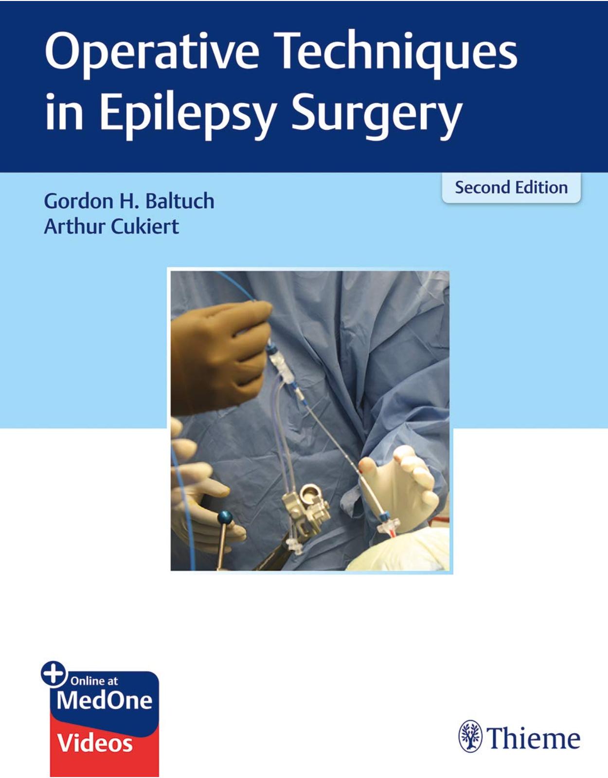
Operative Techniques in Epilepsy Surgery
Livrare gratis la comenzi peste 500 RON. Pentru celelalte comenzi livrarea este 20 RON.
Disponibilitate: La comanda in aproximativ 4 saptamani
Editura: Thieme
Limba: Engleza
Nr. pagini: 520
Coperta: Hardcover
Dimensiuni: 27.94 x 21.59 cm
An aparitie: 18 Oct. 2019
Description:
An indispensable, single-volume resource on state-of-the-art epilepsy procedures from renowned international experts!
Epilepsy is a common neurological disorder affecting an estimated 1% of the population, about 20 to 30% of which experience seizures inadequately controlled by medical therapy alone. Advances in anatomic and functional imaging modalities, stereotaxy, and the integration of neuronavigation during surgery have led to cutting-edge treatment options for patients with medically refractory epilepsy. Operative Techniques in Epilepsy Surgery, Second Edition by Gordon Baltuch, Arthur Cukiert, and an impressive international group of contributors has been updated and expanded, reflecting the newest treatments for pediatric and adult epilepsy.
Seven sections with 30 chapters encompass surgical planning, invasive EEG studies, cortical resection, intraoperative mapping, disconnection, neuromodulation, and further topics. Twelve cortical resection chapters cover surgical approaches such as amygdalohippocampectomy; hippocampal transection; frontal lobe, central region, and posterior quadrant resections; and microsurgery versus endoscopy for hypothalamic hamartomas. Disconnection procedures discussed in section five include corpus callosotomy, hemispherectomy, and endoscopic-assisted approaches. Well-established procedures such as vagus nerve and deep brain stimulation are covered in the neuromodulation section, while the last section discusses radiosurgery for medically intractable cases.
Key Highlights:
Chapters new to this edition include endoscopic callosotomy, laser-induced thermal therapy (LITT), and focused ultrasound
High-quality illustrations, superb operative and cadaver photographs, radiologic images, and tables enhance understanding of impacted anatomy and specific techniques
The addition of videos provides insightful step-by-step procedural guidance
This is an essential reference for fellows and residents interested in epilepsy and functional neurosurgery, and an ideal overview for neurosurgeons, neurologists, and neuroradiologists in early career stages who wish to pursue this subspecialty.
Table of Contents:
Part I: Surgical Planning
1 Collaborative Planning in Epilepsy Surgery
1.1 Introduction
1.2 Epileptology Phase: Establishing the Plan
1.2.1 Computer Processing of Neuroimaging Data
1.2.2 Planning Using Regions of Interest
1.3 Neurosurgical Phase: Building the Plan
1.3.1 The Vascular Network during Trajectory Planning
1.3.2 Virtual Reality during the Planning Process
1.4 Revision Phase: Locking the Plan
1.5 Implantation Phase: Executing the Plan
1.5.1 Frame-Based Implantation Approaches
1.5.2 Frameless Implantation Approaches
1.5.3 Robot-Assisted Implantation Approaches
1.5.4 Augmented-Reality Approaches
1.6 Validation Phase: Evaluating the Plan
1.7 Collaborative Workflow Systems: Planning Together
1.7.1 SYLVIUS: A VR-CWS for Epilepsy Surgery
1.8 Conclusion
2 Intraoperative Neuronavigation
2.1 Introduction
2.2 Frame Based
2.3 Frameless
2.4 Robotics
Part II: Invasive EEG Studies
3 Invasive EEG Studies: Peg, Strip, and Grid Implantation
3.1 Introduction
3.2 History
3.3 Patient Selection
3.4 Electrode Types
3.5 Planning Electrode Placement
3.6 Surgical Technique
3.7 Postimplant Management
3.8 Outcomes/Complications
3.9 Conclusion
4 Depth Electrodes
4.1 Introduction
4.2 Preoperative Planning
4.3 Operative Process
4.4 Bone Fiducial Placement
4.5 Frame Placement and Positioning
4.6 Bolt Placement and Preparation for Electrode Insertion
4.7 Frameless Stereotactic Insertion Method
4.8 Frame-Based Methods
4.9 Electrode Insertion
4.10 Postoperative Care
4.11 Complications
4.12 Epilepsy Monitoring Unit Stay
4.13 Explant
4.14 Postdischarge Care
Part III: Cortical Resection
5 Radiofrequency Lesions through Depth Electrodes
5.1 Introduction
5.2 Technical Details of RF-TC
5.2.1 Side-Effects and Morbidity of RF-TC
5.3 Illustrative Case
5.3.1 RF-TC Tips and Tricks
5.4 Conclusions
6 Temporal Lobectomy and Amygdalohippocampectomy
6.1 Introduction
6.2 Layout of the Operating Room and Positioning the Patient
6.3 Surface Anatomy and Designing the Incision for Right Temporal Lobectomy
6.4 Incision and Extracranial Dissection
6.5 Durotomy and Surface Exposure
6.6 Lateral Lobectomy
6.7 Microsurgical Resection of the Mesial Temporal Lobe
6.8 Closure
6.9 Conclusion
7 Selective Amygdalohippocampectomy
7.1 Introduction
7.2 Surgical Anatomy of the Temporal Lobe with Emphasis on the Mesial Structures
7.3 Surgery for Temporal Lobe Epilepsy
7.3.1 Temporal Lobectomy
7.3.2 Cortico amygdalohippocampectomy
7.3.3 Cortico amygdalectomy
7.3.4 Selective Amygdalohippocampectomy
7.4 Surgical Technique of Selective Amygdalohippocampectomy
7.4.1 Keyhole Approach in Selective Amygdalohippocampectomy
7.5 Results of Selective Amygdalohippocampectomy on Seizure Tendency
7.6 Conclusion
8 Parahippocampectomy: A New Surgical Technique for Temporal Lobe Epilepsy
8.1 Introduction
8.2 Historical Evolution of Some Surgical Techniques for SAH
8.3 Proposed Approach: Parahippocampectomy
8.3.1 Rationale
8.3.2 Surgical Technique
8.3.3 Indications for Parahippocampectomy
8.3.4 Contraindications for Parahippocampectomy
8.4 Concluding Remarks
9 Hippocampal Transection
9.1 Introduction
9.2 Surgical Procedure
9.2.1 Position, Incision, and Extent of Craniotomy
9.2.2 Opening the Sylvian Fissure and Approach to the Inferior Horn of the Lateral Ventricle
9.2.3 Resection of the Amygdala and Hippocampal Recording
9.2.4 Incision of the Alveus and Transection of the Pyramidal Cell Layer
9.2.5 Transection of the Parahip-pocampal Gyrus and Entorhinal Area
9.3 Memory Outcome
9.4 Conclusion
10 Frontal Lobe Resection in Refractory Epilepsy
10.1 Introduction
10.2 Patient Workup and Outcome
10.3 Technical Issues
10.4 Complications
10.5 Conclusion
11 Cortical Resection: Central Region
11.1 Introduction
11.2 Anatomical Considerations
11.3 Seizures of the Central Region
11.4 Etiology of Central Seizures
11.5 Presurgical Evaluation
11.6 Clinical Features
11.6.1 Neuroimaging
11.7 Electroencephalographic Investigation
11.7.1 Neuropsychological Testing
11.8 Psychosocial Assessment
11.9 Diagnostic Surgical Options
11.10 Therapeutic Surgical Options
11.11 Surgical Results of Central Resections
11.12 Conclusions
12 Posterior Quadrant Resections
12.1 Introduction
12.2 Indications for Surgery
12.3 Surgical Technique
12.3.1 Temporo-Parieto-Occipital (Posterior Quadrant) Resection
12.3.2 Outcome
12.3.3 Complications
13 Surgery of the Insula
13.1 Introduction
13.2 Insular Seizure Semiology
13.3 Insular Anatomy and Function
13.4 Vascular Anatomy and the Insular Lobe
13.5 Risks of Surgical Approaches to the Insula
13.6 Insular Tumors Presenting with Epilepsy
13.6.1 Tumors: Surgical Technique
13.6.2 Nonlesional Epilepsy Surgery and the Insula
13.7 Investigations and Workup
13.7.1 Noninvasive Studies
13.7.2 Invasive Studies
13.8 Resective Surgery
13.8.1 Technical Considerations
13.8.2 Alternatives to Open Resection
13.9 Where to Go from Here: Clinical Perspectives
13.10 Conclusions and Future Perspectives
14 Treatment Strategies for Hypothalamic Hamartomas: Microsurgery versus Endoscopy
14.1 Introduction
14.2 Pathophysiology
14.3 Clinical Features
14.4 Diagnosis and Neuroimaging
14.5 Indications for Intervention
14.6 Surgical Technique
14.6.1 Open Surgery—Craniotomy (Interhemispheric or Orbitozygomatic)
14.6.2 Endoscopic Resection
14.7 Alternative Approaches
14.7.1 Stereotactic Laser Ablation
14.8 Specific Considerations
14.9 Surgical Pearls
14.10 Surgical Risks
14.11 Outcome and Prognosis
14.12 Stereotactic Laser Thermoablation
14.13 Conclusion
15 Multiple Subpial Transections
15.1 Introduction
15.2 From Bench to Operating Theater
15.3 Surgical Technique
15.4 Preoperative Evaluation
15.5 Specific Indications
15.6 Outcome Data
15.7 Future Considerations
16 Laser-Induced Thermal Therapy for Medically Refractory Epilepsy
16.1 Introduction
16.2 History
16.3 Relevant Pathophysiology
16.4 Physics and Hardware Considerations
16.5 Surgical Steps
16.6 Outcomes
16.6.1 Mesial Temporal Lobe Epilepsy
16.6.2 Developmental Anomalies/Lesional Epilepsy
16.6.3 Pediatric Epilepsy Syndromes
16.7 Complications
16.8 Future Directions
16.9 Conclusion
Part IV: Intraoperative Mapping
17 Motor, Sensory, and Language Mapping in Epilepsy Surgery
17.1 Introduction
17.2 History
17.3 Patient Selection and Preoperative Functional Localization
17.4 Intraoperative Stimulation Technique
17.5 Language Task Selection and Patterns of Language Function
17.6 Clinical Outcomes following Resection
17.7 Limitations
17.8 Conclusions
Part V: Disconnection
18 Cortical Disconnections
18.1 Introduction
18.2 Preoperative Evaluation Workup
18.3 Indications for Surgery
18.4 Surgical Anatomy
18.4.1 Cerebral Cortex
18.4.2 White Matter Pathways
18.5 Disconnective Procedures
18.5.1 Surgical Adjuncts
18.5.2 Frontal Disconnection
18.5.3 Temporo-Parieto-Occipital (Posterior Quadrantic) Disconnection
18.5.4 Parieto-Occipital Disconnection
18.5.5 Combined Disconnection/Subtotal Hemispherectomy
18.5.6 Extension to Hemispheric Disconnection
18.6 Complications
18.7 Outcome
19 Corpus Callosotomy
19.1 Introduction
19.2 Open Classic Corpus Callosotomy
19.3 Other Techniques as Described in the Literature
19.3.1 Endoscopy-Assisted Procedures
19.3.2 The Use of Laser Techniques in Performing Corpus Callosotomy
19.3.3 Corpus Callosotomy Using Radiosurgery
19.4 Comments and Discussion
20 Endoscopic Callosotomy and Hemispherotomy: Bimanual Endoscopic Technique
20.1 Evolution of Endoscopic Surgical Techniques
20.1.1 Endoscopic Approaches to Disconnective Surgery
20.2 Equipment/Medications
20.2.1 Operating Room Setup
20.3 Anterior Interhemispheric Endoscopic Complete Callosotomy: Precoronal
20.4 Posterior Interhemispheric Endoscopic Complete Corpus Callosotomy
20.4.1 Procedure
20.5 Precoronal Endoscopic (Hemispherotomy) Technique
20.5.1 Procedure
20.6 Benefits and Results
20.7 Conclusion and Future Directions
21 Anatomical Hemispherectomy
21.1 Introduction
21.2 Background
21.3 Surgical Technique
21.4 Postoperative Care
21.5 Complications
21.6 Conclusions
22 Functional Hemispherectomy and Peri-insular Hemispherotomy
22.1 Introduction
22.2 Indications
22.3 Operative Techniques
22.3.1 Functional Hemispherectomy
22.3.2 Resection of the Central Region
22.3.3 Peri-insular Hemispherotomy
22.4 Conclusion
23 Endoscopic-Assisted Hemispherotomy
23.1 Introduction
23.2 Principles
23.3 Surgical Technique
23.4 Outcomes following Surgery
23.5 Summary with Important Points to Note
24 Vertical Parasagittal Hemispherotomy
24.1 Introduction
24.2 Historical Background
24.3 Description of the Surgical Technique of VPH
24.3.1 Approach to the Ventricle: Posterior Frontal Resection
24.3.2 Posterior Callosotomy
24.3.3 Interrupting the Hippocampal Tail
24.3.4 Laterothalamic Disconnection
24.3.5 Anterior Callosotomy
24.3.6 Frontal Disconnection
24.4 Results
24.5 Complications
24.6 Incomplete Disconnection
24.7 Conclusion
Part VI: Neuromodulation
25 Vagus Nerve Stimulation
25.1 Preclinical Studies
25.2 Surgical Indications
25.3 Device
25.4 Surgical Anatomy
25.5 Surgical Implantation
25.6 Stimulation Programming
25.7 Battery Change and System Removal
25.8 Adverse Events and Complications
25.9 Conclusion
26 Hippocampal Deep Brain Stimulation in Refractory Temporal Lobe Epilepsy
26.1 Introduction
26.2 Rationale
26.3 Development Timeline
26.4 Patient Selection
26.5 Targeting
26.6 Hip-DBS Efficacy and Paradigms
26.7 Effects on Memory
27 Deep Brain Stimulation: Stimulation of the Anterior Nucleus of the Thalamus
27.1 General Anatomy
27.2 ANT as a Stereotactic Target
27.3 Surgical Trajectory to the ANT
28 Responsive (Closed-Loop) Stimulation
28.1 Introduction
28.2 Localization of Cortical Function
28.3 Deep Brain Stimulation for Epilepsy
28.4 Cortical Stimulation for Epilepsy
28.5 External Responsive Neurostimulation
28.6 The Implantable RNS System
28.6.1 System Components
28.6.2 Surgical Implantation
28.7 Clinical Trials
28.7.1 Feasibility Clinical Trial
28.7.2 RNS System Pivotal Clinical Investigation in Epilepsy
28.7.3 RNS System: After the Pivotal Trial
28.8 Case Examples
28.8.1 Case 1
28.8.2 Case 2
28.9 Conclusions
Part VII: Further Topics
29 Radiosurgery for Medically Refractory Epilepsy
29.1 Introduction
29.2 Developments Permitting Noninvasive Techniques
29.3 Basic Principles of Radiobiology
29.4 Vascular Effects and Seizure Control
29.5 Effects of Radiation in the Epileptic Focus
29.6 Radiosurgery in Mesial Temporal Epilepsy
29.7 Radiosurgery for Hypothalamic Hamartoma
29.8 Other Focal Epilepsy Syndromes Treated by Radiosurgery
29.9 Conclusion
30 Focused Ultrasound as a Surgical Treatment for Epilepsy
30.1 Introduction
30.2 Barriers in Transcranial MR-Guided FUS for Epilepsy Treatments
30.2.1 Therapeutic Envelope of Current Systems
30.2.2 Volume Mismatch in Acoustic Focus and Epileptic Target
30.3 Potential Applications
30.3.1 Hypothalamic Hamartoma
30.3.2 Malformations of Cortical Development
30.3.3 Mesial Temporal Sclerosis
30.3.4 Disconnection Procedures
30.3.5 Other Focal Lesions Causing Epilepsy
30.4 Conclusions
Index
Additional MedOne Information
| An aparitie | 18 Oct. 2019 |
| Autor | Gordon H. Baltuch, Arthur Cukiert |
| Dimensiuni | 27.94 x 21.59 cm |
| Editura | Thieme |
| Format | Hardcover |
| ISBN | 9781626238183 |
| Limba | Engleza |
| Nr pag | 520 |






Clientii ebookshop.ro nu au adaugat inca opinii pentru acest produs. Fii primul care adauga o parere, folosind formularul de mai jos.