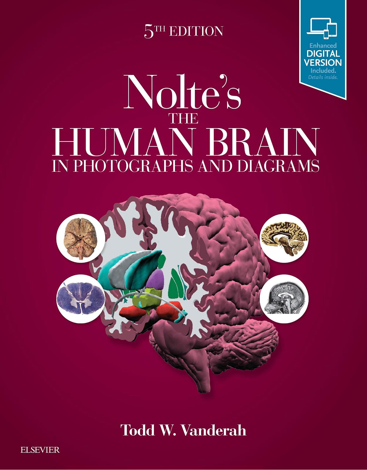
Nolte's The Human Brain in Photographs and Diagrams, 5e: With STUDENT CONSULT Online Access
Livrare gratis la comenzi peste 500 RON. Pentru celelalte comenzi livrarea este 20 RON.
Disponibilitate: La comanda in aproximativ 4-6 saptamani
Autor: Todd Vanderah
Editura: Elsevier
Limba: Engleza
Nr. pagini: 294
Coperta: Paperback
Dimensiuni: 21.59 x 1.27 x 27.43 cm
An aparitie: 4 Jan. 2019
|
Description: In the 5th Edition of this highly accessible atlas, Dr. Todd Vanderah continues the mission of his esteemed colleague, Dr. John "Jack" Nolte, to clearly depict and explain the challenging subject of neuroanatomy. Designed to promote a rapid understanding of complex concepts, Nolte's The Human Brain in Photographs and Diagrams combines easy-to-digest coverage of the brain, spinal cord, and brainstem with carefully selected visuals to cover all aspects of the information needed for success in coursework, on exams, and in clerkships and clinical practice. |
||
|
Table Of Contents: |
||
|
Contents Chapter 1 External Anatomy of the Brain Introduction Brain and Spinal Cord Multiple Views of a Single Brain Lateral and Superior Surfaces Inferior Surface Medial and Inferior Surfaces Insula Cerebellum Brainstem Chapter 2 Transverse Sections of the Spinal Cord Introduction Guided Tour Enlargements of Selected Levels S4 L2 T10 C8 C3 Chapter 3 Transverse Sections of the Brainstem Introduction Schematic Cross Sections Blood Supply Cranial Nerve Nuclei Guided Tour Enlargements of Selected Levels Spinomedullary junction Caudal medulla Rostral medulla Pontomedullary junction Caudal pons Midpons Rostral pons Caudal midbrain Rostral midbrain Far rostral midbrain Chapter 4 Building a Brain: Three-Dimensional Reconstructions Introduction Step-by-Step Reconstruction Chapter 5 Coronal Sections Introduction Guided Tour Enlargements of Selected Levels Striatum, anterior limb of the internal capsule Anterior commissure Anterior thalamus, ansa lenticularis Amygdala, mammillary bodies Anterior hippocampus, subthalamic nucleus Posterior thalamus, red nucleus Posterior thalamus, geniculate nuclei Posterior thalamus, pulvinar Atrium of the lateral ventricle Chapter 6 Axial Sections Introduction Guided Tour Enlargements of Selected Levels Uncus, optic chiasm Hypothalamus, superior cerebellar peduncle Striatum, deep cerebellar nuclei Anterior commissure, subthalamic nucleus Interventricular foramen, posterior Chapter 7 Sagittal Sections Introduction Guided Tour Enlargements of Selected Levels Putamen, hippocampus Hippocampus, amygdala Globus pallidus, internal capsule, thalamus Caudate nucleus, dentate nucleus Mammillothalamic tract, superior cerebellar Chapter 8 Functional Systems Detailed Contents Long Tracts of the Spinal Cord and Brainstem Sensory Systems of the Brainstem and Chapter 9 Clinical Imaging Introduction Computed Tomography Without contrast With contrast Pathology Magnetic Resonance Imaging (MRI) Introduction Coronal T1-weighted MRIs Axial T1-weighted MRIs Sagittal and parasaggital T1-weighted MRIs Pathology Angiography Introduction Internal carotid angiograms Vertebral-basilar angiograms Pathology Chapter 10 Neuropathology Imaging [Add headings] Glossary Contents Chapter 1 External Anatomy of the Brain Introduction Brain and Spinal Cord Multiple Views of a Single Brain Lateral and Superior Surfaces Inferior Surface Medial and Inferior Surfaces Insula Cerebellum Brainstem Chapter 2 Transverse Sections of the Spinal Cord Introduction Guided Tour Enlargements of Selected Levels S4 L2 T10 C8 C3 Chapter 3 Transverse Sections of the Brainstem Introduction Schematic Cross Sections Blood Supply Cranial Nerve Nuclei Guided Tour Enlargements of Selected Levels Spinomedullary junction Caudal medulla Rostral medulla Pontomedullary junction Caudal pons Midpons Rostral pons Caudal midbrain Rostral midbrain Far rostral midbrain Chapter 4 Building a Brain: Three-Dimensional Reconstructions Introduction Step-by-Step Reconstruction Chapter 5 Coronal Sections Introduction Guided Tour Enlargements of Selected Levels Striatum, anterior limb of the internal capsule Anterior commissure Anterior thalamus, ansa lenticularis Amygdala, mammillary bodies Anterior hippocampus, subthalamic nucleus Posterior thalamus, red nucleus Posterior thalamus, geniculate nuclei Posterior thalamus, pulvinar Atrium of the lateral ventricle Chapter 6 Axial Sections Introduction Guided Tour Enlargements of Selected Levels Uncus, optic chiasm Hypothalamus, superior cerebellar peduncle Striatum, deep cerebellar nuclei Anterior commissure, subthalamic nucleus Interventricular foramen, posterior Chapter 7 Sagittal Sections Introduction Guided Tour Enlargements of Selected Levels Putamen, hippocampus Hippocampus, amygdala Globus pallidus, internal capsule, thalamus Caudate nucleus, dentate nucleus Mammillothalamic tract, superior cerebellar Chapter 8 Functional Systems Detailed Contents Long Tracts of the Spinal Cord and Brainstem Sensory Systems of the Brainstem and Chapter 9 Clinical Imaging Introduction Computed Tomography Without contrast With contrast Pathology Magnetic Resonance Imaging (MRI) Introduction Coronal T1-weighted MRIs Axial T1-weighted MRIs Sagittal and parasaggital T1-weighted MRIs Pathology Angiography Introduction Internal carotid angiograms Vertebral-basilar angiograms Pathology Chapter 10 Neuropathology Imaging [Add headings] |
||
| An aparitie | 4 Jan. 2019 |
| Autor | Todd Vanderah |
| Dimensiuni | 21.59 x 1.27 x 27.43 cm |
| Editura | Elsevier |
| Format | Paperback |
| ISBN | 9780323598163 |
| Limba | Engleza |
| Nr pag | 294 |


Clientii ebookshop.ro nu au adaugat inca opinii pentru acest produs. Fii primul care adauga o parere, folosind formularul de mai jos.