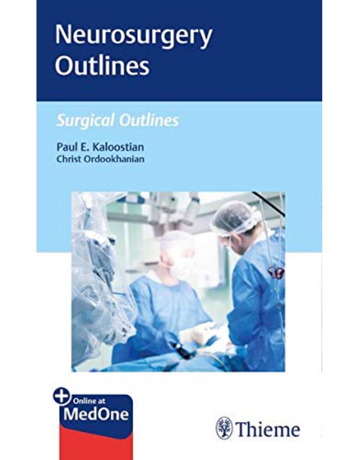
Neurosurgery Outlines
Livrare gratis la comenzi peste 500 RON. Pentru celelalte comenzi livrarea este 20 RON.
Disponibilitate: La comanda in aproximativ 4-6 saptamani
Autor: Paul Kaloostian
Editura: Thieme
Limba: Engleza
Nr. pagini: 348
Coperta: Paperback
Dimensiuni: 20.32 x 12.7 cm
An aparitie: 30 April 2020
Description:
Pocket-size, user-friendly roadmap outlines most common surgical procedures in neurosurgery!
Surgery requires a combination of knowledge and skill acquired through years of direct observation, mentorship, and practice. The learning curve can be steep, frustrating, and intimidating for many medical students and junior residents. Too often, books and texts that attempt to translate the art of surgery are far too comprehensive for this audience and counterproductive to learning important basic skills to succeed. Neurosurgery Outlines by neurosurgeon Paul E. Kaloostian is the neuro-surgical volume in the Surgical Outlines series of textbooks that offer a simplified roadmap to surgery.
This unique resource outlines key steps for common surgeries, laying a solid foundation of basic knowledge from which trainees can easily build and expand. The text serves as a starting point for learning neurosurgical techniques, with room for adding notes, details, and pearls collected during the journey. The chapters are systematically organized and formatted by subspecialty, encompassing spine, radiosurgery, brain tumors and vascular lesions, head trauma, functional neurosurgery, epilepsy, pain, and hydrocephalus. Each chapter includes symptoms and signs, surgical pathology, diagnostic modalities, differential diagnosis, treatment options, indications for surgical intervention, step-by-step procedures, pitfalls, prognosis, and references where applicable.
Key Features:
Provides quick procedural outlines essential for understanding procedures and assisting attending neurosurgeons during rounds
Spine procedures organized by cervical, thoracic, lumbar, sacral, and coccyx regions cover traumatic, elective, and tumor/vascular-related interventions
Cranial topics include lesion resection for brain tumors and cerebrovascular disease and TBI treatment
This is an ideal, easy-to-read resource for medical students and junior residents to utilize during the one-month neurosurgery rotations and for quick consultation during the early years of neurosurgical practice. It will also benefit operating room nurses who need a quick guide on core neurosurgical procedures.
Table of Contents:
Section I: Spine
1. Cervical
1.1 Trauma
1.1.1 Anterior Cervical Fusion/Posterior Cervical Fusion
1.2 Elective
1.2.1 Anterior Cervical Fusion/Posterior Cervical Fusion
1.2.2 Posterior Cervical Foraminotomy/Posterior Cervical Decompression
1.3 Tumor/Vascular
1.3.1 Cervical Tumor Resection (Vertebral Pathology)
1.3.2 Cervical Vascular Lesion Treatment for Arteriovenous Malformation (AVM) (Vertebral Pathology)
1.3.3 Cervical Anterior and Posterior Techniques for Tumor Resection (Spinal Canal Pathology)
2 Thoracic
2.1 Trauma
2.1.1 Thoracic Decompression/Thoracic Fusion
2.1.2 Thoracic Corpectomy and Fusion
2.1.3 Transthoracic Approaches for Decompression and Fusion/Transsternal Approaches for Decompression and Fusion
2.2 Elective
2.2.1 Thoracic Decompression/Thoracic Fusion
2.2.2 Thoracic Corpectomy and Fusion
2.2.3 Transthoracic Approaches for Decompression and Fusion/Transsternal Approaches for Decompression and Fusion
2.3 Tumor/Vascular
2.3.1 Anterior and Posterior Approaches for Thoracic Tumor Resection (Vertebral Pathology)
2.3.2 Thoracic Anterior and Posterior Approaches for Vascular Lesion Resection (Vertebral Pathology)
2.3.3 Thoracic Anterior and Posterior Techniques for Tumor Resection (Spinal Canal Pathology)
3 Lumbar
3.1 Trauma
3.1.1 Lumbar Decompression with Foraminotomy/Lumbar Diskectomy/Lumbar Fusion
3.2 Elective
3.2.1 Lumbar Laminectomy/Decompression/Lumbar Diskectomy/Lumbar Synovial Cyst Resection/Lumbar Fusion Posterior
3.2.2 Anterior Lumbar Fusion
3.2.3 Anterolateral Lumbar Fusion
3.3 Tumor/Vascular
3.3.1 Lumbar Posterior Techniques for Tumor Resection (Vertebral Tumor)
3.3.2 Lumbar Anterior and Posterior Techniques for Vascular Lesion Treatment (Vertebral)
3.3.3 Lumbar Anterior and Posterior Techniques for Tumor Resection (Spinal Canal Pathology)
4 Sacral
4.1 Trauma
4.1.1 Sacral Fusion
4.1.2 Tarlov Cyst Treatment
4.2 Elective
4.2.1 Sacral Decompression
4.2.2 Sacral Fusion
4.3 Tumor/Vascular
4.3.1 Anterior/Posterior Sacral Tumor Resection
5 Coccyx
5.1 Trauma
5.1.1 Coccyx Fracture Repair/Resection
5.2 Elective
5.2.1 Coccyx Resection
Section II: Radiosurgery/CyberKnife Treatment
6 Spinal Radiosurgery
6.1 Symptoms and Signs (Broad)
6.2 Surgical Pathology
6.3 Diagnostic Modalities
6.4 Differential Diagnosis
6.5 Treatment Options
6.6 Indications for Surgical Intervention
6.7 Noninvasive CyberKnife Procedure for Posterior Spine
6.8 Pitfalls
6.9 Prognosis
7 Cranial Radiosurgery
7.1 Symptoms and Signs (Broad)
7.2 Surgical Pathology
7.3 Diagnostic Modalities
7.4 Differential Diagnosis
7.5 Treatment Options
7.6 Indications for Radiosurgical Intervention
7.7 Noninvasive CyberKnife Procedure for Cranium
7.8 Pitfalls
7.9 Prognosis
Section III: Cranial Lesion Resection (Brain Tumor, Vascular Lesions)
8 Brain Tumors
8.1 Meningioma Convexity/Olfactory Groove Meningioma
8.1.1 Symptoms and Signs
8.1.2 Surgical Pathology
8.1.3 Diagnostic Modalities
8.1.4 Differential Diagnosis
8.1.5 Treatment Options
8.1.6 Indications for Surgical Intervention
8.1.7 Surgical Procedure for Bifrontal Craniotomy (Pre-op Embolization Can Be Done Prior to Surgery)
8.1.8 Pitfalls
8.1.9 Prognosis
8.2 Metastatic Tumor Resection
8.2.1 Symptoms and Signs (Broad)
8.2.2 Surgical Pathology
8.2.3 Diagnostic Modalities
8.2.4 Differential Diagnosis
8.2.5 Treatment Options
8.2.6 Indications for Surgical Intervention
8.2.7 Surgical Procedure for Craniotomy
8.2.8 Pitfalls
8.2.9 Prognosis
8.3 Posterior Fossa Tumor Resection
8.3.1 Symptoms and Signs
8.3.2 Surgical Pathology
8.3.3 Diagnostic Modalities
8.3.4 Differential Diagnosis
8.3.5 Treatment Options
8.3.6 Indications for Surgical Intervention
8.3.7 Surgical Procedure for Posterior Fossa Craniectomy
8.3.8 Pitfalls
8.3.9 Prognosis
8.4 Pituitary Tumor Resection
8.4.1 Symptoms and Signs
8.4.2 Surgical Pathology
8.4.3 Diagnostic Modalities
8.4.4 Differential Diagnosis
8.4.5 Treatment Options
8.4.6 Indications for Surgical Intervention
8.4.7 Surgical Procedure for Transsphenoidal Endoscopic Pituitary
8.4.8 Pitfalls
8.4.9 Prognosis
8.5 Superficial Tumor Resection (Arising from Skull)
8.5.1 Symptoms and Signs (Broad)
8.5.2 Surgical Pathology
8.5.3 Diagnostic Modalities
8.5.4 Differential Diagnosis
8.5.5 Treatment Options
8.5.6 Indications for Surgical Intervention
8.5.7 Surgical Procedure for Craniotomy
8.5.8 Pitfalls
8.5.9 Prognosis
8.6 Epidural Tumor Resection
8.6.1 Symptoms and Signs (Broad)
8.6.2 Surgical Pathology
8.6.3 Diagnostic Modalities
8.6.4 Differential Diagnosis
8.6.5 Treatment Options
8.6.6 Indications for Surgical Intervention
8.6.7 Surgical Procedure for Retrosigmoid Craniotomy (Meningiomas or Vestibular Schwannomas)
8.6.8 Pitfalls
8.6.9 Prognosis
9 Cerebral Vascular Lesions
9.1 Cerebral Aneurysms/Cerebral AVM Resection/Cerebral Cavernous Angioma Resection
9.1.1 Symptoms and Signs
9.1.2 Surgical Pathology
9.1.3 Diagnostic Modalities
9.1.4 Differential Diagnosis
9.1.5 Treatment Options
9.1.6 Indications for Surgical Intervention
9.1.7 Surgical Procedure for Microsurgical Resection (Vascular Lesions)
9.1.8 Embolization Procedure (Onyx)
9.1.9 Pitfalls
9.1.10 Prognosis
9.2 Endovascular Treatment of Cerebral Aneurysms/AVM
9.2.1 Symptoms and Signs
9.2.2 Surgical Pathology
9.2.3 Diagnostic Modalities
9.2.4 Differential Diagnosis
9.2.5 Treatment Options
9.2.6 Indications for Surgical Intervention
9.2.7 Surgical Procedure for Endovascular Embolization (Coiling)
9.2.8 Pitfalls
9.2.9 Prognosis
Section IV: Cranial Trauma
10 Epidural Hematoma Evacuation/Subdural Hematoma Evacuation/Intracerebral Hemorrhage Evacuation
10.1 Symptoms and Signs
10.2 Surgical Pathology
10.3 Diagnostic Modalities
10.4 Differential Diagnosis
10.5 Treatment Options
10.6 Indications for Surgical Intervention
10.7 Surgical Procedure for Subdural Hematoma/Epidural Hematoma/Intraparenchymal Hematoma Evacuation
10.8 Pitfalls
10.9 Prognosis
11 Hemicraniectomy (Unilateral, Bilateral, Bifrontal versus Frontotemporal)
11.1 Symptoms and Signs
11.2 Surgical Pathology
11.3 Diagnostic Modalities
11.4 Differential Diagnosis
11.5 Treatment Options
11.6 Indications for Surgical Intervention
11.7 Surgical Procedure for Unilateral Frontotemporoparietal Craniectomy
11.8 Surgical Procedure for Bifrontal Craniectomy
11.9 Pitfalls
11.10 Prognosis
12 Cranioplasty
12.1 Symptoms and Signs
12.2 Surgical Pathology
12.3 Diagnostic Modalities
12.4 Differential Diagnosis
12.5 Treatment Options
12.5.1 Acute Pain Control with Medications and Pain Management
12.5.2 Therapy and Rehabilitation
12.5.3 If Symptomatic
12.6 Indications for Surgical Intervention
12.7 Surgical Procedure for Cranioplasty
12.8 Pitfalls
12.9 Prognosis
13 Placement of External Ventricular Drain/Placement of Intraparenchymal ICP Monitor
13.1 Symptoms and Signs
13.2 Surgical Pathology
13.3 Diagnostic Modalities
13.4 Differential Diagnosis
13.5 Treatment Options
13.5.1 Acute Pain Control with Medications and Pain Management
13.5.2 Therapy and Rehabilitation
13.5.3 If Symptomatic
13.6 Indications for Surgical Intervention
13.7 Surgical Procedure for EVD Placement
13.8 Surgical Procedure for Intraparenchymal ICP Monitor Placement
13.9 Pitfalls
13.10 Prognosis
Section V: Functional Neurosurgery
14 Deep Brain Stimulation
14.1 Symptoms and Signs
14.2 Surgical Pathology
14.3 Diagnostic Modalities
14.4 Differential Diagnosis
14.5 Treatment Options
14.6 Indications for Surgical Intervention
14.7 Surgical Procedure for Deep Brain Stimulation
14.8 Pitfalls
14.9 Prognosis
Section VI: Epilepsy
15 Subdural Grid Placement
15.1 Symptoms and Signs
15.2 Surgical Pathology
15.3 Diagnostic Modalities
15.4 Differential Diagnosis
15.5 Treatment Options
15.6 Indications for Surgical Intervention
15.7 Surgical Procedure for Subdural Grid Placement
15.8 Pitfalls
15.9 Prognosis
16 Amygdalohippocampectomy
16.1 Symptoms and Signs
16.2 Surgical Pathology
16.3 Diagnostic Modalities
16.4 Differential Diagnosis
16.5 Treatment Options
16.5.1 Antiepileptic Medication (Neurological Evaluation)
16.5.2 Therapy and Rehabilitation
16.5.3 If Symptomatic and No Improvement after Nonoperative Management
16.6 Indications for Surgical Intervention
16.7 Surgical Procedure for Transcortical Selective Amygdalohippocampectomy (SAH)
16.8 Pitfalls
16.9 Prognosis
Section VII: Pain Management Strategies
17 Spinal Cord Stimulator
17.1 Symptoms and Signs
17.2 Surgical Pathology
17.3 Diagnostic Modalities
17.4 Differential Diagnosis
17.5 Treatment Options
17.6 Indications for Surgical Intervention
17.7 Surgical Procedure for Spinal Cord Stimulation
17.8 Pitfalls
17.9 Prognosis
18 Baclofen Pump/Morphine Pump
18.1 Symptoms and Signs
18.2 Surgical Pathology
18.3 Diagnostic Modalities
18.4 Differential Diagnosis
18.5 Treatment Options
18.6 Indications for Surgical Intervention
18.7 Surgical Procedure for Intrathecal Baclofen Pump Insertion
18.8 Pitfalls
18.9 Prognosis
Section VIII: Hydrocephalus Treatment
19 Ventriculoperitoneal Shunt
19.1 Symptoms and Signs
19.2 Surgical Pathology
19.3 Diagnostic Modalities
19.4 Differential Diagnosis
19.5 Treatment Options
19.5.1 Surgery if Deemed Suitable Candidate
19.6 Indications for Surgical Intervention
19.7 Surgical Procedure for Ventriculoperitoneal (VP) Shunt
19.8 Pitfalls
19.9 Prognosis
20 Lumboperitoneal Shunt
20.1 Symptoms and Signs
20.2 Surgical Pathology
20.3 Diagnostic Modalities
20.4 Differential Diagnosis
20.5 Treatment Options
20.5.1 Surgery if Deemed Suitable Candidate
20.6 Indications for Surgical Intervention
20.7 Surgical Procedure for Lumboperitoneal (LP) Shunt
20.8 Pitfalls
20.9 Prognosis
21 Ventriculopleural Shunt
21.1 Symptoms and Signs
21.2 Surgical Pathology
21.3 Diagnostic Modalities
21.4 Differential Diagnosis
21.5 Treatment Options
21.5.1 Surgery if Deemed Suitable Candidate
21.6 Indications for Surgical Intervention
21.7 Surgical Procedure for Ventriculopleural (VLP) Shunt
21.8 Pitfalls
21.9 Prognosis
22 Ventriculoatrial Shunt
22.1 Symptoms and Signs
22.2 Surgical Pathology
22.3 Diagnostic Modalities
22.4 Differential Diagnosis
22.5 Treatment Options
22.5.1 Surgery if Deemed Suitable Candidate
22.6 Indications for Surgical Intervention
22.7 Surgical Procedure for Ventriculoatrial (VA) Shunt
22.8 Pitfalls
22.9 Prognosis
Index
| An aparitie | 30 April 2020 |
| Autor | Paul Kaloostian |
| Dimensiuni | 20.32 x 12.7 cm |
| Editura | Thieme |
| Format | Paperback |
| ISBN | 9781684201426 |
| Limba | Engleza |
| Nr pag | 348 |





Clientii ebookshop.ro nu au adaugat inca opinii pentru acest produs. Fii primul care adauga o parere, folosind formularul de mai jos.