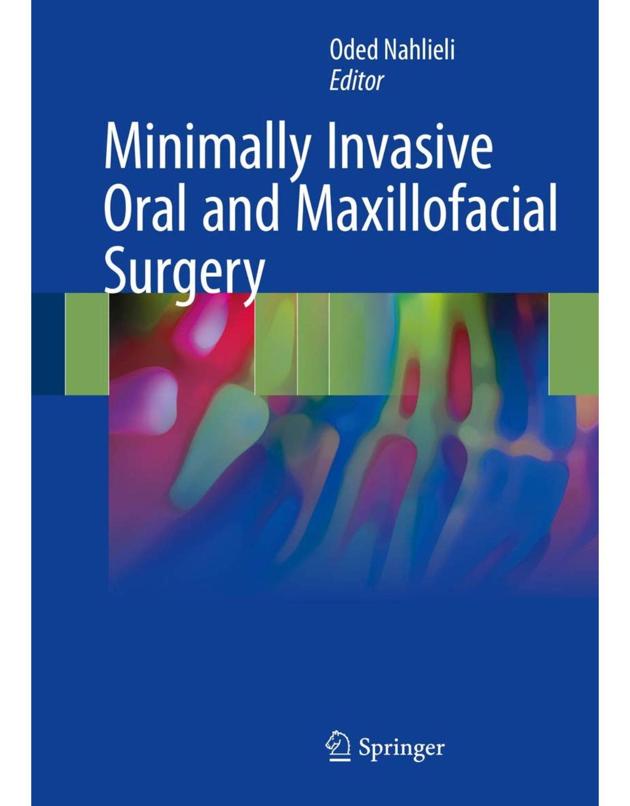
Minimally Invasive Oral and Maxillofacial Surgery
Livrare gratis la comenzi peste 500 RON. Pentru celelalte comenzi livrarea este 20 RON.
Disponibilitate: La comanda in aproximativ 4 saptamani
Autor: Oded Nahlieli
Editura: Springer
Limba: Engleza
Nr. pagini: 203
Coperta: Hardcover
Dimensiuni: 18.29 x 2.03 x 25.65 cm
An aparitie: 5 Jan. 2018
Description:
Minimally invasive techniques, designed to reduce morbidity and risk while simultaneously improving outcomes, are increasingly being used in oral and maxillofacial surgery. This book covers the most recent technological developments and the advanced techniques used when performing such minimally invasive surgery in patients with common and rare oral and maxillofacial pathologies. The relevant basic science is reviewed, but the principal focus is on the surgical techniques themselves. These are described step by step with the aid of numerous superb color illustrations that will help the clinician to gain a full understanding of the technology and the procedures. In addition, still emerging techniques of endoscopy, navigation, and minimally invasive surgery are well covered. This text will be a premier resource for physicians who diagnose and treat oral and maxillofacial pathologies and injuries.
Table of Contents:
Contents
Contributors
1: The History of Minimally Invasive Approach in Oral and Maxillofacial Surgery
1.1 Introduction
1.2 The Development of Endoscopy
1.3 What About the Maxillofacial Area?
1.4 Intraoperative Navigation in Oral and Maxillofacial Surgery
1.5 Distraction Osteogenesis and Tissue Engineering
References
2: Modern Temporomandibular Joint Arthroscopy: Operative Single-Cannula Arthroscopy
2.1 History and Goals
2.2 Anatomy of the Temporomandibular Joint Region
2.3 Temporomandibular Joint Disorders
2.3.1 Diagnosis
2.3.2 Classifications
2.3.3 Conservative Treatment
2.4 Arthroscopy
2.4.1 Indications for Arthroscopy
2.4.2 Contraindications for Arthroscopy
2.4.3 Armamentarium
2.4.3.1 The Arthroscope
2.4.3.2 Cannulas
2.4.3.3 Probes
2.4.3.4 Graspers and Biopsy Forceps
2.4.3.5 Spinal Needles
2.4.3.6 Laser
2.5 The Operative Single-Cannula Arthroscopy Technique
2.5.1 One-Track Arthrocentesis
2.5.2 Standard Arthrocentesis Under Diagnostic Visualization
2.5.3 Visually Guided OSCA
2.5.4 Surgical Interventions Using the OSCA Technique
2.5.4.1 Anterior and Posterior Recess Adhesion Release
2.5.4.2 Synovectomy
2.5.4.3 Anterior Release
2.5.4.4 Posterior Scarification/Contracture
2.5.5 Advantages and Disadvantages of OSCA
2.5.6 Intra-articular Delivery of Medications via OSCA
2.5.6.1 Steroids
2.5.6.2 Botulinum Toxin A
2.5.6.3 Hyaluronic Acid
2.5.6.4 Platelet Concentrates
2.5.7 Post-OSCA Patient Management
2.5.7.1 General Anesthesia Considerations
2.5.7.2 Anti-inflammatory and Pain Management
2.5.7.3 Antibiotics
2.5.7.4 Diet
2.5.8 Complications of Traditional Arthroscopy and OSCA
2.5.8.1 Scuffing of Fibrocartilage
2.5.8.2 Damage to the VII Cranial Nerve
2.5.8.3 Damage to the V Cranial Nerve
2.5.8.4 Damage to the VIII Cranial Nerve
2.5.8.5 Damage to Vessels and Hamartosis
2.5.8.6 Perforation of the Glenoid Fossa
2.5.8.7 Instrument Failure
References
3: Arthrocentesis: A Minimally Invasive Approach to the Temporomandibular Joint
3.1 Introduction
3.2 The Incentives for Using the Minimally Invasive Approach for Temporomandibular Disorders
3.2.1 TMJ Integrity
3.2.2 TMJ Lubrication
3.2.3 TMJ Load Attenuation
3.2.4 TMJ Condyle Remodeling Potential
3.3 Mechanical Challenges to Joint Integrity
3.4 Temporomandibular Joint Arthrocentesis: Technique
3.5 Mode of Action
3.6 Patient Evaluation
3.6.1 Patient Self-Assessment and Clinical Examination
3.6.2 Imaging
3.7 The Role of Arthrocentesis in Various TMJ Disorders
3.7.1 The Clicking Joint
3.7.2 Limited Mouth Opening
3.7.2.1 Disc Displacement Without Reduction
3.7.2.2 Anchored Disc Phenomenon (Figs. 3.13 and 3.14)
3.7.3 Symptomatic Osteoarthritis
3.7.4 Open Lock
3.7.5 Hemarthrosis
3.7.6 Autoimmune Inflammatory Arthritis
References
4: Endoscopic Approach to Temporomandibular Fractures
4.1 Introduction
4.2 History
4.3 Technique
4.3.1 Transoral Approach
4.3.2 Transcervical Technique
4.3.3 Transoral vs. Transcervical Approaches: Technical Advantages and Disadvantages
4.4 Case Selection
4.4.1 Complications
References
5: Minimally Invasive Approach to Orbital Trauma
5.1 Introduction
5.2 Surgical Anatomy of the Orbit
5.3 Indications for Surgery After the Orbital Trauma
5.4 Surgical Approaches and Reconstruction Materials
5.4.1 Reconstruction Materials
5.4.2 The Minimally Invasive Revolution
5.5 The Endoscopic Reconstruction of Orbital Floor Fractures
5.5.1 The Operative Technique
5.5.2 Specific Approaches to Various Cases
5.5.2.1 Illustrative Case 1
5.5.2.2 Illustrative Case 2
References
6: Applications of Distraction Osteogenesis in Oral and Maxillofacial Surgery
6.1 Introduction
6.2 Mandibular Distraction
6.3 Maxilla and Midface Distraction
6.4 Deficient Alveolar Bone
References
7: Minimally Invasive Orthognathic Surgery
7.1 Introduction
7.2 Minimally Invasive Orthognathic Surgery
7.3 Navigation-Assisted Le Fort I Osteotomy
References
8: Salivary Gland Endoscopy and Minimally Invasive Surgery
8.1 Introduction
8.1.1 Anatomical Considerations
8.1.2 Salivary Gland Disorders for Minimally Invasive Surgery
8.1.3 The Tool #1: Sialoendoscope
8.1.4 The Tool #2: Extracorporeal Shock-Wave Lithotripter
8.2 The Types and the Technique of the Minimally Invasive Interventions
8.2.1 Sialoendoscopic Removal of the Stones
8.2.2 Irrigation During Sialoendoscopy
8.3 Techniques of Endoscopic Sialolithotomy
8.3.1 Endoscopy-Assisted Intraoral Surgery
8.3.2 Ductal Stretching
8.3.3 Extraoral Approach to Parotid Stones
8.3.4 Endoscopic Sialolithotomy Face-Lift Approach
8.3.5 The Skin Incision Technique
8.3.5.1 Extracorporeal Shock-Wave Lithotripsy
8.3.6 Combination of ESWL and Sialoendoscopic Approach
8.4 The Author’s Approaches to Various Cases
8.5 Non-calculus-Related Obstruction Pathology
8.6 Complications of Sialoendoscopy and How to Avoid Them
References
9: Extracapsular Dissection for Benign Parotid Tumours
9.1 History of Parotid Surgery
9.2 Incidence of Parotid Tumours
9.3 Surgical Technique Approaches and Traditional Nerve Dissection
9.4 Extracapsular Dissection
9.4.1 Indications: Case Selection
9.4.2 ECD Technique
9.4.3 Results and Meta-Analysis
9.5 Recurrence in Pleomorphic Adenoma
References
10: Minimally Invasive Implant Surgery with and Without Sinus Floor Elevation
10.1 Introduction
10.2 The Tools: The Dental Endoscope and the DIVA Smart Implant
10.2.1 The Dental Endoscope
10.2.2 The DIVA Smart Implant (Upheal Dental Ltd., Netanya, Israel)
10.3 Endoscopy in the Implant Surgery Without Sinus Floor Elevation
10.3.1 Combined Endoscopic and Computerized Guided Implant Surgery
10.3.2 Future Perspective
10.4 When and Why the Maxillary Sinus Floor Elevation/Augmentation Is Needed?
10.4.1 Anatomy and Physiology of the Maxillary Sinuses
10.4.2 The Bone Quantity/Bone Quality Issues
10.5 Indications and Contraindications for the Sinus Floor Elevation
10.6 Endoscopy and the DIVA Implant in the Implant Surgery with the Maxillary Sinus Floor Ele
10.6.1 Techniques and Modifications of Sinus Lift Surgery
10.7 Endoscopic-Assisted SFE
10.7.1 The Dynamic Implant Valve Approach
10.8 Preoperative Planning
10.9 The Crestal Incision
10.10 Osteotome Technique (OSFE)/Transalveolar Approach/Crestal
10.11 Direct Endoscopic Evaluation (Optional)
10.12 Implant Insertion and Sinus Membrane Elevation
10.13 Injection of Grafting Material as Needed
10.14 Postoperative Care and Follow-Up
10.15 Second-Stage Surgery and Prosthetic Loading
10.15.1 Complications
10.16 Alternatives to Performing a Sinus Lift
10.16.1 Short Implants
10.16.2 Tilted Implants
10.16.3 Zygomatic and Pterygoid Implants
10.16.4 Onlay Bone Graft
References
11: Tissue Engineering as a Minimally Invasive Method
11.1 Introduction
11.2 The Concept of Cell Homing and Endogenous Tissue Engineering
11.3 Current Applications of Therapeutic Tissue Regeneration by Endogenous Stem Cell Homing
11.3.1 Periodontal Regeneration
11.3.2 Musculoskeletal Tissue Regeneration
11.3.3 Treatment of Cardiovascular Diseases
11.4 Cell Homing Devices and In Vitro Design
11.4.1 Bioscaffolds for Cell Homing
11.4.2 Navigation Cues and Niche Signals
11.4.3 Release Technology
11.5 Novel Biomaterials
11.5.1 Modified Hydrogels
11.5.2 Shape Memory
11.5.3 Extracellular Matrix Derivatives
11.6 Endoscopic and Arthroscopic Techniques for Cell Delivery in TE/RM
11.7 Transplantation of Organ Germs: Salivary Glands
References
Index
| An aparitie | 5 Jan. 2018 |
| Autor | Oded Nahlieli |
| Dimensiuni | 18.29 x 2.03 x 25.65 cm |
| Editura | Springer |
| Format | Hardcover |
| ISBN | 9783662545904 |
| Limba | Engleza |
| Nr pag | 203 |



Clientii ebookshop.ro nu au adaugat inca opinii pentru acest produs. Fii primul care adauga o parere, folosind formularul de mai jos.