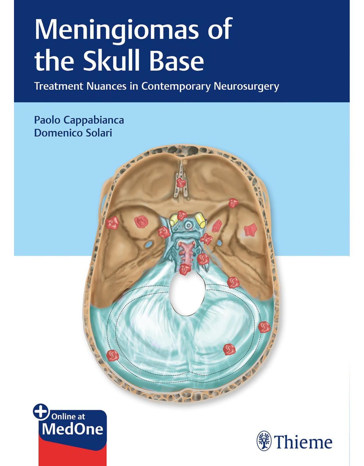
Meningiomas of the Skull Base: Treatment Nuances in Contemporary Neurosurgery
Livrare gratis la comenzi peste 500 RON. Pentru celelalte comenzi livrarea este 20 RON.
Disponibilitate: La comanda in aproximativ 4-6 saptamani
Editura: Thieme
Limba: Engleza
Nr. pagini: 234
Coperta: Hardcover
Dimensiuni: 26.92 x 19.56 cm
An aparitie: 10 Aug. 2018
Description:
Meningiomas, the second most frequent of intracranial tumors, are characterized by a protean range of possible locations and appearances, due to their origin from the extensive and intricately formed meninges. As such, a wide variety of differential diagnoses is typical, and the therapies chosen are necessarily highly variable.
The introductory chapters of this book cover the pathology of these tumors, the evolution of special surgical methods, instrumentation, intraoperative monitoring, and the role of radiosurgery. Ten surgical chapters cover the individual regions of occurrence, including the sphenoid wing, olfactory groove, cerebellopontine angle, etc., all of which require a specialized approach and therapeutic strategy.
Key Features:
Discussion of pathology and therapy organized by anatomic location of the lesions with the goal of providing best patient outcomes
New WHO meningioma classification system based on most recent research in growth patterns, gene sequencing, and molecular patterns of development
Important updates on the newest developments in treatment modalities for meningioma, including the lesser invasive radiotherapy and radiosurgery for the smaller lesions and to avoid the necessity of performing radical surgery
Meningiomas of the Skull Base: Treatment Nuances in Contemporary Neurosurgery is an essential reference guide for neurosurgeons and neurologists (in training and in practice) and will also be welcomed by skull base surgeons and otolaryngologists.
Table of Contents:
1 Introduction
1.1 Essential “Arrows” in the Technical Quiver: Fragments of Personal Memoirs
1.2 Imaging (1973)
1.2.1 The Operating Microscope (1955)
1.2.2 Modern Endoscopy (1974)
1.2.3 Imaging Directed Stereotaxy and Navigation (1977)
1.2.4 Bipolar Forceps (1955)
1.2.5 Ultrasonic Aspirator (1978)
1.2.6 Microanatomy (1973)
2 The Evolution of Surgery—the Soul of Neurosurgery
2.1 Introduction: Early History
2.2 Nomenclature
2.3 Stages of Surgery for Meningiomas
2.4 Initial Surgical Progress
2.5 Extracranial Approaches to Skull Base Lesions
2.6 Microneurosurgery
2.7 Refined Skull Base Approaches and Anatomical Studies
2.8 Allied Advances in Meningioma Management
2.9 Modern Surgical Management
References
3 Inside the Pathology
3.1 Skull Base: Elements of Anatomy and Embryology
3.2 Surgical Resectability
3.3 Meningiomas
3.3.1 Definition and Epidemiology
3.3.2 Histopathology
3.3.3 Immunohistochemistry
3.3.4 Molecular Features
3.3.5 Prognostic Factors
3.4 Solitary Fibrous Tumor/Hemangiopericytoma
3.5 Meningiomas of the Skull Base
3.5.1 Skull Base Meningiomas in Pediatric Age
3.6 Future Perspectives
References
4 Exogenous Factors Affecting Meningiomas
4.1 Introduction
4.2 Ionizing Radiation
4.3 Exogenous Hormones
4.3.1 Link between Cyproterone Acetate and Meningiomas
4.4 Radiofrequency Electromagnetic Fields
4.5 Metabolic Syndrome and Obesity
4.6 Occupational Exposures
4.7 Smoking
4.8 Immunity
4.9 Trauma
4.10 Conclusion
References
5 Instrumentation (Micro, Endo, IGS, MRI Application)
5.1 Introduction
5.2 The Role of the Microscopy in Skull Base Surgery
5.2.1 Positioning
5.2.2 Operating Microscope
5.2.3 Microsurgical Instruments and Techniques
5.3 Endoscopy
5.3.1 Introduction
5.3.2 The Endoscope
5.3.3 Pure Endoscopic Approaches
5.3.4 Endoscope-Assisted Microneurosurgery
5.4 Image-Guided Surgery
5.5 Reconstruction Materials
5.5.1 Duraplasty and Hemostasis
References
6 Intraoperative Neurophysiologic Monitoring during Surgery
6.1 Introduction
6.2 Intraoperative Neurophysiologic Monitoring Techniques
6.2.1 Mapping Techniques to Identify Cranial Motor Nerves
6.2.2 Monitoring Techniques
6.2.3 Anesthetic Considerations
6.3 Clinical Application in Skull Base Surgery
6.3.1 IONM for Sellar and Cavernous Sinus Surgery
6.3.2 IONM for Cerebellopontine Angle Surgery
6.3.3 IONM for Jugular Foramen—Clival and Foramen Magnum Surgery
6.4 IONM Changes and Surgical Strategy
6.5 Conclusions
References
7 Role of Stereotactic Radiosurgery
7.1 Introduction
7.2 Radiobiology of Skull Base Meningiomas
7.3 Outcomes of Stereotactic Radiosurgery for Skull Base Meningiomas
7.3.1 Radiological Response
7.3.2 Cranial Neuropathy
7.3.3 Brainstem Toxicity
7.3.4 Other Complications
7.4 Management of Skull Base Meningiomas and Indications for Stereotactic Radiosurgery
7.4.1 General Indications
7.4.2 Specific Tumor Locations
7.4.3 Primary, Residual, and Recurrent Disease
7.4.4 Atypical and Anaplastic Meningiomas
7.4.5 Case Example
7.5 Conclusions
References
8 Sphenoid Wing Meningiomas
8.1 General Considerations and Definition
8.2 Lateral or Pterional Sphenoid Wing Meningiomas
8.2.1 General Aspects and Clinical Presentation
8.2.2 Evaluation
8.2.3 Indications for Procedure
8.2.4 Surgical Approach
8.3 Medial Sphenoid Wing Meningioma
8.3.1 General Aspects and Clinical Presentation
8.3.2 Evaluation
8.3.3 Indications for Procedure
8.3.4 Surgical Procedure
8.4 Final Considerations
References
9 Clival Meningiomas
9.1 Preoperative Definition of Lesion Features
9.1.1 Definition
9.1.2 Anatomical Considerations
9.2 Surgical Indications
9.3 Surgical Techniques
9.3.1 Preoperative Assessment
9.3.2 Approach Selection
9.4 Complications
9.4.1 Vascular
9.4.2 Cranial Nerve
9.4.3 Brainstem
9.4.4 CSF Leak
9.4.5 Cholesteatoma
9.5 Early and Long-term Postoperative Management
References
10 Petroclival Meningiomas
10.1 Introduction
10.2 Anatomical Considerations
10.3 Clinical Presentation
10.4 Preoperative Evaluation
10.5 Surgical Indications
10.6 Surgical Techniques
10.6.1 Retrosigmoid Approach
10.6.2 Transpetrosal Approaches
10.6.3 Anterior Petrosal Approach
10.6.4 Posterior Petrosal Approach
10.7 Complications
10.8 Early and Long-term Postoperative Management
10.9 Conclusion
References
11 Olfactory Groove Meningiomas
11.1 Introduction
11.2 Preoperative Definition of the Lesion Features
11.2.1 Anatomical Consideration
11.2.2 Clinical Presentation and Workup
11.3 Surgical Indications
11.3.1 Planning the Surgical Strategy
11.3.2 Preoperative Embolization
11.4 Surgical Techniques
11.4.1 Subfrontal Approach with Unilateral or Bilateral Frontal Craniotomy
11.4.2 Transbasal Approach
11.4.3 Pterional Approach
11.4.4 Frontolateral Approach
11.4.5 Supraorbital Keyhole Approach
11.4.6 Endoscopic Endonasal Approach
11.4.7 Algorithm for Surgical Management of Olfactory Groove Meningiomas (Endoscopic Endonasal Approaches vs. Craniotomy vs. Two-stages Strategy)
11.5 Illustrative Cases
11.5.1 Case 1
11.5.2 Case 2
11.5.3 Case 3
11.6 Complications
11.7 Early and Long-term Follow-up
11.7.1 Functional Outcomes
11.7.2 Recurrence
References
12 Middle Fossa Floor Meningiomas
12.1 Introduction
12.2 Preoperative Definition of Lesion Features
12.2.1 Surgical Anatomy of the Middle Cranial Fossa
12.2.2 Middle Cranial Fossa Floor Meningiomas
12.3 Preoperative Management
12.4 Surgical Indications
12.5 Surgical Approaches
12.5.1 Pterional Approach
12.5.2 Fronto-temporo-(orbito)-zygomatic Approach
12.5.3 Temporal Approach
12.5.4 Operative Techniques
12.6 Complications
12.6.1 How to Avoid Complications?
12.7 Long-term Outcome
12.8 Conclusion
12.9 Acknowledgment
References
13 Cerebellopontine Angle Meningiomas
13.1 Introduction
13.2 Clinical Presentation and Preoperative Evaluation
13.3 Neuroradiological Evaluation
13.4 Patient Evaluation and Decision-making
13.5 Preoperative Surgical Planning and Complication Avoidance
13.6 Surgical Approaches
13.6.1 Anterior Petroesectomy Approach
13.7 Posterior and Combined Petrosal Approaches
13.7.1 Retrolabyrinthine Approach
13.7.2 Translabyrinthine Approach
13.7.3 Combined Petrosal Approach
13.7.4 Closure of Posterior and Combined Petrosal Approaches
13.8 Retrosigmoid Approach for the Treatment of Cerebellopontine Angle Meningiomas
13.9 Postoperative Complications Avoidance
13.10 Radiosurgery for Cerebellopontine Angle Meningiomas
13.11 Conclusion
References
14 Foramen Magnum Meningioma
14.1 Introduction
14.2 History
14.3 Surgical Anatomy
14.3.1 Occipital Condyle
14.3.2 Vertebral Artery
14.4 Epidemiology, Clinical Presentation, and Imaging
14.5 Classification of Foramen Magnum Meningioma
14.6 Operative Approaches
14.6.1 Posterior Suboccipital Approach
14.6.2 Far Lateral Approach
14.6.3 Extreme Lateral Approach
14.7 Controversies in Surgical Approaches
14.7.1 Need for Condyle Resection
14.7.2 Need for C1 Laminectomy
14.7.3 Need of Vertebral Artery Mobilization
14.8 Choosing the Right Approach
14.9 Complications
14.10 Outcome
14.11 Role of Radiosurgery
14.12 Personal Experience
14.13 Conclusion
References
15 Tuberculum Sellae/Planum Meningiomas
15.1 Introduction
15.2 Preoperative Definition of Lesion Features
15.3 Surgical Indications
15.3.1 Indications for the Endoscopic Endonasal Approach
15.3.2 Indications for the Supraorbital Approach
15.4 Surgical Techniques
15.4.1 Endoscopic Endonasal Approach
15.4.2 Supraorbital Eyebrow Approach
15.5 Complications
15.6 Surgical Results
15.6.1 Endoscopic Endonasal Surgery
15.6.2 Supraorbital Surgery
15.7 Early- and Long-term Postoperative Management
15.8 Conclusion
References
16 Cavernous Sinus Meningiomas
16.1 Introduction
16.2 Anatomy
16.3 Development
16.4 Symptoms
16.5 Diagnosis
16.6 Treatment
16.7 Surgical Approaches
16.7.1 Preoperative Preparation
16.7.2 Positioning and Incision
16.7.3 Fronto-Orbitozygomatic Osteotomy
16.7.4 Drilling of the Sphenoid Ridge
16.7.5 Identification and Dissection of the Meningoorbital Band
16.7.6 Anterior Clinoidectomy
16.7.7 Unroofing the Optic Nerve
16.7.8 Extradural Exposure of the Cavernous Sinus
16.7.9 Cavernous Sinus Exploration
16.8 Surgery and Radiosurgery
16.9 Acknowledgments
References
17 Reconstruction of the Skull Base
17.1 Introduction
17.2 Reconstruction Techniques and Materials
17.2.1 Free Autografts
17.2.2 Nonautologous Materials
17.2.3 Vascularized Flaps
17.3 Current Reconstruction Techniques
17.3.1 Transcranial Approaches
17.3.2 Endoscopic Endonasal Approaches
17.4 Discussion and Conclusions
References
18 Management of Recurrent Skull Base Meningiomas
18.1 Introduction
18.2 Recurrence
18.2.1 Presentation
18.2.2 Extent of Resection
18.2.3 Location
18.2.4 Histopathology
18.2.5 Radiation-induced Meningiomas
18.2.6 Adjuvant Therapy of De Novo Meningiomas
18.2.7 Multiple Recurrences
18.3 Management
18.3.1 Surveillance
18.3.2 Surgical Resection
18.3.3 Radiation
18.3.4 Chemotherapy
18.4 Conclusion
References
19 Natural History and Adjunctive Modalities of Treatment
19.1 Introduction
19.2 Natural History
19.2.1 WHO Grade I
19.2.2 WHO Grade II
19.2.3 WHO Grade III
19.3 Adjunct Treatment Modalities
19.3.1 Radiation
19.3.2 Brachytherapy
19.3.3 Medical Therapies
19.4 Conclusion
References
20 Complications in the Management of Skull Base Meningioma
20.1 Overview
20.2 Preoperative Factors for Prediction of Complications
20.2.1 Clinicoradiological Factors
20.2.2 Multidisciplinary Approach
20.2.3 Surgeon Factors
20.2.4 Patient Factors
20.3 Perioperative, Operative Complications and Prevention
20.3.1 Anesthesia Considerations
20.3.2 Operative Complications
20.4 Postoperative Complications and Prevention
20.5 Approach-related Problems
20.5.1 Transcranial Approaches (Table 20.1, Table 20.2, and Table 20.3)
20.5.2 Transnasal Endoscopic Skull Base Approaches (Table 20.4)
20.6 Location-related Complications
20.6.1 Olfactory Groove
20.6.2 Planum and Tuberculum Sellae
20.6.3 Cavernous Sinus/Parasellar
20.6.4 Sphenoid Wing Meningioma
20.6.5 Middle Cranial Fossa Meningioma
20.6.6 Petroclival Meningioma
20.6.7 Petrous-tentorial
20.6.8 Foramen Magnum Meningioma
20.7 Conclusion
20.8 Acknowledgment
References
Index
| An aparitie | 10 Aug. 2018 |
| Autor | Paolo Cappabianca, Domenico Solari |
| Dimensiuni | 26.92 x 19.56 cm |
| Editura | Thieme |
| Format | Hardcover |
| ISBN | 9783132412866 |
| Limba | Engleza |
| Nr pag | 234 |


Clientii ebookshop.ro nu au adaugat inca opinii pentru acest produs. Fii primul care adauga o parere, folosind formularul de mai jos.