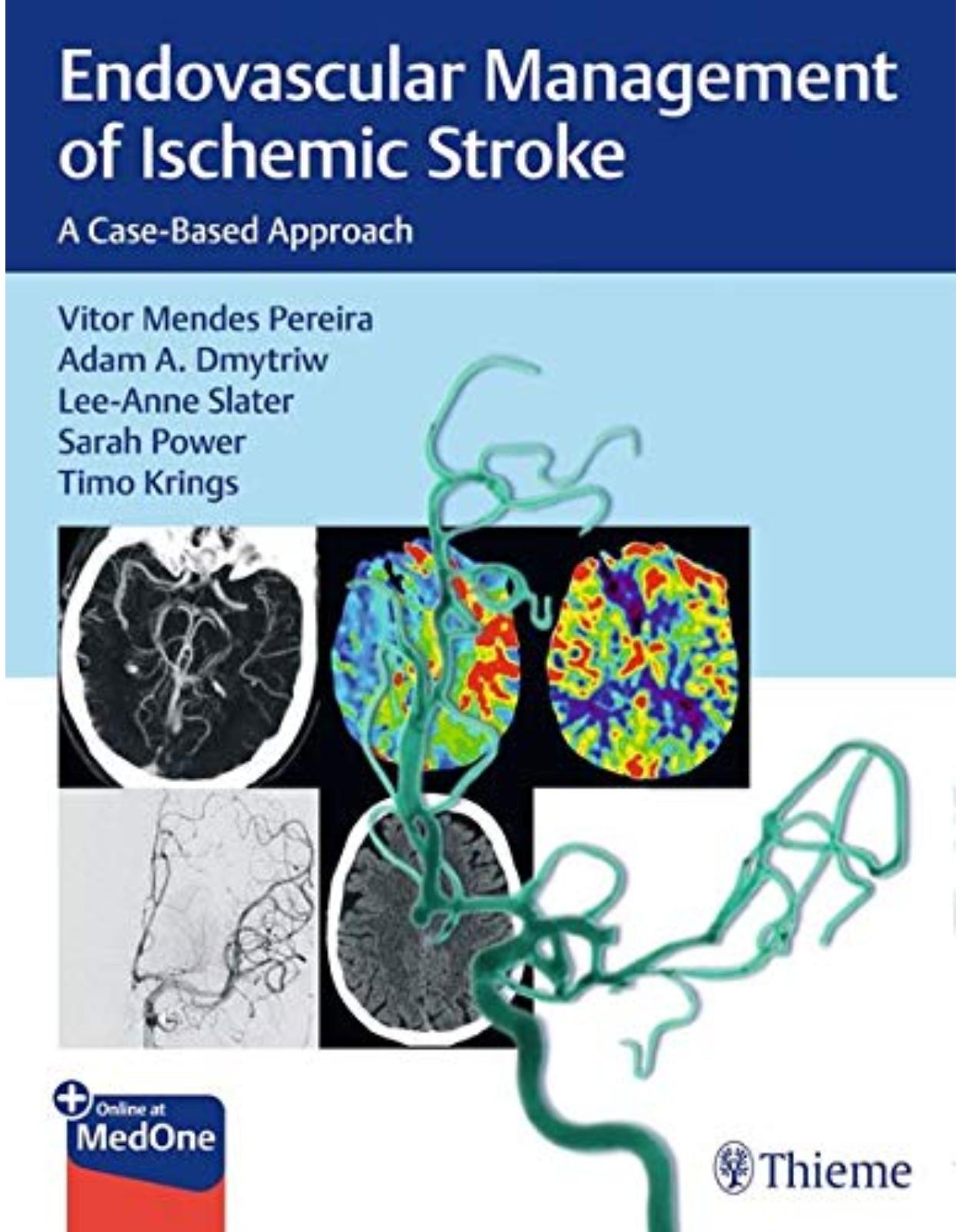
Endovascular Management of Ischemic Stroke: A Case-Based Approach
Livrare gratis la comenzi peste 500 RON. Pentru celelalte comenzi livrarea este 20 RON.
Disponibilitate: La comanda in aproximativ 4-6 saptamani
Autor: Vitor Mendes Pereira (Author), Adam A. Dmytriw (Author), Lee-Anne Slater (Author), Sarah Power
Editura: Thieme
Limba: Engleza
Nr. pagini: 300
Coperta: Paperback
Dimensiuni: 27.94 x 21.59 cm
An aparitie: 08 Decembrie 2021
Description:
A case-based guide to the interventional management of stroke from leading international experts!
Stroke is the most prevalent cerebrovascular emergency, impacting an estimated 15 million people worldwide every year. Endovascular treatment (EVT) of ischemic stroke has expanded at an unforeseen pace, with EVT the most common neurointerventional procedure performed at most large centers. Endovascular Management of Ischemic Stroke: A Case-Based Approach by renowned stroke pioneer Vitor Mendes Pereira and distinguished co-editors features contributions from a "who's who" of global experts. This practical resource provides straightforward guidance for clinicians who need to learn and master state-of-the-art endovascular interventions reflecting the new, evidenced-based treatment paradigm for acute stroke.
This carefully crafted reference takes readers on a journey from the early building blocks that led to modern stroke interventions to meticulous step-by-step descriptions of the latest approaches. Fifty high-yield cases mirror real-life scenarios trainees and professionals are likely to encounter in clinical practice. Seven sections encompass a full spectrum of diverse patient presentations, anatomical variations, advanced techniques, complex pathologies, complications, and stroke mimics.
Key Highlights
Discussion of emerging techniques likely to stand the test of time such as SAVE, ARTS, transradial access, and transcarotid access
Stroke mimics important for differential diagnoses, including hemiplegic migraine, MELAS, RCVS, seizure, and more
An appendix that covers fundamental terms, trials, and tools
This cutting-edge resource is essential reading for trainee and early-career interventionalists, as well as seasoned practitioners in interventional radiology, neuroradiology, endovascular neurosurgery, and interventional neurology.
Table of Contents:
Section I Evolution of Endovascular Management
1 Intra-arterial Tissue Plasminogen Activator: The First Step
1.1 Case Description
1.1.1 Clinical Presentation
1.1.2 Imaging Workup and Investigations
1.1.3 Diagnosis
1.1.4 Management
1.1.5 Technique
1.1.6 Outcome
1.1.7 Discussion
1.2 References
2 Transcatheter MERCI Clot Retrieval: The Early Generation
2.1 Case Description
2.1.1 Clinical Presentation
2.1.2 Imaging Workup and Investigations
2.1.3 Diagnosis
2.1.4 Management
2.1.5 Technique
2.1.6 Outcome
2.1.7 Discussion
2.2 References
3 Penumbra Clot Aspiration Technique: The Dark Days
3.1 Case Description
3.1.1 Clinical Presentation
3.1.2 Imaging Workup and Investigations
3.1.3 Diagnosis
3.1.4 Management
3.1.5 Technique
3.1.6 Outcome
3.1.7 Discussion
3.1.8 Further Attempts and Critiques
3.2 References
4 Trevo Stent-Retriever Thrombectomy: Light on the Horizon
4.1 Case Description
4.1.1 Clinical Presentation
4.1.2 Imaging and Workup
4.1.3 Diagnosis
4.1.4 Technique
4.1.5 Outcome
4.1.6 Discussion
4.2 References
5 Solitaire Stent-Retriever Thrombectomy: Building the Evidence
5.1 Case Description
5.1.1 Clinical Presentation
5.1.2 Imaging and Workup
5.1.3 Diagnosis
5.1.4 Technique
5.1.5 Outcome
5.1.6 Discussion
5.2 References
Section II Case Selection
6 Timing in Stroke and the Tissue Clock
6.1 Case Description
6.1.1 Clinical Presentation
6.1.2 Imaging Workup and Investigations
6.1.3 Diagnosis
6.1.4 Treatment
6.1.5 Outcome
6.2 Companion Case
6.2.1 Clinical Presentation
6.2.2 Imaging Workup and Investigations
6.2.3 Diagnosis
6.2.4 Treatment
6.2.5 Endovascular
6.2.6 Outcome
6.2.7 Discussion
6.2.8 Pearls and Pitfalls
6.3 References
7 Role of Leptomeningeal Collaterals
7.1 Case Description
7.1.1 Clinical Presentation
7.1.2 Imaging Workup and Investigations
7.1.3 Diagnosis
7.1.4 Treatment
7.2 Postprocedure Care/Outcome
7.3 Companion Case
7.3.1 Clinical Presentation
7.3.2 Imaging Workup and Investigations
7.3.3 Diagnosis
7.3.4 Treatment
7.4 Postprocedure Care/Outcome
7.4.1 Discussion
7.4.2 Workup and Diagnosis
7.4.3 Imaging Findings
7.4.4 Decision-Making Process
7.4.5 Management
7.4.6 Postprocedural Care
7.4.7 Literature Synopsis
7.4.8 Pearls and Pitfalls
7.5 Further Reading
8 Importance of Clot Burden and Clot Location
8.1 Case Description
8.1.1 Clinical Presentation
8.1.2 Imaging Workup and Investigations
8.1.3 Diagnosis
8.1.4 Treatment
8.1.5 Discussion
8.1.6 Workup and Diagnosis
8.1.7 Imaging Findings
8.1.8 Decision-Making Process
8.1.9 Postprocedural Care
8.1.10 Pearls and Pitfalls
8.2 References
9 ASPECTS: When Not to Treat
9.1 Case Description
9.1.1 Clinical Presentation
9.1.2 Imaging Workup and Investigations
9.1.3 Diagnosis
9.1.4 Management
9.1.5 Endovascular Treatment
9.1.6 Postprocedural Care
9.1.7 Outcome
9.1.8 Background
9.1.9 Discussion
9.1.10 Pearls and Pitfalls
9.2 References
10 Microbleeds Are Not a Contraindication to Thrombolysis in Acute Stroke
10.1 Case Description
10.1.1 Clinical Presentation
10.1.2 Imaging Workup and Investigations
10.2 Diagnosis
10.3 Treatment
10.3.1 Initial Management
10.3.2 Endovascular Management
10.3.3 Outcome
10.4 Discussion
10.4.1 Background
10.4.2 Workup and Diagnosis
10.4.3 Decision-Making Process
10.5 Literature Synopsis
10.6 Pearls and Pitfalls
10.7 References
10.8 Further Reading
11 Stroke Etiologies: Hemodynamic, Embolic, and Perforator Stroke
11.1 Case Description
11.1.1 Clinical Presentation
11.1.2 Imaging Workup and Investigations
11.1.3 Diagnosis
11.1.4 Treatment
11.2 Companion Case
11.2.1 Clinical Presentation
11.2.2 Imaging Workup and Investigations
11.2.3 Diagnosis
11.2.4 Treatment
11.3 Additional Companion Case
11.3.1 Clinical Presentation
11.3.2 Imaging Findings
11.3.3 Diagnosis
11.3.4 Treatment
11.4 Discussion
11.4.1 Background
11.4.2 Workup and Diagnosis
11.4.3 Imaging Findings
11.4.4 Decision-Making Process
11.4.5 Literature Synopsis
11.4.6 Pearls and Pitfalls
11.5 Further Reading
12 Endovascular Therapy in a Patient with a Proximal MCA Occlusion and No Neurological Deficits
12.1 Case Description
12.1.1 Clinical Presentation
12.1.2 Imaging Workup and Investigations
12.1.3 Diagnosis
12.1.4 Treatment
12.1.5 Outcome
12.2 Discussion
12.2.1 Background
12.3 Pearls and Pitfalls
12.4 References
13 MRI in Stroke (Core Size, Mismatch, and New Advances)
13.1 Case Description
13.1.1 Clinical Presentation
13.1.2 Imaging Workup and Investigations
13.1.3 Diagnosis
13.1.4 Treatment
13.1.5 Outcome
13.2 Companion Case
13.2.1 Clinical Presentation
13.2.2 Imaging Workup and Investigations
13.2.3 Diagnosis
13.2.4 Treatment
13.2.5 Outcome
13.3 Discussion
13.3.1 Background
13.3.2 Early Signs
13.4 Imaging Sequences
13.4.1 Diffusion-Weighted Imaging
13.4.2 Fluid-Attenuated Inversion Recovery
13.4.3 Susceptibility-Weighted Imaging
13.4.4 Time of Flight Angiography
13.4.5 MR Perfusion-Weighted Imaging
13.4.6 Evolution of Stroke on MR Imaging
13.4.7 CT versus MRI in Stroke
13.5 References
Section III Fundamentals and Standard Approaches
14 The Classical Setup: Carotid-T with Balloon
14.1 Case Description
14.1.1 Clinical Presentation
14.1.2 Imaging Workup and Investigations
14.1.3 Diagnosis
14.1.4 Treatment
14.2 Discussion
14.2.1 Pearls and Pitfalls
14.3 References
15 M1 Anatomy and Role of Perforators in Outcome
15.1 Case Description
15.1.1 Clinical Presentation
15.1.2 Imaging Workup and Investigations
15.1.3 Diagnosis
15.1.4 Treatment
15.2 Companion Case
15.2.1 Clinical Presentation
15.2.2 Imaging Workup and Investigations
15.2.3 Diagnosis
15.2.4 Treatment
15.3 Discussion
15.3.1 Background
15.3.2 Workup and Diagnosis
15.3.3 Imaging Findings
15.3.4 Decision-Making Process
15.3.5 Management
15.3.6 Literature Synopsis
15.3.7 Pearls and Pitfalls
15.4 Further Readings
16 Basilar Artery Occlusion
16.1 Case Description
16.1.1 Clinical Presentation
16.1.2 Imaging Workup and Investigations
16.1.3 Diagnosis and Treatment
16.1.4 Technique
16.1.5 Postprocedural Care and Outcome
16.2 Companion Case
16.2.1 Clinical Presentation
16.2.2 Imaging Workup and Investigations
16.2.3 Diagnosis and Treatment
16.2.4 Technique
16.2.5 Postprocedural Care and Outcome
16.2.6 Background
16.2.7 Discussion
16.2.8 Pearls and Pitfalls
16.3 References
17 ADAPT Technique for Acute Ischemic Stroke Thrombectomy
17.1 Case Description
17.1.1 Clinical Presentation
17.1.2 Imaging Workup and Investigations
17.1.3 Diagnosis
17.1.4 Treatment
17.1.5 Materials and Endovascular Treatment
17.1.6 Outcome
17.1.7 Discussion
17.1.8 Pearls and Pitfalls
17.2 Further Reading
18 General Anesthesia in Thrombectomy
18.1 Case Description
18.1.1 Clinical Presentation
18.1.2 Imaging Workup and Investigations
18.1.3 Diagnosis
18.1.4 Treatment
18.1.5 Postprocedure Care/Outcome
18.2 Discussion
18.2.1 Early Retrospective Studies
18.2.2 Prospective Studies and Randomized Controlled Trials
18.2.3 Meta-analyses
18.2.4 Decision Making
18.2.5 Airway Protection
18.2.6 Compliance and Patient Movement
18.2.7 Time from Symptom Onset to Effective Treatment
18.2.8 Hemodynamic Complications under General Anesthesia
18.2.9 Monitoring of Complications
18.3 Conclusion
18.4 Pearls and Pitfalls
18.5 References
19 Conscious Sedation in Thrombectomy
19.1 Case Description
19.1.1 Clinical Presentation
19.1.2 Imaging Workup and Investigations
19.1.3 Diagnosis
19.1.4 Treatment
19.2 Companion Case
19.2.1 Clinical Presentation
19.2.2 Imaging Workup and Investigations
19.2.3 Outcome
19.2.4 Case Discussion: Anesthesia Choices in Stroke Intervention
19.2.5 Pearls and Pitfalls
19.3 References
20 Endovascular Therapy for Patients with an Isolated M2 Occlusion
20.1 Case Description
20.1.1 Clinical Presentation
20.1.2 Imaging Workup and Investigations
20.1.3 Diagnosis
20.1.4 Management
20.1.5 Endovascular Treatment
20.1.6 Technique
20.1.7 Postprocedural Care
20.1.8 Outcome
20.1.9 Tips and Tricks
Section IV Advanced Techniques
21 Percutaneous Radial Arterial Access for Thrombectomy
21.1 Case Description
21.1.1 Clinical Presentation
21.1.2 Imaging Workup and Investigations
21.1.3 Diagnosis
21.1.4 Treatment
21.1.5 Postprocedural Care
21.1.6 Outcome
21.1.7 Background
21.1.8 Discussion
21.1.9 Pearls and Pitfalls
21.2 References
22 Percutaneous Transcarotid Endovascular Thrombectomy for Acute Ischemic Stroke
22.1 Case Description
22.1.1 Clinical Presentation
22.1.2 Imaging Workup and Investigations
22.1.3 Diagnosis
22.1.4 Treatment
22.1.5 Postprocedure Care
22.1.6 Outcome
22.2 Background
22.3 Discussion
22.3.1 Pearls and Pitfalls
23 Distal Access Catheter Technique without Balloon Assistance
23.1 Case Description
23.1.1 Clinical Presentation
23.1.2 Imaging Workup and Investigations
23.1.3 Diagnosis
23.1.4 Treatment
23.1.5 Progress
23.1.6 Discussion
23.2 References
Section V Complex Cases
24 Tandem Lesions
24.1 Case Description
24.1.1 Clinical Presentation
24.1.2 Imaging Workup and Investigations
24.1.3 Diagnosis
24.1.4 Treatment
24.2 Companion Case
24.2.1 Clinical Presentation
24.2.2 Discussion
24.2.3 Workup and Diagnosis
24.2.4 Imaging Findings
24.2.5 Decision-Making Process
24.2.6 Management
24.2.7 Postprocedure Care
24.2.8 Literature Synopsis
24.2.9 Pearls and Pitfalls
24.3 Further Readings
25 Pseudo-occlusion
25.1 Case Description
25.1.1 Clinical Presentation
25.1.2 Imaging Workup and Investigations
25.1.3 Diagnosis
25.1.4 Treatment
25.2 Companion Case
25.2.1 Clinical Presentation
25.2.2 Imaging Workup and Investigations
25.2.3 Diagnosis
25.2.4 Treatment
25.3 Discussion
25.3.1 Background
25.3.2 Workup and Diagnosis
25.3.3 Imaging Findings
25.3.4 Decision-Making Process
25.3.5 Management
25.3.6 Postprocedure Care
25.3.7 Literature Synopsis
25.3.8 Pearls and Pitfalls
25.4 Further Readings
26 Underlying Intracranial Stenosis
26.1 Case Description
26.1.1 Clinical Presentation
26.1.2 Imaging Workup and Investigations
26.1.3 Diagnosis
26.1.4 Management
26.1.5 Endovascular Treatment
26.1.6 Postprocedure Care
26.1.7 Outcome
26.2 Companion Case
26.2.1 Clinical Presentation
26.2.2 Imaging Workup and Investigations
26.2.3 Management
26.2.4 Endovascular Treatment
26.2.5 Postprocedure Care
26.2.6 Outcome
26.3 Discussion
26.3.1 Background
26.3.2 Discussion
26.3.3 Pearls and Pitfalls
26.4 References
27 Intracranial Dissection
27.1 Case Description
27.1.1 Clinical Presentation
27.1.2 Imaging Workup and Investigations
27.1.3 Diagnosis
27.1.4 Treatment
27.1.5 Posttreatment Management
27.1.6 Comments
27.2 Discussion
27.3 References
28 Hyperacute Extracranial Angioplasty and Stenting: When and How
28.1 Case Description
28.1.1 Clinical Presentation
28.1.2 Imaging Workup and Investigations
28.1.3 Diagnosis
28.1.4 Treatment
28.2 Discussion
28.2.1 Background
28.2.2 Workup and Diagnosis
28.2.3 Imaging Findings
28.2.4 Decision-Making Process
28.2.5 Management
28.2.6 Postprocedure Care
28.2.7 Literature Synopsis
28.2.8 Pearls and Pitfalls
28.3 Further Readings
29 Failed Mechanical Thrombectomy: What to Do Next
29.1 Case Description
29.1.1 Clinical Presentation
29.1.2 Imaging Workup and Investigations
29.1.3 Diagnosis
29.1.4 Treatment
29.2 Discussion
29.2.1 Postprocedure Care
29.2.2 Pearls and Pitfalls
29.3 References
30 Rescue Permanent Stenting
30.1 Case Description
30.1.1 Clinical Presentation
30.1.2 Imaging Workup and Investigations
30.1.3 Diagnosis
30.1.4 Management
30.1.5 Endovascular Treatment
30.1.6 Postprocedure Care
30.1.7 Outcome
30.1.8 Background
30.1.9 Pearls and Pitfalls
30.2 References
31 Acute Dissection with Hemodynamic Infarctions
31.1 Case Description
31.1.1 Clinical Presentation
31.1.2 Imaging Studies
31.1.3 Diagnosis
31.1.4 Treatment
31.2 Outcome
31.3 Discussion
31.4 References
32 The SAVE Technique
32.1 Case Description
32.1.1 Clinical Presentation
32.1.2 Imaging Workup and Investigations
32.1.3 Diagnosis
32.1.4 Management
32.1.5 Endovascular Treatment
32.1.6 Postprocedure Care
32.1.7 Outcome
32.1.8 Background
32.1.9 Discussion
32.1.10 Pearls and Pitfalls
32.2 References
33 Aspiration-Retriever Technique for Stroke (ARTS)
33.1 Case Description
33.1.1 Clinical Presentation
33.1.2 Imaging
33.1.3 Management
33.1.4 Endovascular Devices
33.1.5 Outcome
33.2 Background
33.2.1 Discussion
33.3 References
Section VI Complications
34 Clot Migration with Emboli to Distal Territories
34.1 Case Description
34.1.1 Clinical Presentation
34.1.2 Imaging Workup and Investigations
34.1.3 Discussion
34.2 References
35 Endothelial Damage
35.1 Case Description
35.1.1 Clinical Presentation
35.1.2 Imaging Workup and Investigations
35.1.3 Diagnosis
35.1.4 Treatment
35.2 Discussion
35.2.1 Background
35.2.2 Workup and Diagnosis
35.2.3 Decision-Making Process
35.2.4 Management
35.2.5 Postprocedure Care
35.2.6 Literature Synopsis
35.3 Suggested Readings
36 Management of Vessel Perforation during Stroke Intervention
36.1 Case Description:
36.1.1 Clinical Presentation
36.1.2 Imaging Workup and Investigations
36.1.3 Diagnosis
36.1.4 Treatment
36.2 Companion Case
36.2.1 Clinical Presentation
36.2.2 Imaging Workup and Investigations
36.2.3 Diagnosis
36.2.4 Treatment
36.2.5 Progress
36.2.6 Further Treatment
36.2.7 Progress
36.2.8 Case Discussion
36.3 References
37 Arterial Access Complications
37.1 Case Description
37.1.1 Clinical Presentation
37.1.2 Mini-Case A: Active Extravasation Round the Vascular Sheath
37.1.3 Mini-Case B: Femoral Artery Pseudoaneurysm
37.1.4 Mini-Case C: Hematomas in Anticoagulated Patients
37.1.5 Mini-Case D: Distal Embolic Occlusion
37.1.6 Mini-Case E: Vessel Dissection
37.1.7 Mini-Case F: Delayed Thrombus Formation on the Angio-Seal Foot Plate
37.1.8 Mini-Case G: Acute Loss of Pulse
37.1.9 Pearls and Pitfalls
38 Intraprocedural Vasospasm during Thrombectomy
38.1 Case Description
38.1.1 Clinical Presentation
38.1.2 Imaging Workup and Investigations
38.1.3 Diagnosis
38.1.4 Treatment
38.1.5 Progress
38.2 Discussion
38.2.1 Incidence and Clinical Sequelae
38.2.2 Intraprocedural Management
38.3 References
39 Hemorrhagic Transformation after Endovascular Stroke Therapy
39.1 Case Description
39.1.1 Clinical Presentation
39.1.2 Imaging Workup and Investigations
39.1.3 Diagnosis
39.1.4 Treatment
39.1.5 Endovascular Treatment
39.1.6 Postprocedure Care
39.1.7 Outcome
39.2 Companion Case
39.2.1 Case
39.2.2 Clinical Presentation
39.2.3 Imaging Workup and Investigations
39.2.4 Diagnosis
39.2.5 Treatment
39.2.6 Endovascular Treatment
39.2.7 Postprocedure Care
39.2.8 Outcome
39.3 Discussion
39.4 Pearls and Pitfalls
39.5 References
40 Endovascular Treatment of Cerebral Venous Thrombosis
40.1 Case Description
40.1.1 Clinical Presentation
40.1.2 Baseline Imaging
40.2 Diagnosis
40.3 Treatment
40.3.1 Outcome
40.4 Discussion
40.5 References
41 Endovascular Thrombectomy for Pediatric Acute Ischemic Stroke
41.1 Case Description
41.1.1 Clinical Presentation
41.1.2 Imaging Workup and Investigations
41.2 Diagnosis
41.2.1 Treatment
41.2.2 Postprocedure Care
41.2.3 Outcome
41.2.4 Background
41.3 Discussion
41.3.1 Pearls and Pitfalls
41.4 References
Section VII Stroke Mimics and Rare Causes
42 Hemiplegic Migraine
42.1 Case Description
42.1.1 Clinical Presentation
42.1.2 Imaging Workup and Investigations
42.1.3 Diagnosis
42.1.4 Treatment
42.2 Discussion
42.2.1 Background
42.2.2 Workup and Diagnosis
42.2.3 Decision-Making Process
42.2.4 Management
42.2.5 Literature Synopsis
42.2.6 Pearls and Pitfalls
42.3 References
43 Intra-arterial Contrast Injection during Computed Tomography Angiography
43.1 Case Description
43.1.1 Clinical Presentation
43.1.2 Imaging Workup and Investigations
43.1.3 Diagnosis
43.1.4 Treatment
43.2 Discussion
43.2.1 Background
43.2.2 Workup and Diagnosis
43.2.3 Decision-Making Process
43.2.4 Management
43.2.5 Literature Synopsis
43.2.6 Pearls and Pitfalls
43.3 References
44 Mitochondrial Encephalomyopathy, Lactic Acidosis, and Stroke-Like Episodes (MELAS)
44.1 Case Description
44.1.1 Clinical Presentation
44.1.2 Imaging Workup and Investigations
44.1.3 Diagnosis
44.1.4 Treatment
44.2 Discussion
44.2.1 Background
44.2.2 Workup and Diagnosis
44.2.3 Decision-Making Process
44.2.4 Management
44.2.5 Literature Synopsis
44.2.6 Pearls and Pitfalls
44.3 Further Readings
45 Reversible Cerebral Vasoconstriction Syndrome
45.1 Case Description
45.1.1 Clinical Presentation
45.1.2 Imaging Workup and Investigations
45.1.3 Diagnosis
45.1.4 Treatment and Follow-up
45.1.5 Outcome
45.2 Discussion
45.2.1 Background
45.2.2 Pathology
45.2.3 Presentation
45.2.4 Diagnosis
45.2.5 Differential Diagnosis
45.2.6 Clinical Course
45.2.7 Management and Prognosis
45.2.8 Pearls and Pitfalls
45.3 References
46 Acute Ischemic Stroke Secondary to Cardiac Myxoma Embolus
46.1 Case Description
46.1.1 Clinical Presentation
46.1.2 Imaging Workup and Investigations
46.1.3 Diagnosis
46.1.4 Treatment
46.2 Companion Case
46.2.1 Clinical Presentation
46.2.2 Imaging Workup and Investigations
46.2.3 Diagnosis
46.2.4 Treatment
46.3 Discussion
46.3.1 Background
46.3.2 Pearls and Pitfalls
46.4 References
47 Seizure
47.1 Case Description
47.1.1 Clinical Presentation
47.1.2 Investigations
47.1.3 Clinical Reassessment
47.1.4 Diagnosis
47.1.5 Treatment
47.2 Discussion
47.2.1 Background
47.2.2 Workup and Diagnosis
47.2.3 Decision-Making Process
47.2.4 Management
47.2.5 Literature Synopsis
47.2.6 Pearls and Pitfalls
47.3 References
48 Cerebral Amyloid Angiopathy
48.1 Case Description
48.1.1 Case 1: Patient with Seizures and Cognitive Impairment
48.1.2 Investigations and Imaging
48.1.3 Treatment and Outcome
48.1.4 Final Diagnosis
48.2 Case 2: Patient with Rapidly Progressive Cognitive Decline
48.2.1 Clinical Presentation
48.2.2 Investigations and Imaging
48.2.3 Treatment and Outcome
48.2.4 Final Diagnosis
48.3 Discussion
48.3.1 Clinical Presentations of CAA
48.3.2 Diagnostic Criteria for CAA
48.3.3 Inflammatory Variants of CAA
48.3.4 Imaging Investigations for CAA
48.3.5 Biochemical Markers of CAA
48.3.6 Treatment and Prognosis
48.4 Summary
48.5 Suggested Readings
49 Contrast Staining Masquerading as Hemorrhage
49.1 Case
49.2 Case Description
49.2.1 Clinical Presentation
49.2.2 Imaging Workup and Investigations
49.2.3 Diagnosis
49.2.4 Treatment
49.3 Discussion
49.4 Pearls and Pitfalls
49.5 References
50 HaNDL
50.1 Case Description
50.1.1 Clinical Presentation
50.1.2 Imaging Workup and Investigations
50.1.3 Diagnosis
50.1.4 Treatment and Follow-up
50.2 Companion Case
50.2.1 Clinical Presentation
50.2.2 Imaging Workup and Investigations
50.2.3 Diagnosis
50.2.4 Treatment
50.3 Discussion
50.3.1 Background
50.3.2 Workup and Diagnosis
50.3.3 Decision-Making Process
50.3.4 Management
50.3.5 Literature Synopsis
50.3.6 Pearls and Pitfalls
50.4 References
51 Appendix: Essential Terms, Trials, and Tools
51.1 Introduction
51.2 Recanalization and Reperfusion Scoring
51.2.1 Reperfusion Scales
51.2.2 Recanalization Scale: The Arterial Occlusive Lesion Scoring System
51.3 The National Institute of Health Stroke Scale
51.4 The Alberta Stroke Program Early CT Score
51.5 Common CT/CTA Protocols
51.5.1 Noncontrast Head Computed Tomographic Scan
51.5.2 CT Angiography
51.5.3 CT Perfusion
51.6 European Cooperative Acute Stroke Study II Trial Hemorrhage Classification
51.7 Collateral Circulation Score
51.8 Thrombectomy Devices
51.8.1 Stent Retrievers
51.8.2 Aspiration Devices
51.8.3 Permanent Stents
51.9 Tool Terminology Glossary
51.9.1 Flexibility
51.9.2 Internal Diameter (ID)
51.9.3 Kink Resistance
51.9.4 Lubricity (Coefficient of Friction)
51.9.5 Outer Diameter (OD)
51.9.6 Shore Hardness
51.9.7 Tensile Strength
51.9.8 Torque
51.10 Curves of Catheters and Selective Catheters Types
51.11 Modern Inclusion/Eligibility Criteria
51.12 Main Trials
51.13 References
Index
| An aparitie | 08 Decembrie 2021 |
| Autor | Vitor Mendes Pereira (Author), Adam A. Dmytriw (Author), Lee-Anne Slater (Author), Sarah Power |
| Dimensiuni | 27.94 x 21.59 cm |
| Editura | Thieme |
| Format | Paperback |
| ISBN | 9781626232754 |
| Limba | Engleza |
| Nr pag | 300 |




Clientii ebookshop.ro nu au adaugat inca opinii pentru acest produs. Fii primul care adauga o parere, folosind formularul de mai jos.