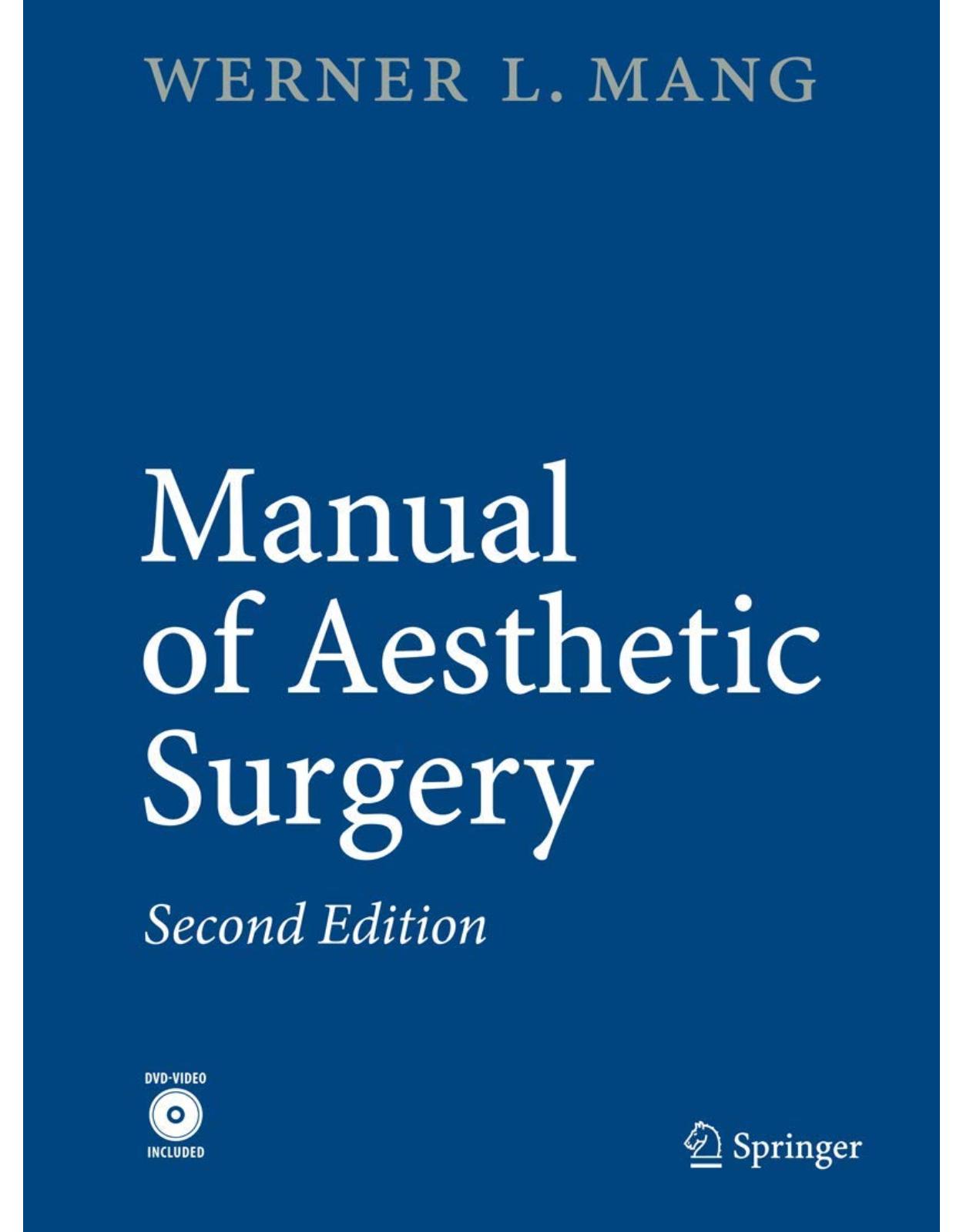
Manual of Aesthetic Surgery
Livrare gratis la comenzi peste 500 RON. Pentru celelalte comenzi livrarea este 20 RON.
Disponibilitate: La comanda in aproximativ 4-6 saptamani
Autor: Werner Mang
Editura: Springer
Limba: Engleza
Nr. pagini: 710
Coperta: Paperback
Dimensiuni: 19.3 x 26.92 cm
An aparitie: 5 May 2017
Description:
At the request of Springer-Verlag, Heidelberg, I have written with pride and pleasure the new edition of the Manual of Aesthetic Surgery. Volumes1(headand neckregion)and 2(body)havebeen integrated into one volume, which has been revised and extended with the addition of the topics “Cosmetic Aesthetic Surgery,” “Breast Surgery,” “Mini Lift,” “Mini Abdomen,” “Buttock Lift,” and “Tumescence Li- suction with the MicroAire® System.” The current trend is towards gentle surgical methods. The ‘Mang School’ has as its motto: Less is more! You should not see cosmetic s- gery. Aesthetic surgery is feel-good surgery and not altering surgery. That should be the philosophy of this book. The first editions of both volumes of the Manual of Aesthetic Surgery had high print runs and were translated into many languages, including Spanish, Russian, and Chinese. The new edition bridges a few gaps, namely breast lifting and breast reduction. These operations are described in detail using illustrations and videos, in order to provide also plastic and aesthetic surgeons with standards. Standards are of crucial importance in aesthetic surgery. Results must be reproducible. Every aesthetic surgeon will then be able to build on this manual and refine his or her methods.
Table of Contents:
A General Remark
The Standard Procedures
Rhinoplasty
Rhytidectomy Using the Tumescence Technique – Mini Lift
Upper Eyelid Surgery
Lower Eyelid Surgery
Otoplasty
Breast Augmentation
Breast Lifting and Breast Reduction
Brachioplasty
Abdominoplasty – Mini Abdominoplasty
Thigh and Buttock Lift – New Methods
Liposuction – Removal of Fat with the Tumescence Technique (Mang’s Solution) – MicroAire® Sys
Hair Transplantation
Spacelift
Adjuvant Therapies
Acknowledgements
History – Vita
Ten Rules
Table of Contents
1 Photographic Documentation in Aestheticand Plastic Surgery
Examples in Photo Documentation
1. Breast Procedures
2. Facial Procedures
3. Body Contouring Procedures: Abdomen
2 Informed Consent in Aesthetic and Plastic Surgery
Guiding Principles in the Informed Consent Discussion
1. Persons Obliged to Obtain Informed Consent
2. Informed Consent in Non-medically Indicated Procedures(Cosmetic Procedures)
3. Timing of the Informed Consent Discussion
4. Informed Consent for Foreign-Language Speakers
5. Extent of the Informed Consent Discussion
3 Rhinoplasty
Introduction
Anatomical Overview (Fig. 3.1)
Instruments and Medication (Fig. 3.2–3.4)
Duplicate Patient Instruction
Nasal Examination
Photographic Documentation
Surgical Planning
Tumescence Injection Technique
Disinfection
Suction and Surgical Planning
Incision Line
Decollement
Correction of the Nasal Tip with the Eversion Method (Fig. 3.15–3.18)
Nasal Shortening (Fig. 3.19, 3.20)
Bump Ablation (Fig. 3.21–3.25)
Reshaping of the Tip (Fig. 3.26, 3.27)
Suturing of the Mucosal Incisions
Resection of the “Mang Triangle” (Fig. 3.30)
Osteotomies (Fig. 3.32)
External Dressing
Postoperative Medication and Precautions
Results
Tips and Tricks
4 Rhytidectomy (Cervicobuccal Plasty)
Introduction
Anatomical Overview (Fig. 4.1)
Instruments and Medication (Fig. 4.3–4.5)
1. With local anesthesia
2. With endotracheal anesthesia
Duplicate Patient Instruction
Photographic Documentation
Surgical Planning
Premedication
Anesthesia with Hypotension
Tumescence of the Face and Neck
The Mang Method of Tumescence Rhytidectomy
Liposuction and Undermining with the Suction Instruments (Fig. 4.8, 4.9)
Endoscopic Brow Lift (Temporal Tightening) (Fig. 4.10)
Stage 1 Rhytidectomy (30 – 40 Age Group) (Fig. 4.11)
Stage 2 Rhytidectomy (40 – 45 Age Group) (Fig. 4.12)
Stage 3 Rhytidectomy (45 – 50 Age Group) (Fig. 4.13–4.14)
Stage 4 Rhytidectomy (50-Plus Age Group) – Standard Facelift (ESP Lift)(Fig. 4.15–4.53)
Incision Lines
Dissection of the Lipocutaneous Flap (Fig. 4.19–4.28)
Dissection of the Cheeks and Neck (Fig. 4.25, 4.26)
Deep Dissection and Exposure of the Platysma (Fig. 4.27)
Wound Trimming andWound Sealing with Fibrin Adhesive (Fig. 4.31)
Skin Tightening (Fig. 4.32, 4.33)
Skin Incision and Placement of the Key Sutures (Fig. 4.34–4.43)
Subcutaneous Wound Closure (Fig. 4.44, 4.45)
Temporal Flap Resection and Sutures (Fig. 4.46–4.47)
PeriauricularWound Closure
Retroauricular Skin Resection, Redon Drain, andWound Closure (Fig. 4.51–4.53)
Identical Approach on the Contralateral Side
Special Bandaging Technique
Postoperative Care and Precautions
Results
Mini Lift
Disinfection
Premedication
Anesthesia (Fig. 4.60)
Preliminary Marking of Incision Lines (Maximum Dissection) (Fig. 4.61)
Incision (Fig. 4.62)
Dissection (Fig. 4.63, 4.64)
Resection (Fig. 4.65)
Wound Revision, Hemostasis, Cutaneous Suturing (Fig. 4.66–4.68)
Results
Tips and Tricks: Rhytidectomy
Tips and Tricks: M-Lifting
5 Eyelid Surgery – Blepharoplasty
Upper Eyelid Surgery – Blepharoplasty
Introduction
Anatomical Overview (Fig. 5.1)
Instruments and Medication (Fig. 5.2)
Duplicate Patient Instruction
Ophthalmological Status
Photographic Documentation
Surgical Planning
Preliminary Marking of Incision Lines (Fig. 5.3)
Local Anesthesia (Fig. 5.4)
Disinfection
Type of Incision (Fig. 5.5)
Skin Resection Under Tension (Fig. 5.6, 5.7)
Medial and Intermediate Lipectomy (Fig. 5.8–5.12)
Removal of a Strip of Connective Tissue and Muscle (Fig. 5.13)
Cutaneous Sutures (Fig. 5.15)
Postoperative Treatment and Precautions
Results
Lower Eyelid Surgery – Blepharoplasty
Introduction
Anatomical Overview (Fig. 5.19)
Instruments and Medication (Fig. 5.20)
Duplicate Patient Instruction
Ophthalmological Status
Surgical Planning
Preliminary Marking of Incision Lines (Fig. 5.21)
Local Anesthesia (Fig. 5.22)
Disinfection
Type of Incision (Fig. 5.23)
Excision of the Musculocutaneous Flap (Fig. 5.26)
Medial, Intermediate, and Lateral Lipectomy (Fig. 5.29–5.32)
Removal of a Muscular Strip From the Orbicular Muscle of the Eye
Skin Resection
Sutures and Dressing
Postoperative Treatment and Precautions
Results
Tips and Tricks
6 Otoplasty
Introduction
Anatomical Overview (Fig. 6.1)
Instruments and Medication (Fig. 6.2)
Photographic Documentation
Preliminary Examination of the Ear
Surgical Planning
Disinfection
Preliminary Marking of Incision Lines (Fig. 6.3)
Local Anesthesia (Fig. 6.4)
Incision (Fig. 6.5)
Skin Resection (Fig. 6.6–6.7)
Exposure of the Dorsal Surface of the Auricular Cartilage (Fig. 6.9)
Preparation of the Auricular Concha
1. Marking with Fine Needles (Fig. 6.10)
2. Marking of Incision Lines (Fig. 6.11)
3. Incision of Conchal Cartilage (Fig. 6.12)
4. Blunt Cartilage Dissection (Fig. 6.13)
Resection of the Concha (Fig. 6.14)
Reshaping the Anthelix (Fig. 6.15–6.22)
Cartilage Sutures (Fig. 6.21, 6.22)
Wound Closure (Fig. 6.23, 6.24)
Identical Approach on the Contralateral Side
Dressing
Postoperative Treatment and Precautions
Results
Tips and Tricks
7 BreastSurgery
7.1 Breast Augmentation
Introduction
Breast Implants*
Anatomical Overview (Fig. 7.1)
Instruments (Fig. 7.2–7.5)
Duplicate Patient Information
Preliminary Examinations
Photographic Documentation
Surgical Planning (Fig. 7.6)
Incision Line in the Case of Inframammary Access
Definition of the Subsequent Breast or Implant Size by Establishing the Distance Between the Lower M
Positioning of the Patient, Disinfection of the Surgical Area
Tumescence (Fig. 7.9)
Inframammary Incision (Fig. 7.10)
7.1.1 Supramuscular Access
Dissection, Step 1 (Fig. 7.11)
Dissection, Step 2: PreciseDemonstration of the Caudal,Medial, and Lateral Borders of Pectoralis Maj
Deep, Blunt Dissection
Wound Revision and Hemostasis Using the Illuminated Retractor and Bipolar Tweezers (Fig. 7.14)
Determining the Size and Shape of the Implant (Fig. 7.15)
Fitting the Final Implant (Fig. 7.16)
Exact Positioning of the Implant (Fig. 7.17)
Insertion of the Redon Drain (Size 10) (Fig. 7.18)
Deep Wound Closure (Fig. 7.19)
Two-Layer, Atraumatic Wound Closure Using 4.0 Monocryl (Fig. 7.20)
Dressing (Fig. 7.21)
Aftercare
7.1.2 Submuscular Access (Fig. 7.22–7.27)
Submuscular Implant
Dual Plane Dissection
Results
7.2 Breast Reduction/Breast Lifting
Tips and Tricks
Results
8 Brachioplasty
Introduction
Upper-Arm Tightening
Anatomical Overview (Fig. 8.1)
Instruments (Fig. 8.2–8.4)
Duplicate Patient Information
Preliminary Examinations
Photographic Documentation
Surgical Planning
Preliminary Marking of Incision Lines (Fig. 8.5)
Mang’s Fish-Mouth Technique (Fig. 8.6)
Positioning, Disinfection
Tumescence (Fig. 8.7)
Incision (Fig. 8.8)
Superficial Dissection (Fig. 8.9)
Deep Dissection, Hemostasis (Fig. 8.10)
Incision of the Dissected Dermofat Flap in Stages (Fig. 8.11)
Fixing of the Skin Flap with 3.0 Monocryl Key Sutures (Fig. 8.12)
Resection in Stages (Fig. 8.13)
Two-Layer Skin Closure (Fig. 8.14)
Cutaneous Sutures: Running or Intracutaneous 4.0 Monocryl (Fig. 8.15)
Dressing (Fig. 8.16)
Aftercare
Results
Tips and Tricks
9 Abdominoplasty
Introduction
Tightening of the Abdominal Wall
Anatomical Overview (Fig. 9.1)
Instruments* (Fig. 9.2–9.4)
Duplicate Patient Information
Preliminary Examinations
Photographic Documentation
Surgical Planning
Postoperative Treatment
Typical Findings: Indications for Tightening the Abdominal Wall
Positioning, Disinfection
Marking the Individual Incision Line (Fig. 9.6)
Tumescence (Fig. 9.7)
Incision (Fig. 9.8)
Dissection of the Lower Abdomen (Fig. 9.9)
Incision Around the Navel (Fig. 9.10)
Mobilization and Dissection of the Navel (Fig. 9.11)
Vertical Splitting of the Dermofat Flap as Far as the Base of the Navel (Fig. 9.12)
Complete Mobilization of the Umbilical Stalk (Fig. 9.13)
Dissection of the Upper Abdomen (Fig. 9.14)
Doubling of the Rectus Abdominis Fascia (Fig. 9.15)
Defining the Resection Boundaries with Upper Body Flexed at 30°
Repositioning of the Navel Using a V-Shaped Incision (Fig. 9.17)
Pulling the Navel Out of the V-Shaped Incision with Curved Forceps (Fig. 9.18)
Positioning the Navel (Fig. 9.19)
Trimming of the Skin of the Navel and Adaptation to the V-Shaped Incision (Fig. 9.20)
Closure of the Navel in Three Layers (Fig. 9.21)
Fixation of the Surplus Sections of Skin with 2.0 Monocryl Key Sutures
Resection of the Skin in Stages
Insertion of Redon Drains
Wound Closure in Three Layers
Dressing
Fitting the Abdominal Belt
Results
Mini Abdominoplasty
Tips and Tricks
Results
10 Thigh and Buttock Lift
Introduction
Anatomical Overview (Fig. 10.2)
Instruments (Fig. 10.3–10.5)
Duplicate Patient Information
Preliminary Examinations
Photographic Documentation
Surgical Planning
Positioning, Disinfection
Tumescence (Fig. 10.7)
Incision of the Skin (Fig. 10.8)
Dissection (Fig. 10.9)
Deep Dissection and Hemostasis (Fig. 10.10)
Definition of Resection Boundaries (Fig. 10.11)
Skin Resection (Fig. 10.12)
Fixation Suture on the Pubic Bone with 2.0 Monocryl (Fig. 10.13)
Second Fixation Suture on the Inguinal Ligament (Fig. 10.14)
Deep Wound Closure and Insertion of a Redon Drain (No. 10) (Fig. 10.15)
Intracutaneous Skin Closure (Fig. 10.16)
Dressing (Fig. 10.17)
Buttock Lift: Positioning, Disinfection
Tumescence (Fig. 10.18)
Incision Line (Fig. 10.18)
Incision (Fig. 10.19)
Wedge-Shaped Dermolipectomy (Fig. 10.20, 10.21)
Transverse Incision Through the Resected Area (Fig. 10.22)
Hemostasis, Deep Wound Closure, Insertion of a Redon Drain (Fig. 10.23)
Two-Layer, Tension-Free Wound Closure (Fig. 10.24)
Dressing (Fig. 10.25)
Postoperative Treatment: Course of Action After the Operation; Precautionary Measures
Results
NewProcedure for Buttock Lifting with Re-shaping of the Gluteal Fold (Fig. 10.27–10.30)
Surgical Planning
Tumescence
Incision, De-epithelialization
Dissection
Fixation
Insertion of a Redon drain
Two-layer, tension-free wound closure
Dressing
Postoperative Treatment
Results
Tips and Tricks
11 Liposuction
Introduction
Instruments (Fig. 11.1, 11.2)
Duplicate Patient Information
Preliminary Examination
Surgical Planning
Anatomy of Liposuction of the Abdomen, Hips, Thighs (Fig. 11.3)
Anatomical Overview (Fig. 11.3)
Anatomy of Liposuction of the Hips, Back, Thighs, Buttocks (Body Contouring) (Fig. 11.4)
Anatomical Overview (Fig. 11.4)
Anatomy of Liposuction of the Axilla, Chest, Hips, Lateral Side of the Thighs (Fig. 11.5)
Anatomical Overview (Fig. 11.5)
Anatomy of Liposuction of the Medial/Lateral Side of the Thighs, Knee(Fig. 11.6)
Anatomical Overview (Fig. 11.6)
Anatomy of Liposuction of the Medial Part of the Thighs, Knees, Calves, Ankles
Anatomical Overview (Fig. 11.7a)
Anatomy of Liposuction of the Calf and Ankles
Anatomical Overview (Fig. 11.7b)
Anatomy of Liposuction of the Breast, Axilla, Upper Arms (Fig. 11.8)
Anatomical Overview (Fig. 11.8)
Anatomy of Liposuction of a Double Chin (Fig. 11.9)
Anatomical Overview (Fig. 11.9)
Mechanical and Manual Tumescence (Fig. 11.10)
Location of the Incision Sites from the Rear (Fig. 11.11a)
Location of the Incision Sites from the Front (Fig. 11.11b)
Schematic Diagram of Mang’s Tumescent Liposuction Technique
Cross-Section of the Liposuction Technique (Fig. 11.14)
Cross-Section of the Tissue 6 Months After Liposuction with Preservation of the Infrastructural Conn
Technique
Disinfection
Manual Liposuction
Mechanical Liposuction
Procedure
Dressing
Aftercare
Results
MicroAire System
12 Hair Transplantation
Introduction
Instruments
Basic Instrument Set (Sterilizable)* (Fig. 12.2)
Instruments for Graft/Follicular Unit Preparation (Fig. 12.3)
Instruments for Micropunch Technique (Fig. 12.4)
Instruments for Microslit Technique (Fig. 12.5)
Preparation of the Patient, Hairline Design (Fig. 12.6)
Donor Area
Local Anesthesia, Tumescence (Fig. 12.7)
Donor Strip Harvesting (Fig. 12.8)
Skin Closure with Continuous Sutures (Fig. 12.9)
Follicular Unit Dissection (Minigrafts, Micrografts, Single Hairs) (Fig. 12.10)
Recipient Area, Holes, and Slits (Fig. 12.11)
Transplantation Channels (Fig. 12.12)
Transplantation (Fig. 12.13)
Aftercare
Postoperative Precautions
Results
Tips and Tricks
13 Adjuvant Therapies, Including Laser Surgery
Introduction
Local Anesthesia
Nerve Exit Points, Supraorbital Nerve, Infraorbital Nerve, Mental Nerve
1. Biological Implants
Report – Technique
Collagen and Hyaluronic Acid
Results
Application and Absorption of Collagen
Application Examples (Fig. 13.10)
Glabella Wrinkles
Eye Wrinkles
Nasolabial Folds (Fig. 13.11)
Lip Augmentation (Fig. 13.12)
Results
Acne Scars
Liquid Lifting
Contouring Using the Mang Method
Results
Liquid Lift According to Mang (Polylactic Acid + Juv´ederm)
Sculptra®: Crystalline polylactic acid
Usage
Hyal System
Introduction
1. Dermal filler
2. Hyal System
Report – Technique
Follow-Up Treatment
2. Mang’s Spacelift (Autologous Fat Injection)
Introduction
History
Indications
Instruments (Fig. 13.21)
Technique
Injection Technique
Possible Complications
Advantages of Lipotransfer
Diagram of the Fat Injection (Fig. 13.24)
Three-Dimensional Diagram of Fat Injection into Subcutaneous Tissue (Fig. 13.25)
Increasing the Density of the Connective Tissue Following Breakdown of Fat Droplets
Results
3. Botulinum Toxin*
Report – Technique
Indications
Results
Anger Wrinkles
Horizontal Forehead Wrinkles (Fig. 13.32)
Neck
Upper Lip Wrinkles
Drooping Corner of the Mouth
4. Dermabrasion
Indications
Technique (Fig. 13.35, 13.36)
Dressing and Follow-Up Treatment
Results
5. Chemical Peeling
Stage 1
Method
Effect
Preparation
Procedure
Follow-Up Treatment
Stage 2
Method
Effect
Preparation
Procedure
Follow-Up Treatment
Risks
Effect
Stage 3
Method
Effect
Results
6. Ultrapulse CO2 Laser Surgery*
Indications
Pretreatment
Anesthesia
Surgical Steps
Follow-Up Treatment
Results
7. Erbium:YAG Laser
Introduction
Report – Technique
Results
14 Aesthetic Surgery: Quo Vadis?
Prospects
15 Bibliography
Further Reading
Chapter 1: Photodocumentation in Aesthetic Plastic Surgery
Chapter 2: Informed Consent in Aesthetic Plastic Surgery
Chapter 3: Rhinoplasty
Chapter 4: Rhytidectomy
Chapter 5: Eylid Surgery
Chapter 6: Otoplasty
Chapter 7: Breast Surgery
Chapter 8: Brachioplasty
Chapter 9: Abdominoplasty
Chapter 10: Thigh and Buttock Lift
Buttock Lift
Thigh Lift
Chapter 11: Liposuction
Chapter 12: Hair Transplantation
Chapter 13: Adjuvant Therapies Including Laser Surgery
16 Subject Index
17 List of Suppliers
| An aparitie | 5 May 2017 |
| Autor | Werner Mang |
| Dimensiuni | 19.3 x 26.92 cm |
| Editura | Springer |
| Format | Paperback |
| ISBN | 9783662517413 |
| Limba | Engleza |
| Nr pag | 710 |
-
2,07800 lei 1,76500 lei
-
1,34300 lei 1,26900 lei




Clientii ebookshop.ro nu au adaugat inca opinii pentru acest produs. Fii primul care adauga o parere, folosind formularul de mai jos.