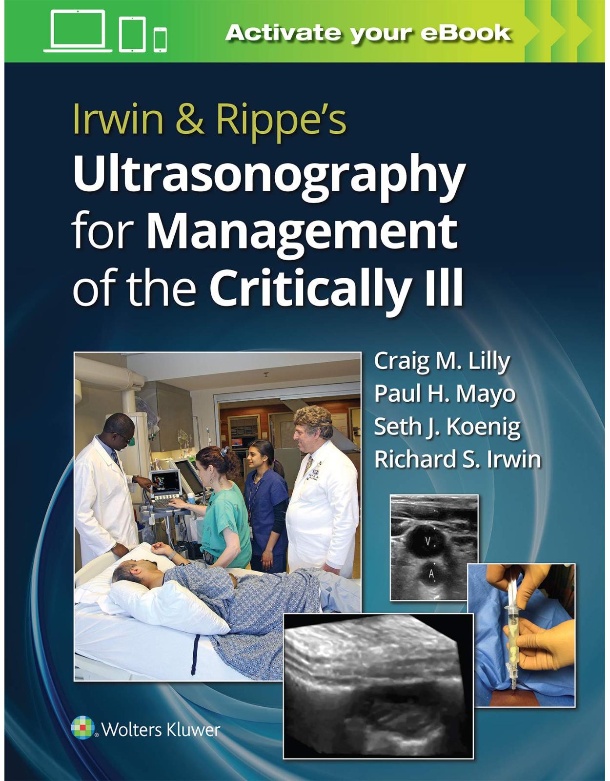
Irwin & Rippe’s Ultrasonography for Management of the Critically Ill
Livrare gratis la comenzi peste 500 RON. Pentru celelalte comenzi livrarea este 20 RON.
Disponibilitate: La comanda in aproximativ 4-6 saptamani
Editura: LWW
Limba: Engleza
Nr. pagini: 480
Coperta: Paperback
Dimensiuni: 17.53 x 1.52 x 25.15 cm
An aparitie: 12 Nov. 2020
Description:
Enhancing and adding to ultrasonographic text and video content in Irwin & Rippe’s Intensive Care Medicine, Eighth Edition, this easy-to-follow volume provides expert guidance on the optimal use of ultrasound in the critical care environment. Irwin & Rippe’s Ultrasonography for Management of the Critically Ill covers a wide variety of critical care procedures, explaining how to perform them and how ultrasound can be used to support evaluation and management services to patients with life-threatening illnesses or injuries.
Table of Contents:
SECTION 1 Introduction to Ultrasonography
1 Point-of-Care for Critical Care Ultrasonography
COMPETENCE
Machine Requirements
Machine Controls and Scanning Technique
Scope of Practice
Limitations of Critical Care Ultrasonography
CONCLUSIONS
REFERENCES
SECTION 2 Neurological Procedures
2 Neurologic Multimodal Monitoring
GOALS OF BRAIN MONITORING
CEREBRAL ISCHEMIA AND BLOOD FLOW
TECHNIQUES OF NEUROLOGIC MONITORING
Neurologic Examination
Systemic Monitoring
Electroencephalography/Electrocorticography
Evoked Potentials
Intracranial Pressure Monitoring
Cerebral Blood Flow Monitoring
Transcranial Doppler Ultrasound
Jugular Venous Bulb Oxygen Saturation
Brain Tissue Oxygen Tension
Neurochemical Monitoring
Near-Infrared Spectroscopy
Neuroimaging
MULTIMODAL MONITORING STRATEGIES
REFERENCES
3 Cerebrospinal Fluid Aspiration
INTRODUCTION
CEREBROSPINAL FLUID ACCESS
Diagnostic Objectives
Therapeutic Interventions
TECHNIQUES OF CEREBROSPINAL FLUID ACCESS
Lumbar Puncture
Utility of Ultrasonography for Performance of Lumbar Puncture
TECHNIQUE FOR ULTRASONOGRAPHIC GUIDANCE OF LUMBAR PUNCTURE
Patient Positioning
Transducer Selection and Indicator Positioning
Localization of the Target Lumbar Interspace for Lumbar Puncture
Summary
Lateral Cervical (C1-C2) Puncture
Cisternal Puncture
Aspiration of Reservoirs and Shunts
VENTRICULOSTOMY
Lumbar Drainage
Summary
REFERENCES
SECTION 3 Airway and Respiratory Procedures
4 Airway Management and Endotracheal Intubation
INTRODUCTION
ANATOMY
Nose
Mouth and Jaw
Nasopharynx
Oropharynx
Hypopharynx
Larynx
Trachea
EMERGENCY AIRWAY MANAGEMENT
Airway Obstruction
Use of Face Mask and Bag Valve Device
Airway Adjuncts
INDICATIONS FOR INTUBATION
Preintubation Evaluation
Education and Intubation Management
EQUIPMENT FOR INTUBATION
Laryngoscopes
Endotracheal Tubes
Endotracheal Tube Cuff
ANESTHESIA BEFORE INTUBATION
TECHNIQUES OF INTUBATION
Specific Techniques and Routes of Endotracheal Intubation
Management of the Difficult Airway
Airway Management in the Intubated Patient
COMPLICATIONS OF ENDOTRACHEAL INTUBATION
Complications During Intubation
Complications While the Tube Is in Place
Complications After Extubation
EXTUBATION
UTILITY OF ULTRASONOGRAPHY FOR AIRWAY MANAGEMENT
Identification of Gastric Fluid
Identification of Endotracheal Tube Position
REFERENCES
5 Tracheostomy
INTRODUCTION
INDICATIONS
CONTRAINDICATIONS
TIMING OF TRACHEOSTOMY
PROCEDURES
Emergency Tracheostomy
Cricothyrotomy
TRACHEOSTOMY PROCEDURES IN THE INTENSIVE CARE UNIT
Open Surgical Tracheostomy
Percutaneous Dilational Techniques
TUBES AND CANNULAS
POSTOPERATIVE CARE
Wound and Dressing Care
Inner Cannulas
Humidification
Suctioning
Tracheostomy Tube Changes
Oral Feeding and Swallowing Dysfunction Associated with Tracheostomies
Discharging Patients With Tracheotomies From the ICU to the General Ward
COMPLICATIONS
Obstruction
Tube Displacement/Dislodgment
Subcutaneous Emphysema
Pneumothorax and Pneumomediastinum
Hemorrhage
Tracheoinnominate Artery Fistula
Misplacement of Tube
Tracheal Cartilage Fracture
Stomal Infections
Tracheoesophageal Fistula
Tracheal Stenosis
Tracheomalacia
Dysphagia and Aspiration
Persistent Tracheocutaneous Fistula
CONCLUSIONS
REFERENCES
6 Lung Ultrasonography
BASIC PRINCIPLES OF LUS
MACHINE REQUIREMENTS
PERFORMANCE OF LUS
IMAGING PATTERNS IN LUS
Main LUS Patterns
CLINICAL APPLICATIONS OF LUS
Clarification of the Ambiguous Chest Radiograph
Differentiation of ARDS from Cardiogenic Pulmonary Edema
Prediction of Extubation Failure
Measurement of Lung Recruitment with PEEP
Diagnosis of Pneumonia
Algorithmic Diagnosis of Respiratory Failure
Estimates of Left Atrial Pressure
Combination of LUS with Echocardiography
Diagnosis of Pulmonary Embolism
Timing of Chest Tube Removal
LUS for Guidance of Procedures
Utility of LUS for Airway Management
SUMMARY
REFERENCES
7 Thoracentesis
INTRODUCTION
INDICATIONS
CONTRAINDICATIONS
COMPLICATIONS
THORACENTESIS PROCEDURE
Steps in the Procedure
TECHNIQUE FOR REMOVAL OF AIR FROM A PTX SPACE
Steps in the Procedure
UTILITY OF ULTRASONOGRAPHY FOR GUIDANCE OF THORACENTESIS
Equipment
Scanning Technique
Interpretation of Pleural Fluid Analysis
TRANSUDATES VERSUS EXUDATES
SELECTED TESTS THAT ARE POTENTIALLY HELPFUL TO ESTABLISH ETIOLOGY FOR A PLEURAL EFFUSION
pH
Glucose
Protein, Lactate Dehydrogenase, and Protein Gradient
Amylase
Triglyceride and Cholesterol
Adenosine Deaminase
Cell Counts and Differential
Cultures, Polymerase Chain Reaction, and Stains
Cytologic Evaluation
REFERENCES
8 Chest Tube Insertion and Care
PLEURAL ANATOMY AND PHYSIOLOGY
CHEST TUBE PLACEMENT
Indications
CONTRAINDICATIONS
TECHNIQUE
COMPLICATIONS
CHEST TUBE MANAGEMENT AND CARE
CHEST TUBE REMOVAL
RELATED SYSTEMS
Utility of Ultrasonography for Chest Tube Insertion and Care
Pneumothorax
SUMMARY
REFERENCES
9 Discontinuation of Mechanical Ventilation
INTRODUCTION
UNDERSTANDING THE PROBLEM
Who Are the Patients and What Are Their Outcomes?
What Is Wrong with Patients on Prolonged Ventilator Support?
What Factors Lead to Respiratory Muscle Fatigue and Weakness?
PREDICTING SUCCESSFUL DISCONTINUATION
When Is It Appropriate to Begin the Discontinuation Process?
Predictive Indices for Total Discontinuation of Mechanical Ventilation (Weaning Indices)
When Is It Appropriate to Extubate the Patient?
How Long Should Discontinuation Trials Last?
MANAGING DISCONTINUATION FAILURE
Protocol-Based Weaning
Once-Daily Attempts at Liberation from Mechanical Ventilation
Addressing Factors That Perpetuate Respiratory Muscle Fatigue
Utility of Ultrasonography for Assessment of Diaphragmatic Function for Discontinuation of Mechanical Ventilation
Clinical Applications of Ultrasonography Assessment of Diaphragmatic Function
REFERENCES
SECTION 4 Cardiac Procedures
10 Echocardiography for the Evaluation of Shock and Cardiac Arrest
EFFICACY
EXPERIMENTAL AND ALTERNATIVE TECHNIQUES OF CARDIOPULMONARY RESUSCITATION
INFECTIOUS DISEASES AND CARDIOPULMONARY RESUSCITATION
Implications for Rescuers With Known or Potential Infection
STANDARD PROCEDURES AND TEAM EFFORT
BASIC LIFE SUPPORT FOR ADULTS WITH AN UNOBSTRUCTED AIRWAY
Respiratory Arrest
Cardiac Arrest
Assessment and Determination of Unresponsiveness and Alerting of Emergency Medical Services
Opening the Airway and Determining Breathlessness
Rescue Breathing
Chest Compressions
Two-Rescuer Cardiopulmonary Resuscitation
Complications of Basic Life Support Procedures
Monitoring the Effectiveness of Basic Life Support
PEDIATRIC RESUSCITATION
OBSTRUCTED AIRWAY
ADVANCED CARDIAC LIFE SUPPORT IN ADULTS
Airway and Ventilatory Support
Circulatory Support
Advanced Cardiac Life Support: Defibrillation and Rhythm-Based Therapies
Defibrillation
Ventricular Fibrillation and Pulseless VT
Pulseless Electric Activity and Asystole
Correction of Hypoxia
Correction of Excessive Hydrogen Ion (Acidosis)
Hypovolemia
Venous Access
DRUG THERAPY
Sympathomimetic Drugs and Vasopressors
Antiarrhythmic Agents
Lidocaine
Other Agents
CLINICAL SETTINGS
Special Situations
Termination of ACLS and Post-ACLS Care
UTILITY OF ULTRASONOGRAPHY FOR CARDIOPULMONARY RESUSCITATION
CONCLUSIONS
REFERENCES
11 Critical Care Echocardiography
BASIC CRITICAL CARE ECHOCARDIOGRAPHY
Equipment Requirements
Technical Challenges
Training in Basic Critical Care Echocardiography
The Basic Critical Echocardiography Examination
Clinical Applications of Goal-Directed Echocardiography
Limitations of Basic Critical Care Echocardiography
ADVANCED CRITICAL CARE ECHOCARDIOGRAPHY
Training Requirements
Transesophageal Echocardiography
Limitations of Advanced Critical Care Echocardiography
REFERENCES
12 Resuscitation From Shock Following Hemorrhage
PHYSIOLOGIC RESPONSES TO HEMORRHAGE
Coagulopathy
Hemodynamics
Metabolic
Immunologic
SHOCK RECOGNITION
Utility of Ultrasonography for Diagnosis of Hemorrhagic Shock
HEMORRHAGE CONTROL
HISTORY OF HEMORRHAGIC SHOCK RESUSCITATION
INITIAL RESUSCITATION
ONGOING RESUSCITATION
Endpoints of Resuscitation
Coagulation Endpoints
Monitoring
Fluids and Component Therapy
Vasopressor and Inotropic Support
Additional Therapies
SUMMARY
REFERENCES
13 Mechanical Complications of Myocardial Infarction
CONSEQUENCES OF ISCHEMIA
DIAGNOSIS, TREATMENT, AND OUTCOME OF SHOCK DUE TO LEFT VENTRICULAR PUMP FAILURE
RIGHT VENTRICULAR INFARCTION
MYOCARDIAL RUPTURE
Papillary Muscle Rupture
Ventricular Septal Rupture
Free Wall Rupture
LEFT VENTRICULAR REMODELING
UTILITY OF ULTRASONOGRAPHY FOR DIAGNOSIS OF MECHANICAL COMPLICATIONS OF MYOCARDIAL INFARCTION
Papillary Muscle Rupture
Ventricular Wall Rupture
Free Wall Rupture
REFERENCES
14 Pericardiocentesis
INTRODUCTION
INDICATIONS FOR PERICARDIOCENTESIS
ANATOMY
PROCEDURE
SHORT-TERM AND LONG-TERM MANAGEMENT
Utility of Ultrasonography for Management of Pericardial Effusion
SUMMARY
REFERENCES
15 Temporary Cardiac Pacing
INTRODUCTION
INDICATIONS FOR TEMPORARY CARDIAC PACING
Bradyarrhythmias
Tachyarrhythmias
DIAGNOSIS OF RAPID RHYTHMS
ACUTE MYOCARDIAL INFARCTION
EQUIPMENT AVAILABLE FOR TEMPORARY PACING
Transvenous Pacing Catheters
Utility of Ultrasonography for Transvenous Pacemaker Insertion
Esophageal Electrode
Transcutaneous External Pacemakers
Epicardial Pacing
Pulse Generators for Temporary Pacing
CHOICE OF PACING MODE
PROCEDURE TO ESTABLISH TEMPORARY PACING
COMPLICATIONS OF TEMPORARY PACING
REFERENCES
16 Pulmonary Artery Catheters
INTRODUCTION
PHYSIOLOGIC RATIONALE FOR USE OF PULMONARY ARTERY CATHETER
CONTROVERSIES REGARDING USE OF PULMONARY ARTERY CATHETER
INDICATIONS FOR USE OF PULMONARY ARTERY CATHETER
CATHETER FEATURES AND CONSTRUCTION
Pressure Transducers
INSERTION TECHNIQUES
General Considerations
Typical Catheter Insertion Procedure
Special Considerations
Utility of Ultrasonography for Pulmonary Artery Catheter Insertion
PHYSIOLOGIC DATA
Pressures
Cardiac Output
Derived Parameters
CLINICAL APPLICATIONS OF THE PULMONARY ARTERY CATHETER
Normal Resting Hemodynamic Profile
COMPLICATIONS
Complications Associated with Central Venous Access
Balloon Rupture
Knotting
Pulmonary Infarction
Pulmonary Artery Perforation
Thromboembolic Complications
Rhythm Disturbances
Intracardiac Damage
Infections
Other Complications
GUIDELINES FOR SAFE USE OF PULMONARY ARTERY CATHETERS
SUMMARY
REFERENCES
17 Management of Implantable Pacemakers, Defibrillators, and Cardiac Resynchronization Devices and Cardiac Devices in the ICU
INTRODUCTION
GENERAL DEVICE MANAGEMENT
Normal Device Function and Special Considerations
Pacemaker Malfunction
DEVICE-SPECIFIC CONSIDERATIONS
Implantable Cardioverter Defibrillators
Cardiac Resynchronization Therapy (Biventricular Pacing)
SUMMARY
REFERENCES
SECTION 5 Vascular and Hematological Procedures
18 Arterial Line Placement and Care
INTRODUCTION
INDICATIONS FOR ARTERIAL CANNULATION
EQUIPMENT, MONITORING, TECHNIQUES, AND SOURCES OF ERROR
TECHNIQUE OF ARTERIAL CANNULATION
Site Selection
Radial Artery Cannulation
Evaluation of Collateral Circulation of the Hand
Percutaneous Insertion
Dorsalis Pedis Artery Cannulation
Brachial Artery Cannulation
Femoral Artery Cannulation
Axillary Artery Cannulation
Utility of Ultrasonography for Arterial Catheterization
COMPLICATIONS OF ARTERIAL CANNULATION
Thrombosis
Cerebral Embolization
Diagnostic Blood Loss
Other Mechanical and Technical Complications
Infections
RECOMMENDATIONS
REFERENCES
19 Central Venous Catheters
INTRODUCTION
INDICATIONS AND SITE SELECTION
GENERAL CONSIDERATIONS
Informed Consent
Patient Comfort and Safety
Mobile Catheter Cart
Catheter Tip Location
Vascular Erosions
Complications
ROUTES OF CENTRAL VENOUS CANNULATION
Antecubital Approach
Internal Jugular Approach
External Jugular Vein Approach
Femoral Vein Approach
Subclavian Vein Approach
REFERENCES
20 Venous Thromboembolism and Associated Prothrombotic Disorders in the Intensive Care Unit
INCIDENCE
RISK FACTORS
Inherited Hypercoagulable Disorders
Acquired Hypercoagulable Disorders
Diagnostic Approach to Hypercoagulable Conditions
PATHOPHYSIOLOGY
PREVENTION
DIAGNOSIS
Symptoms and Signs
Clinical Prediction Models
D-Dimer
Diagnosis of Acute Deep Venous Thrombosis
Diagnosis of Acute Pulmonary Embolism
An Integrated Approach to Venous Thromboembolism Diagnosis
Risk Stratification
TREATMENT
Anticoagulation
Thrombolytic Therapy
Inferior Vena Cava Interruption
Pulmonary Embolectomy
Adjunctive Care for Massive Pulmonary Embolism
SPECIAL THERAPEUTIC CONSIDERATIONS
Central Venous Catheter–Associated Thrombosis
Pregnancy-Associated VTE
SEQUELAE OF VENOUS THROMBOEMBOLISM
Duration of Anticoagulation
Complications of Anticoagulation
Utility of Ultrasonography for Evaluation of Venous Thromboembolism
ACKNOWLEDGMENT
REFERENCES
21 Therapeutic Apheresis: Technical Considerations and Indications in Critical Care
TECHNICAL RATIONALE AND INSTRUMENTS
PHYSIOLOGIC PRINCIPLES
VASCULAR ACCESS FOR THERAPEUTIC APHERESIS
ANTICOAGULATION AND FLUID REPLACEMENT
VASCULAR ACCESS
LIMITATIONS AND POTENTIAL ADVERSE EVENTS
INDICATIONS IN CRITICAL CARE
Therapeutic Plasma Exchange
Red Blood Cell Exchange
Leukapheresis
Plateletpheresis
APHERESIS CONSULTATION
REFERENCES
SECTION 6 Gastrointestinal and Abdominal Procedures
22 Endoscopic Placement of Feeding Tubes
INDICATIONS FOR ENTERAL FEEDING
ACCESS TO THE GASTROINTESTINAL TRACT
TECHNIQUES
Nasoenteric Route
Percutaneous Route
Fluoroscopic Technique
Complications
SURGICAL PROCEDURES
Gastrostomy
Needle-Catheter Jejunostomy
Transgastric Jejunostomy
DELIVERING THE TUBE-FEEDING FORMULA
MEDICATIONS
COMPLICATIONS
Nasopulmonary Intubation
Aspiration
Gastrointestinal Intolerance
Metabolic Complications
Bacterial Contamination
Occluded Feeding Tubes
Utility of Ultrasonography for Feeding Tube Insertion
REFERENCES
23 Gastrointestinal Endoscopy
PATIENT SELECTION
Evaluation of the Upper Gastrointestinal Tract
Evaluation of the Pancreaticobiliary Tract
Evaluation of the Mid-gastrointestinal Tract (Jejunum and Ileum)
Evaluation of the Lower Gastrointestinal Tract
PREPROCEDURAL CARE
INTRAPROCEDURAL CARE
Upper Gastrointestinal Endoscopy
PANCREATICOBILIARY ENDOSCOPY
Lower Gastrointestinal Endoscopy
POSTPROCEDURAL CARE
FUTURE DIRECTIONS
REFERENCES
24 Gastroesophageal Balloon Tamponade for Acute Variceal Hemorrhage
HISTORICAL DEVELOPMENT
ROLE OF BALLOON TAMPONADE FOR THE MANAGEMENT OF BLEEDING ESOPHAGEAL VARICES
INDICATIONS AND CONTRAINDICATIONS
TECHNICAL AND PRACTICAL CONSIDERATIONS
Airway Control
Hypovolemia, Shock, and Coagulopathy
Clots and Gastric Decompression
Infection, Ulceration, and Encephalopathy
Balloons, Ports, and Preparation
Insertion and Placement of the Tube
Ultrasonography for Tube Insertion
Fixation and Traction Techniques
Maintenance, Monitoring, and Care
Removal of the Tube
COMPLICATIONS
REFERENCES
25 Paracentesis and Diagnostic Peritoneal Lavage
ABDOMINAL PARACENTESIS
Indications
Techniques
Needle Technique
Catheter Technique
Complications
DIAGNOSTIC PERITONEAL LAVAGE
Indications
Techniques
Interpretation of Results
Complications
REFERENCES
26 Ultrasound-Guided Procedures in the ICU
ULTRASOUND GUIDANCE FOR PERCUTANEOUS TRACHEOSTOMY
Technique for Use of Ultrasound for Percutaneous Dilatational Tracheostomy
Real-Time Ultrasound Guidance—Evidence and Technique
SUPERFICIAL LYMPH NODE BIOPSY
CHEST WALL AND PLEURAL-BASED MASS BIOPSY
Technical Considerations
CONSIDERATION OF PERCUTANEOUS DRAINAGE OF LUNG ABSCESSES
EVOLVING USES OF ULTRASOUND FOR CRITICALLY ILL PATIENTS
Real-Time Guidance of Thoracentesis and Chest Tube Placement
Transthoracic Ultrasound-Guided Transbronchial Lung Biopsy in the ICU
CONCLUSIONS
REFERENCES
SECTION 7 Genitourinary Procedures
27 Renal Replacement Therapy in the ICU
INTRODUCTION
PRINCIPLES OF SOLUTE CLEARANCE AND FLUID REMOVAL BY DIALYTIC TECHNIQUES
OVERVIEW OF DIALYSIS MODALITIES
Intermittent Hemodialysis
Peritoneal Dialysis
Continuous Renal Replacement Therapies
Continuous Arteriovenous Hemofiltration, Hemodialysis, and Hemodiafiltration
Continuous Venovenous Hemofiltration, Hemodialysis, and Hemodiafiltration
Slow Continuous Ultrafiltration
TECHNICAL CONSIDERATIONS FOR RENAL REPLACEMENT THERAPY
Anticoagulation
Blood and Dialysate Flow Rates
Dialyzer Membrane
Dialysate Composition
Dialysis Access
Arteriovenous Fistula and Graft
Peritoneal Dialysis Catheters
HEMODIALYSIS CATHETERS AND INSERTION TECHNIQUES
Temporary Hemodialysis Catheter Placement
INDICATIONS FOR AND TIMING OF INITIATION OF RENAL REPLACEMENT THERAPY
Early Versus Late Initiation of RRT
Dialysis Dose
MODALITY SELECTION
IHD Versus CRRT
Recommendations
Discontinuation of Therapy
COMPLICATIONS OF RRT
Infection
Electrolyte and Acid-Base Disorders
Access Thrombosis
Hypotension
REFERENCES
28 Ultrasonography for the Evaluation of the Urinary System
EQUIPMENT
SCANNING TECHNIQUE FOR THE KIDNEY
ULTRASONOGRAPHIC ANATOMY OF THE KIDNEY
SCANNING TECHNIQUES FOR IMAGING THE BLADDER
CLINICAL APPLICATION OF ULTRASONOGRAPHY OF THE URINARY SYSTEM
LIMITATIONS OF ULTRASONOGRAPHY FOR EVALUATION OF AKI
REFERENCES
SECTION 8 Peripheral Procedures
29 Aspiration of the Knee and Synovial Fluid Analysis
INTRODUCTION
INDICATIONS
CONTRAINDICATIONS
COMPLICATIONS
TECHNIQUE
UTILITY OF ULTRASONOGRAPHY FOR KNEE JOINT ASPIRATION
Identification of Knee Joint Effusion
Guidance of Knee Aspiration
SYNOVIAL FLUID ANALYSIS
GROSS EXAMINATION
Color
Clarity
Viscosity
CELL COUNT AND DIFFERENTIAL
CRYSTALS
GRAM STAIN AND CULTURE
REFERENCES
SECTION 9 Patient Comfort During Procedures
30 Anesthesia for Bedside Procedures
INTRODUCTION
COMMON PAIN MANAGEMENT PROBLEMS IN ICU PATIENTS
Dosing of Agents
Selection of Agents
CHARACTERISTICS OF SPECIFIC AGENTS USED FOR BEDSIDE PROCEDURES
Hypnotics
Opioids
NEUROMUSCULAR BLOCKING AGENTS
PRACTICAL CONSIDERATIONS FOR TIVA
REFERENCES
31 Therapeutic Paralysis
PHARMACOLOGY OF NEUROMUSCULAR BLOCKING AGENTS
THE NICOTINIC ACETYLCHOLINE RECEPTOR
DEPOLARIZING NEUROMUSCULAR BLOCKERS
NONDEPOLARIZING NMBAS
Atracurium
Cisatracurium
Rocuronium
Vecuronium
Pancuronium
Doxacurium
Pipecuronium
THERAPEUTIC PARALYSIS
DRUG INTERACTIONS
MONITORING OF NMBAS
ADVERSE EFFECTS OF DEPOLARIZING AND NONDEPOLARIZING NMBAS IN CRITICALLY ILL PATIENTS
INTENSIVE CARE UNIT–ACQUIRED WEAKNESS
Critical Illness Polyneuropathy
Critical Illness Myopathy
SUMMARY AND RECOMMENDATIONS
REFERENCES
Index
| An aparitie | 12 Nov. 2020 |
| Autor | Craig M. Lilly MD, Paul H. Mayo, Seth J. Koenig, Richard S. Irwin MD |
| Dimensiuni | 17.53 x 1.52 x 25.15 cm |
| Editura | LWW |
| Format | Paperback |
| ISBN | 9781975144951 |
| Limba | Engleza |
| Nr pag | 480 |
-
 1,10300 lei 95000 lei
 -
-


Clientii ebookshop.ro nu au adaugat inca opinii pentru acest produs. Fii primul care adauga o parere, folosind formularul de mai jos.