Introduction to Vascular Ultrasonography
Livrare gratis la comenzi peste 500 RON. Pentru celelalte comenzi livrarea este 20 RON.
Disponibilitate: La comanda in aproximativ 4-6 saptamani
Editura: Elsevier
Limba: Engleza
Nr. pagini: 882
Coperta: Hardcover
Dimensiuni: 19.05 x 3.18 x 26.67 cm
An aparitie: 4 Dec. 2019
Description:
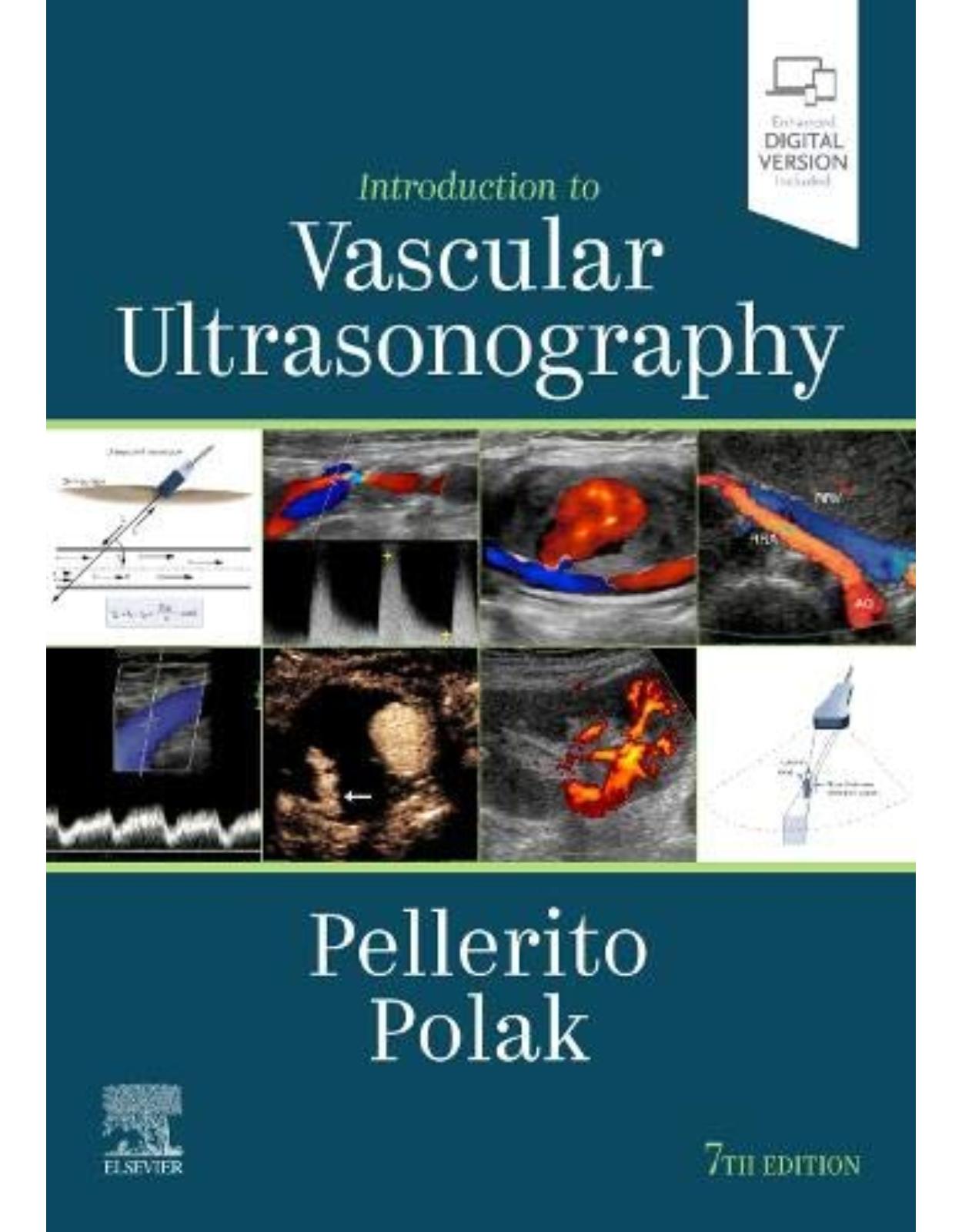
Focused content, an easy-to-read writing style, and abundant illustrations make Introduction to Vascular Ultrasonography the definitive reference on arterial and venous ultrasound. Trusted by radiologists, interventional radiologists, vascular and interventional fellows, residents, and sonographers through six outstanding editions, the revised 7th Edition covers all aspects of ultrasound vascular diagnosis, including peripheral veins and arteries, carotid and vertebral arteries, abdominal vessels, and transcranial Doppler. Step-by-step explanations, all highly illustrated, walk you through the full spectrum of ultrasound sonography practice, including all that’s new in this quickly evolving field.
Organizes sections with quick reference in mind: clinical rationale, anatomy, examination technique, findings, and interpretation.
Includes 2,100 clinical ultrasound images and anatomic line drawings, including over 1,000 in full color.
Features new coverage of noninvasive image-guided procedures, robotic embolization, laser therapy, new Doppler ultrasound and color images, and guidance on promoting patient relationships.
Takes a clear, readable, and practical approach to interventions and underlying rationales for a variety of complex IR principles, such as the physics of Doppler ultrasound and hemodynamics of blood flow.
Contains extensive tables, charts, and graphs that clearly explain examination protocols, normal values, diagnostic parameters, and ultrasound findings.
Table of Contents:
Section 1 Basics
1 The Hemodynamics of Vascular Disease
Abstract
Overview
Introduction
Physiologic Factors Governing Blood Flow and Its Characteristics
Effects of Arterial Obstruction
Venous Hemodynamics
Summary
References
2 Principles and Instruments of Ultrasonography
Introduction
Ultrasound Principles
Transducers
Sonographic Instruments
Advanced Features
Doppler Principles
References
3 Doppler Flow Imaging and Spectral Analysis
Introduction
Blood Flow Patterns
Listening to the Auditory Frequency Spectrum
What You Need to Know About Waveform Analysis
Diagnosis of Arterial Obstruction
Color Flow Ultrasound Imaging
Three-Dimensional Vascular Imaging
B-Mode Flow Imaging
References
Section 2 Cerebral Vessels
4 Anatomy of the Cerebral Structure
Abstract
Introduction
Vascular Anatomy
Cerebral Hemodynamics
Collateralization
Summary
References
5 Carotid Sonography
Abstract
B-Mode Ultrasound of the Normal Carotid Artery Wall
Normal Blood Flow Characteristics
Vessel Identity
Examination Protocol
The Examination Sequence
References
6 Evaluating Carotid Plaque and Carotid Intima-Media Thickness
Abstract
Introduction
Pathogenesis of Carotid Atherosclerosis
Imaging Protocol: IMT and Plaque
Plaque Characterization
Plaque Classification Schemes
Plaque Surface Features
Plaque Neovascularity
Perspective on Plaque Characterization
Paradigm Shift: Intima-Media Thickness
Carotid Plaques: Extent, Severity, and Follow-Up
References
7 Ultrasound Assessment of Carotid Stenosis
Abstract
Introduction
Clinical Background
Imaging of Stenosis
Diagnostic Criteria
Plaque Characterization
Technical Considerations and Pitfalls
The Common Carotid Artery
The External Carotid Artery
The Innominate Artery
Summary
References
8 How to Assess Difficult and Uncommon Carotid Cases
Introduction
Carotid Occlusion
Carotid Artery Dissection
Carotid Pseudoaneurysm
Carotid Arteriovenous Fistula
Fibromuscular Dysplasia
Carotid Body Tumor (Glomus Tumor)
Difficult Carotid Cases
References
9 Ultrasound Assessment of the Vertebral Arteries
Abstract
Introduction
Examination Technique
Vertebral Artery Hemodynamics: Qualitative Assessment
Vertebral Artery Hemodynamics: Quantitative Assessment
Correlative Imaging With Magnetic Resonance Angiography and Computed Tomographic Angiography
Treatment of Disease
Summary
References
10 Ultrasound Assessment of the Intracranial Arteries
Introduction
Examination Techniques
Diagnostic Parameters for Specific Clinical Applications
Summary
References
Section 3 Extremity Arteries
11 Anatomy of the Upper and Lower Extremity Arteries
Abstract
Introduction
Upper Extremity
Lower Extremity
References
12 Physiologic Testing of Lower Extremity Arterial Disease
Abstract
Introduction
Instrumentation
Physiologic Testing Procedures
Diagnostic Algorithm
Clinical Applications
Summary
References
13 Assessment of Upper Extremity Arterial Disease
Abstract
Introduction
Basic Anatomy
Obstructive Arterial Disease
Thoracic Outlet Evaluation
Digital Artery Evaluation
Raynaud Disease/Phenomenon
Color Doppler Imaging and Duplex Ultrasound of Vasculitis
Ultrasound Imaging of Upper Extremity Arterial Aneurysms
Upper Extremity Arterial Access
Radial Artery Harvest
Summary
References
14 Ultrasound Evaluation Before and After Hemodialysis Access
Abstract
Introduction
Basic Concepts of Hemodialysis Access
Descriptive Terminology
Vascular Mapping Before Hemodialysis Access Placement
Arteriovenous Fistula Maturity Assessment
Graft Evaluation
Acknowledgments
References
15 Ultrasound Assessment of Lower Extremity Arteries
Abstract
Introduction
Instrumentation
Duplex Ultrasound Technique
Classification of Disease
Clinical Applications
Summary
References
16 Ultrasound Assessment During and After Carotid and Peripheral Interventions
Introduction
Intraprocedural Duplex Ultrasound Assessment
Surveillance After Carotid Intervention
Surveillance After Peripheral Intervention
Infrainguinal Bypass Assessment
Synthetic Grafts
Duplex-Monitored Peripheral Angioplasty/Stent Placement
Post-Procedural Follow-Up
Summary
References
17 Ultrasound in the Assessment and Management of Arterial Emergencies
Abstract
Introduction
Ruptured Abdominal Aortic Aneurysm
Carotid Artery Stenosis/Thrombosis
Carotid Artery Dissection
Acute Lower Extremity Ischemia
Femoral Pseudoaneurysm
Traumatic Arteriovenous Fistula
Penetrating Arterial Trauma
Summary
References
Section 4 Extremity Veins
18 Extremity Venous Anatomy and Technique for Ultrasound Examination
Introduction
Anatomy of the Lower Extremity
Venous Duplex Imaging Examination Technique and Protocol
The Venous Anatomy and Examination Protocol
Anatomy of the Upper Extremity
Upper Extremity Examination Protocol
Characterization of Thrombus
Summary
References
19 Ultrasound Diagnosis of Lower Extremity Venous Thrombosis
Abstract
Introduction
Prevalence, Etiology, and Risk Factors
Venous Anatomy of the Lower Extremities
Technique
Ultrasound Findings
Pitfalls
May-Thurner Syndrome
Alternative Diagnoses/Incidental Findings
Summary
References
20 Risk Factors and the Role of Ultrasound in the Management of Extremity Venous Disease
Abstract
Introduction
Acute Deep Vein Thrombosis Etiology and Risk Factors
Anticoagulation and Thrombolysis in the Management of Venous Thrombosis
Acute Deep Vein Thrombosis of Specific Extremity Veins
Lower Extremity Duplex Scanning in Suspected Pulmonary Embolism
Lower Extremity Superficial Thrombophlebitis
Upper Extremity Venous Thrombosis
Sequelae of Deep Vein Thrombosis
Primary Varicose Veins
Preprocedure Venous Mapping
Summary
References
21 Ultrasound Diagnosis of Venous Insufficiency
Abstract
Introduction
Lower Limb Venous Anatomy
Pathophysiology of Venous Insufficiency
Duplex Diagnosis of Venous Insufficiency
Quantitative Measurement of Venous Incompetence
The Role of Sonography in the Treatment of Chronic Venous Insufficiency
Summary
Acknowledgments
References
22 Nonvascular Findings Encountered During Venous Sonography
Introduction
Soft Tissue Edema
Lymphedema
Hematoma
Muscle Injury
Lymph Nodes
Popliteal (Baker) Cysts
Joint Effusion
Infection
Soft Tissue Tumors
References
Section 5 Abdomen and Pelvis
23 Anatomy and Normal Doppler Signatures of Abdominal Vessels
Abstract
Introduction
Abdominal Aorta
Celiac Artery
Splenic Artery
Hepatic Artery
Superior and Inferior Mesenteric Arteries
Renal Arteries
Portal Venous System
Hepatic Veins
Inferior Vena Cava
Renal Veins
References
24 Ultrasound Assessment of the Abdominal Aorta
Abstract
Introduction
Anatomy
Normal Sizes
Normal Doppler Velocity Profiles
Pathologic States
Abdominal Aortic Aneurysm
Ultrasound Examination
Definition of an Aneurysm: Size Thresholds
Growth Rate of Aneurysms
Common Iliac Artery Aneurysms
Aneurysm Complications
Varied Pathologies
Postsurgical Assessment
Summary
References
25 Ultrasound Assessment Following Endovascular Aortic Aneurysm Repair
Abstract
Introduction
Overview of EVAR
Ultrasound Examination
Endoleak Detection
Aneurysm Size
Endovascular Graft Deformity and Native Artery Complications
Contrast-Enhanced Ultrasound
Aneurysm Sac Pressure Measurement
Surveillance Intervals
Summary
References
26 Doppler Ultrasound of the Mesenteric Vasculature
Abstract
Introduction
Anatomy, Physiology, and Natural History of Bowel Ischemia
Technique
Examination Protocol
Diagnostic Criteria
Role of Duplex Ultrasound in Surveillance Following Mesenteric Revascularization (Stent Angioplasty and Bypass Graft Assessment)
Keys to a Successful Examination
Pitfalls
Other Mesenteric Artery Pathologies
Summary
Acknowledgments
References
27 Ultrasound Assessment of the Hepatic Vasculature
Abstract
Introduction
Technique and Normal Hemodynamics
Portal Hypertension
Portal Vein Thrombosis
Portal Vein Stenosis
Arterioportal Fistulas
Portal Vein Gas
Hepatic Vein Obstruction
Transjugular Intrahepatic Portosystemic Shunts
Congenital Intrahepatic Portosystemic Shunts
Hepatic Artery Stenosis/Occlusion
Hepatic Artery Pseudoaneurysm
References
28 Duplex Ultrasound of Native Renal Vasculature
Abstract
Introduction
Anatomy
Principles of Examination
Technique
Protocol
Vascular Disorders
Summary
Acknowledgments
References
29 Duplex Ultrasound Evaluation of the Uterus and Ovaries
Abstract
Introduction
Technical Issues
Normal Anatomy and Hemodynamics
Current Applications
Ectopic Pregnancy
Pseudogestation
Value of Doppler for Evaluation of Ectopic Pregnancy
Pitfalls
Summary
Acknowledgments
References
30 Duplex Ultrasound Evaluation of the Male Genitalia
Abstract
Introduction
The Scrotum
The Penis
Summary
References
31 Evaluation of Organ Transplants
Abstract
Introduction
Renal Transplantation
Hepatic Transplantation
References
Section 6 Trends in Ultrasound Vascular Imaging
32 Credentialing, Accreditation, and Quality in the Vascular Laboratory
Abstract
Introduction
Credentialing
Accreditation
Quality Improvement
Appropriateness of Vascular Testing
Efforts to Standardize Vascular Testing and Improve Quality at the National Level
Summary
References
33 Ultrasound Screening for Vascular Disease
Abstract
Introduction
Definition and Types of Screening
Screening for Asymptomatic Carotid Stenosis
Abdominal Aneurysm Screening
Screening for Cardiovascular Disease: Risk and Subclinical Cardiovascular Disease
Summary
References
34 Correlative Imaging
Abstract
Overview
Catheter-Based Arteriography
Magnetic Resonance Angiography
Computed Tomographic Angiography
Overview and Correlative Findings
Summary
References
35 Ultrasound Contrast Agents in Vascular Disease
Abstract
Introduction
Technical Aspects of Contrast-Enhanced Ultrasound
Safety of Ultrasound Contrast Agents
Clinical Applications of Ultrasound Contrast Agents
Trauma
Future Considerations
Summary
References
Index
| An aparitie | 4 Dec. 2019 |
| Autor | John Pellerito MD , Joseph F Polak MD MPH |
| Dimensiuni | 19.05 x 3.18 x 26.67 cm |
| Editura | Elsevier |
| Format | Hardcover |
| ISBN | 9780323428828 |
| Limba | Engleza |
| Nr pag | 882 |


Clientii ebookshop.ro nu au adaugat inca opinii pentru acest produs. Fii primul care adauga o parere, folosind formularul de mai jos.