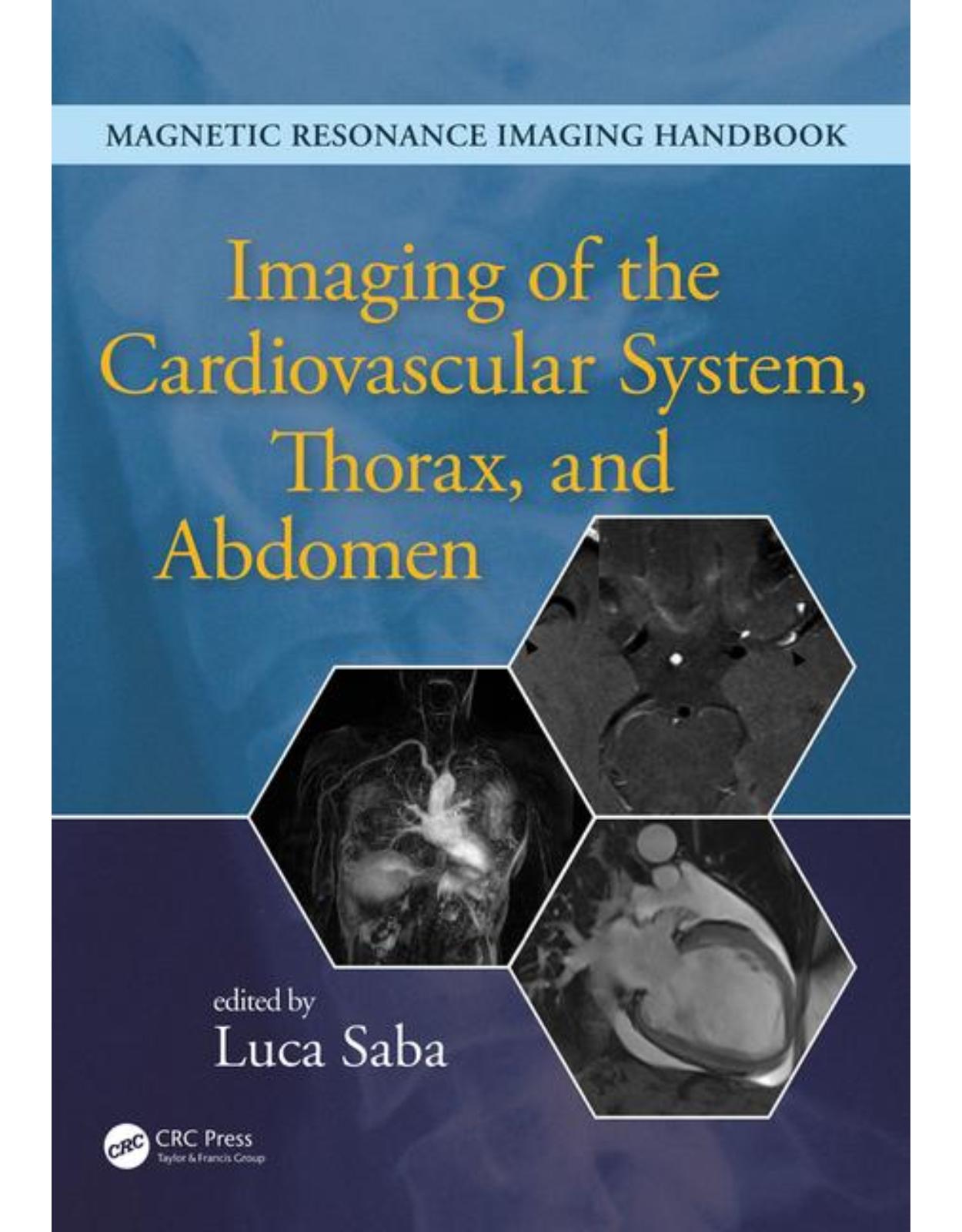
Imaging of the Cardiovascular System, Thorax, and Abdomen
Livrare gratis la comenzi peste 500 RON. Pentru celelalte comenzi livrarea este 20 RON.
Description:
Magnetic resonance imaging (MRI) is a technique used in biomedical imaging and radiology to visualize internal structures of the body. Because MRI provides excellent contrast between different soft tissues, the technique is especially useful for diagnostic imaging of the brain, muscles, and heart. In the past 20 years, MRI technology has improved significantly with the introduction of systems up to 7 Tesla (7 T) and with the development of numerous post-processing algorithms such as diffusion tensor imaging (DTI), functional MRI (fMRI), and spectroscopic imaging. From these developments, the diagnostic potentialities of MRI have improved impressively with an exceptional spatial resolution and the possibility of analyzing the morphology and function of several kinds of pathology. Given these exciting developments, the Magnetic Resonance Imaging Handbook: Imaging of the Cardiovascular System, Thorax, and Abdomen is a timely addition to the growing body of literature in the field. Offering comprehensive coverage of cutting-edge imaging modalities, this book: Discusses MRI of the heart, blood vessels, lungs, breasts, diaphragm, liver, gallbladder, spleen, pancreas, adrenal glands, and gastrointestinal tract Explains how MRI can be used in vascular, posttraumatic, postsurgical, and computer-aided diagnostic (CAD) applications Highlights each organ’s anatomy and pathological processes with high-quality images Examines the protocols and potentialities of advanced MRI scanners such as 7 T systems Includes extensive references at the end of each chapter to enhance further study Thus, the Magnetic Resonance Imaging Handbook: Imaging of the Cardiovascular System, Thorax, and Abdomen provides radiologists and imaging specialists with a valuable, state-of-the-art reference on MRI.
Table of contents:
1. Coronary and Perfusion Imaging with Cardiovascular Magnetic Resonance: Current State of the Art
2. Imaging of the Heart: Myocardial Imaging
3. Computed Topography/Magnetic Resonance Imaging of Pericardial Disease
4. Vascular Imaging of the Head and Neck
5. Magnetic Resonance Imaging: Aorta and Splanchnic Vessels
6. Peripheral Magnetic Resonance Angiography
7. Magnetic Resonance Venography
8. Vascular Malformations
9. Four-Dimensional Flow Imaging
10. Magnetic Resonance Imaging for Trachea, Bronchi, and Lung
11. Clinical Magnetic Resonance Imaging Applications for the Lung
12. Pleura and Diaphragm
13. Functional Magnetic Resonance Imaging of the Lung and Pulmonary Vasculature
14. Breast MRI
15. Liver: Technique and Diffuse Pathology
16. Liver: Focal Pathology
17. Gallbladder
18. Magnetic Resonance Imaging of Biliary Tract
19. Update in Magnetic Resonance Imaging of the Spleen
20. Pancreas
21. The Adrenal Gland
22. Diseases of the Upper Gastrointestinal Tract and Small Bowel
23. MRI of the Retroperitoneum
24. Abdominal Wall and Hernias
25. Diseases of the Colon and Rectum
26. Posttraumatic and Postsurgical Abdomen
Back Cover
ebookshop
| An aparitie | 2016 |
| Autor | Luca Saba |
| Dimensiuni | 28.9 x 3.3 x 22.8 cm |
| Editura | CRC Press |
| Format | Hardback |
| ISBN | 9781482216264 |
| Limba | Engleza |
| Nr pag | 627 |
-

-
-
 1,19500 lei 1,13100 lei
 -

Clientii ebookshop.ro nu au adaugat inca opinii pentru acest produs. Fii primul care adauga o parere, folosind formularul de mai jos.