- 16%
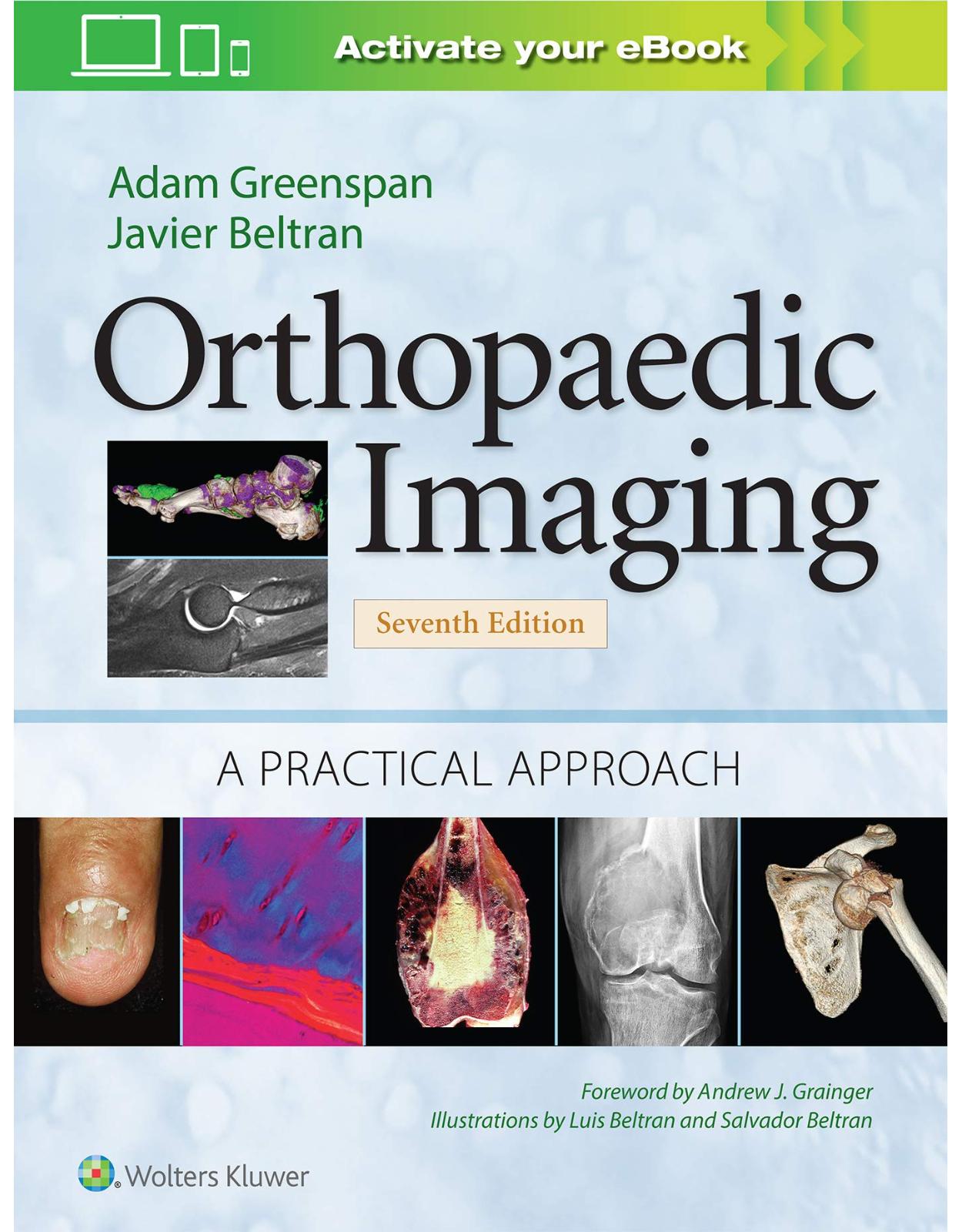
Orthopaedic Imaging: A Practical Approach (Orthopedic Imaging a Practical Approach)
by GREENSPAN
1937 Lei 1628 Lei(TVA inclus)
Livrare gratis la comenzi peste 500 RON. Pentru celelalte comenzi livrarea este 20 RON.
Livrare gratis la comenzi peste 500 RON. Pentru celelalte comenzi livrarea este 20 RON.
Description:
Trusted by both radiologists and orthopaedic surgeons for authoritative, comprehensive guidance on the interpretation of musculoskeletal images, Orthopedic Imaging: A Practical Approach is an ideal resource at every stage of training and practice. The fully revised seventh edition retains the large images, easy-to-read writing style, and careful blend of illustrations and text that clearly depict all relevant imaging modalities and all pathological entities.
- Helps you interpret a full range of findings with nearly 4,000 high-quality conventional radiography, ultrasound, CT, 3D CT, dual-energy CT, MRI, PET, PET/CT, PET/MRI, and other imaging techniques—1/3 new to this edition.
- Contains new coverage of sports injuries and cartilage imaging; clinical features and pathologic correlations of many conditions; and up-to-date references.
- Provides guidance in choosing the best imaging approach for each patient with discussions of each technique’s accuracy, speed, and cost.
- Features informative diagrams and schematics, as well as “Practical Points” summaries at the end of each chapter for quick review.
Enrich Your Digital Reading Experience
- Read directly on your preferred device(s), such as computer, tablet, or smartphone.
- Easily convert to audiobook, powering your content with natural language text-to-speech.
Table of Contents:
- Foreword
- Preface to the First Edition
- Preface
- Acknowledgments
- Part I: INTRODUCTION TO ORTHOPAEDIC IMAGING
- Chapter 1: The Role of the Orthopaedic Radiologist
- SUGGESTED READINGS
- Chapter 2: Imaging Techniques in Orthopaedics
- Choice of Imaging Modality
- Imaging Techniques
- Conventional Radiography
- Magnification Radiography
- Stress Views
- Scanogram
- Fluoroscopy and Videotaping
- Digital (Computed) Radiography
- Tomography
- Computed Tomography
- Arthrography
- Angiography
- Myelography
- Diskography
- Ultrasound
- Scintigraphy (Radionuclide Bone Scan)
- Diphosphonates
- Gallium-67
- Indium
- Nanocolloid
- Immunoglobulins
- Chemotactic Peptides
- Iodine
- Gadolinium
- PET, PET/CT, and PET/MRI
- Magnetic Resonance Imaging
- SUGGESTED READINGS
- Chapter 3: Histology, Formation, and Growth of Bone and Articular Cartilage
- Bone: Histology, Formation, and Growth
- Articular Cartilage: Histology, Formation, and Growth
- SUGGESTED READINGS
- Part II: TRAUMA
- Chapter 4: Imaging Evaluation of Trauma
- Imaging Modalities
- Radiography and Fluoroscopy
- Computed Tomography
- Scintigraphy
- Ultrasound
- Arthrography
- Myelography and Diskography
- Angiography
- Magnetic Resonance Imaging
- Fractures and Dislocations
- Diagnosis
- Radiographic Evaluation of Fractures
- Indirect Signs as Diagnostic Clues
- Radiographic Evaluation of Dislocations
- Monitoring the Results of Treatment
- Fracture Healing and Complications
- Other Complications of Fractures, Dislocations, and Traumatic Insults to the Bones and Soft Tissues
- Stress Fractures
- Injury to Soft Tissues
- Sports Injuries
- Upper Extremity
- Weight Lifter Pectoralis
- Little League Shoulder
- Golfer’s Elbow
- Tennis Elbow
- Little League Elbow
- Baseball Pitcher’s Elbow
- Goalkeeper’s Elbow
- Oarsman’s Wrist
- Cyclist’s Wrist
- Gymnast’s Wrist
- Boxer’s Fracture
- Skier’s Thumb
- Bowler’s Thumb
- Lower Extremity
- Sports Hernia
- Runner’s Knee (Iliotibial Band Friction Syndrome)
- Jumper’s Knee
- Tennis Leg
- Shin Splints
- Footballer’s Ankle (Athlete’s Ankle)
- Snowboarder’s Fracture
- Turf Toe
- PRACTICAL POINTS TO REMEMBER
- SUGGESTED READINGS
- Chapter 5: Upper Limb I: Shoulder Girdle
- Trauma to the Shoulder Girdle
- Anatomic–Radiologic Considerations
- Injury to the Shoulder Girdle
- Fractures About the Shoulder
- Dislocations in the Glenohumeral Joint
- Impingement Syndrome
- Rotator Cuff Tear
- Injury to the Cartilaginous Labrum
- Injury to the Glenohumeral Ligaments
- Miscellaneous Abnormalities
- Compressive and Entrapment Neuropathies of the Shoulder
- Suprascapular Nerve Syndrome
- Quadrilateral Space Syndrome
- Parsonage-Turner Syndrome (Idiopathic Brachial Plexopathy, Neuralgic Amyotrophy)
- Serratus Anterior Paralysis (Scapular Winging)
- The Postoperative Shoulder
- PRACTICAL POINTS TO REMEMBER
- SUGGESTED READINGS
- Chapter 6: Upper Limb II: Elbow
- Trauma to the Elbow
- Anatomic–Radiologic Considerations
- Injury to the Elbow
- Fractures About the Elbow
- Osteochondritis Dissecans of the Capitellum
- Dislocations in the Elbow Joint
- Injury to the Soft Tissues
- Bursitis
- Compressive and Entrapment Neuropathies of the Elbow
- Pronator Teres Muscle Syndrome
- Supinator Muscle Syndrome
- Cubital Tunnel Syndrome
- PRACTICAL POINTS TO REMEMBER
- SUGGESTED READINGS
- Chapter 7: Upper Limb III: Distal Forearm, Wrist, Hand, and Fingers
- Distal Forearm
- Anatomic–Radiologic Considerations
- Injury to the Distal Forearm
- Fractures of the Distal Radius
- Injury to the Soft Tissue at the Distal Radioulnar Articulation
- Wrist and Hand
- Anatomic–Radiologic Considerations
- Injury to the Wrist
- Fractures of the Carpal Bones
- Kienböck Disease
- Hamatolunate Impaction Syndrome
- Dislocations of the Carpal Bones
- Carpal Instability
- Injury to the Bones of the Hand
- Bennett and Rolando Fractures
- Boxer’s Fracture
- Injury to the Soft Tissue of the Hand
- Carpal Tunnel Syndrome
- Guyon Canal Syndrome
- Anterior Interosseous Nerve Syndrome
- Fingers
- Normal Anatomy of the Fingers
- Injury to the Bones and Soft Tissues of the Fingers
- Gamekeeper’s Thumb
- Avulsion Fractures of the Fingers
- Flexor Pulley System Rupture (Climber’s Finger)
- de Quervain Syndrome
- Tendon and Ligament Tears
- PRACTICAL POINTS TO REMEMBER
- SUGGESTED READINGS
- Chapter 8: Lower Limb I: Pelvic Girdle, Sacrum, and Proximal Femur
- Trauma to the Pelvic Girdle
- Anatomic–Radiologic Considerations
- Injury to the Pelvis and Acetabulum
- Classification of Pelvic Fractures
- Fractures of the Pelvis
- Fractures of the Acetabulum
- Injuries of the Acetabular Labrum
- Femoroacetabular Impingement Syndrome
- Injury to the Sacrum
- Trauma to the Proximal Femur
- Fractures of the Proximal Femur
- Intracapsular Fractures
- Extracapsular Fractures
- Dislocations in the Hip Joint
- Tendon and Muscle Lesions
- Compressive and Entrapment Neuropathies
- Piriformis Syndrome
- Iliacus Syndrome
- Obturator Neuropathy
- Lateral Femoral Cutaneous Neuropathy (Meralgia Paresthetica)
- Morel-Lavallée Lesion (Closed Degloving Injury)
- Sports Hernia
- Stress and Insufficiency Fractures
- PRACTICAL POINTS TO REMEMBER
- SUGGESTED READINGS
- Chapter 9: Lower Limb II: Knee
- Trauma to the Knee
- Anatomic–Radiologic Considerations
- Injury to the Knee
- Fractures About the Knee
- Sinding-Larsen-Johansson Disease
- Osgood-Schlatter Disease
- Injuries to the Cartilage of the Knee
- Injury to the Soft Tissues About the Knee
- The Postoperative Knee
- Surgical Management of Meniscal Tears
- Anterior Cruciate Ligament Reconstruction
- Cartilage Repair
- PRACTICAL POINTS TO REMEMBER
- SUGGESTED READINGS
- Chapter 10: Lower Limb III: Ankle and Foot
- Trauma to the Ankle and Foot
- Anatomic–Radiologic Considerations
- Imaging of the Ankle and Foot
- Injury to the Ankle
- Fractures About the Ankle Joint
- Injury to the Soft Tissues About the Ankle Joint and Foot
- Injury to the Foot
- Fractures of the Foot
- Complications
- Dislocations in the Foot
- Miscellaneous Painful Soft-Tissue Abnormalities of the Ankle and Foot
- Tarsal Tunnel Syndrome
- Sinus Tarsi Syndrome
- Posterior Tibialis Tendon Dysfunction
- Chronic Tendinosis of the Posterior Tibialis Tendon
- Tears of the Posterior Tibialis Tendon
- Painful Accessory Navicular Bone Syndrome
- Peroneal Tendinopathies
- Peroneus Brevis Split Tear
- Peroneus Longus Tendinosis and Tear
- Peroneal Tendon Dislocation
- Painful Os Peroneum Syndrome
- Baxter Neuropathy
- Morton Neuroma
- Plantar Fasciitis
- Impingement Syndromes
- PRACTICAL POINTS TO REMEMBER
- SUGGESTED READINGS
- Chapter 11: Spine
- Introduction
- Cervical Spine
- Anatomic–Radiologic Considerations
- Injury to the Cervical Spine
- Fractures of the Occipital Condyles
- Occipitocervical Dislocations
- Fractures of the C1 and C2 Vertebrae
- Fractures of the Mid and Lower Cervical Spine
- Locked Facets
- Thoracolumbar Spine
- Anatomic–Radiologic Considerations
- Injury to the Thoracolumbar Spine
- Fractures of the Thoracolumbar Spine
- Fracture–Dislocations
- Spondylolysis and Spondylolisthesis
- Transitional Lumbosacral Vertebra
- Injury to the Diskovertebral Junction
- Orthopaedic Management
- PRACTICAL POINTS TO REMEMBER
- Cervical Spine
- Thoracolumbar Spine
- SUGGESTED READINGS
- Part III: ARTHRITIDES
- Chapter 12: Clinical, Imaging, and Pathologic Evaluation of the Arthritides and Arthropathies
- Radiologic Imaging Modalities
- Conventional Radiography
- Magnification Radiography
- Tomography and Computed Tomography
- Scintigraphy
- Ultrasound
- Magnetic Resonance Imaging
- The Arthritides
- Diagnosis
- Clinical Information
- Pathology
- Imaging Features
- Management
- Medical Treatment
- Radiotherapy
- Orthopaedic Treatment
- Complications of Surgical Treatment
- PRACTICAL POINTS TO REMEMBER
- SUGGESTED READINGS
- Chapter 13: Degenerative Joint Disease
- Osteoarthritis
- Osteoarthritis of the Large Joints
- Osteoarthritis of the Hip
- Osteoarthritis of the Knee
- Osteoarthritis of Other Large Joints
- Osteoarthritis of the Small Joints
- Primary Osteoarthritis of the Hand
- Secondary Osteoarthritis of the Hand
- Osteoarthritis of the Foot
- Degenerative Diseases of the Spine
- Clinical Features
- Pathology
- Osteoarthritis of the Synovial Joints
- Degenerative Disk Disease
- Spondylosis Deformans
- Diffuse Idiopathic Skeletal Hyperostosis
- Complications of Degenerative Disease of the Spine
- Degenerative Spondylolisthesis
- Spinal Stenosis
- Neuropathic Arthropathy
- PRACTICAL POINTS TO REMEMBER
- SUGGESTED READINGS
- Chapter 14: Inflammatory Arthritides
- Erosive Osteoarthritis
- Clinical Features
- Pathology
- Imaging Features
- Differential Diagnosis
- Treatment
- Rheumatoid Arthritis
- Adult Rheumatoid Arthritis
- Rheumatoid Factors
- Clinical Features
- Imaging Features
- Complications of Rheumatoid Arthritis
- Rheumatoid Nodulosis
- Juvenile Idiopathic Arthritis
- Still Disease
- Polyarticular Juvenile Idiopathic Arthritis
- Juvenile Idiopathic Arthritis with Pauciarticular Onset (Oligoarthritis)
- Arthritis with Enthesitis
- Juvenile Psoriatic Arthritis
- Imaging Features of JIA
- Macrophage Activation Syndrome
- Treatment of Rheumatoid Arthritis
- Seronegative Spondyloarthropathies
- Ankylosing Spondylitis
- Clinical Features
- Pathology
- Imaging Features
- Treatment
- Reactive Arthritis (Reiter Syndrome)
- Clinical Features
- Imaging Features
- Treatment
- Psoriatic Arthritis
- Clinical Features
- Imaging Features
- Treatment
- Enteropathic Arthropathies
- Undifferentiated Spondyloarthritis
- SAPHO Syndrome
- PRACTICAL POINTS TO REMEMBER
- SUGGESTED READINGS
- Chapter 15: Miscellaneous Arthritides and Arthropathies
- Connective Tissue Arthropathies
- Systemic Lupus Erythematosus
- Clinical Features
- Pathology
- Imaging Features
- Treatment
- Scleroderma
- Clinical Features
- Pathology
- Imaging Features
- Treatment
- Polymyositis and Dermatomyositis
- Clinical Features
- Pathology
- Imaging Features
- Treatment
- Mixed Connective Tissue Disease
- Vasculitis
- Metabolic, Endocrine, and Crystal Deposition Arthropathies and Arthritides
- Gout
- Clinical Features
- Hyperuricemia
- Examination of Synovial Fluid
- Pathology
- Imaging Features
- Differential Diagnosis
- Treatment
- Calcium Pyrophosphate Dihydrate Crystal Deposition Disease
- Clinical Features
- Pathology
- Imaging Features
- Differential Diagnosis
- Calcium Hydroxyapatite Crystal Deposition Disease
- Clinical Features
- Imaging Features
- Treatment
- Hemochromatosis
- Clinical Features
- Pathology
- Imaging Features
- Treatment
- Wilson Disease
- Clinical Features
- Imaging Features
- Treatment
- Alkaptonuria (Ochronosis)
- Clinical Features
- Imaging Features
- Treatment
- Hyperparathyroidism
- Clinical Features
- Pathology
- Imaging Features
- Treatment
- Acromegaly
- Miscellaneous Conditions
- Amyloidosis
- Clinical Features
- Pathology
- Imaging Features
- Treatment
- Multicentric Reticulohistiocytosis
- Clinical Features
- Imaging Features
- Pathology
- Treatment
- Sarcoidosis
- Clinical Features
- Pathology
- Imaging Features
- Treatment
- Hemophilia
- Imaging Features
- Jaccoud Arthritis
- Arthritis Associated with AIDS
- Infectious Arthritis
- PRACTICAL POINTS TO REMEMBER
- SUGGESTED READINGS
- Part IV: TUMORS AND TUMOR-LIKE LESIONS
- Chapter 16: Imaging Evaluation of Tumors and Tumor-like Lesions
- Classification of Tumors and Tumor-like Lesions
- Imaging Modalities
- Conventional Radiography
- Computed Tomography
- PET and PET/CT
- Arteriography
- Myelography
- Magnetic Resonance Imaging
- Skeletal Scintigraphy (Radionuclide Bone Scan)
- Interventional Procedures
- Tumors and Tumor-like Lesions of Bone
- Diagnosis
- Clinical Information
- Choice of Imaging Modality
- Radiographic Features of Bone Lesions
- Pathology
- Stains
- Immunohistochemistry
- Electron Microscopy
- Genetics of Bone Tumors
- Management
- Monitoring the Results of Treatment
- Complications
- Soft-Tissue Tumors
- PRACTICAL POINTS TO REMEMBER
- SUGGESTED READINGS
- Chapter 17: Benign Tumors and Tumor-like Lesions I: Bone-Forming Lesions
- Benign Bone-Forming (Osteoblastic, Osteogenic) Lesions
- Osteoma
- Clinical and Imaging Features
- Pathology
- Differential Diagnosis
- Osteoid Osteoma
- Clinical and Imaging Features
- Pathology
- Differential Diagnosis
- Complications
- Treatment
- Osteoblastoma
- Clinical and Imaging Features
- Pathology
- Differential Diagnosis
- Treatment
- PRACTICAL POINTS TO REMEMBER
- SUGGESTED READINGS
- Chapter 18: Benign Tumors and Tumor-like Lesions II: Lesions of Cartilaginous Origin
- Benign Chondroblastic Lesions
- Enchondroma (Chondroma)
- Clinical and Imaging Features
- Pathology
- Differential Diagnosis
- Complications
- Treatment
- Enchondromatosis, Ollier Disease, and Maffucci Syndrome
- Clinical and Imaging Features
- Pathology
- Complications
- Osteochondroma
- Clinical and Imaging Features
- Pathology
- Complications
- Treatment
- Multiple Osteocartilaginous Exostoses
- Clinical and Imaging Features
- Complications
- Treatment
- Bizarre Parosteal Osteochondromatous Proliferation
- Clinical and Imaging Features
- Pathology
- Treatment
- Chondroblastoma
- Clinical and Imaging Features
- Pathology
- Treatment and Complications
- Chondromyxoid Fibroma
- Clinical and Imaging Features
- Pathology
- Differential Diagnosis
- Treatment
- PRACTICAL POINTS TO REMEMBER
- SUGGESTED READINGS
- Chapter 19: Benign Tumors and Tumor-like Lesions III: Fibrous, Fibroosseous, and Fibrohistiocytic Lesions
- Fibrous Cortical Defect and Nonossifying Fibroma
- Clinical and Imaging Features
- Pathology
- Complications and Treatment
- Benign Fibrous Histiocytoma
- Clinical and Imaging Features
- Periosteal Desmoid
- Clinical and Imaging Features
- Pathology
- Differential Diagnosis
- Fibrous Dysplasia
- Monostotic Fibrous Dysplasia
- Clinical and Imaging Features
- Pathology
- Polyostotic Fibrous Dysplasia
- Clinical and Imaging Features
- Pathology
- Complications
- Fibrocartilaginous Dysplasia
- Clinical, Pathologic, and Imaging Features
- Associated Disorders
- McCune-Albright Syndrome
- Mazabraud Syndrome
- Osteofibrous Dysplasia
- Clinical, Imaging, and Histopathologic Features
- Complications and Treatment
- Desmoplastic Fibroma
- Clinical and Imaging Features
- Pathology
- Treatment
- PRACTICAL POINTS TO REMEMBER
- SUGGESTED READINGS
- Chapter 20: Benign Tumors and Tumor-like Lesions IV: Miscellaneous Lesions
- Simple Bone Cyst
- Clinical and Imaging Features
- Pathology
- Complications and Differential Diagnosis
- Treatment
- Aneurysmal Bone Cyst
- Clinical and Imaging Features
- Pathology
- Complications and Differential Diagnosis
- Treatment
- Solid Variant of Aneurysmal Bone Cyst
- Clinical and Imaging Features
- Pathology
- Treatment
- Giant Cell Tumor
- Clinical and Imaging Features
- Pathology
- Differential Diagnosis
- Complications and Treatment
- Fibrocartilaginous Mesenchymoma
- Clinical and Imaging Features
- Pathology
- Hemangioma
- Clinical and Imaging Features
- Pathology
- Differential Diagnosis
- Treatment
- Intraosseous Lipoma
- Clinical and Imaging Features
- Pathology
- Nonneoplastic Lesions Simulating Tumors
- Intraosseous Ganglion
- Brown Tumor of Hyperparathyroidism
- Langerhans Cell Histiocytosis (Eosinophilic Granuloma)
- Clinical and Imaging Features
- Pathology
- Treatment and Prognosis
- Infantile Myofibromatosis
- Erdheim-Chester Disease (Lipogranulomatosis)
- Medullary Bone Infarct
- Myositis Ossificans
- PRACTICAL POINTS TO REMEMBER
- SUGGESTED READINGS
- Chapter 21: Malignant Bone Tumors I: Osteosarcomas and Chondrosarcomas
- Osteosarcomas
- Primary Osteosarcomas
- Conventional Osteosarcoma
- Low-Grade Central Osteosarcoma
- Telangiectatic Osteosarcoma
- Giant Cell–Rich Osteosarcoma
- Small Cell Osteosarcoma
- Fibrohistiocytic Osteosarcoma
- Intracortical Osteosarcoma
- Gnathic Osteosarcoma
- Multicentric (Multifocal) Osteosarcoma
- Surface (Juxtacortical) Osteosarcomas
- Soft-Tissue (Extraskeletal) Osteosarcoma
- Osteosarcomas with Unusual Clinical Presentation
- Secondary Osteosarcomas
- Chondrosarcomas
- Primary Chondrosarcomas
- Conventional Chondrosarcoma
- Clear Cell Chondrosarcoma
- Mesenchymal Chondrosarcoma
- Myxoid Chondrosarcoma
- Dedifferentiated Chondrosarcoma
- Periosteal (Juxtacortical) Chondrosarcoma
- Secondary Chondrosarcomas
- Soft-Tissue (Extraskeletal) Chondrosarcomas
- PRACTICAL POINTS TO REMEMBER
- SUGGESTED READINGS
- Chapter 22: Malignant Bone Tumors II: Miscellaneous Tumors
- Fibrosarcoma and Malignant Fibrous Histiocytoma
- Clinical Features
- Imaging Features
- Pathology
- Differential Diagnosis
- Complications and Treatment
- Ewing Sarcoma
- Clinical Features
- Imaging Features
- Pathology
- Differential Diagnosis
- Treatment and Prognosis
- Malignant Lymphoma
- Clinical Features
- Imaging Features
- Pathology
- Differential Diagnosis
- Treatment and Prognosis
- Myeloma
- Clinical Features
- Imaging Features
- Pathology
- Differential Diagnosis
- Complications, Treatment, and Prognosis
- Adamantinoma of Long Bones
- Clinical and Imaging Features
- Pathology
- Treatment and Prognosis
- Chordoma
- Clinical Features
- Imaging Features
- Pathology
- Complications, Treatment, and Prognosis
- Primary Leiomyosarcoma of Bone
- Clinical Features
- Imaging Features
- Pathology
- Differential Diagnosis
- Hemangioendothelioma and Angiosarcoma
- Clinical Features
- Imaging Features
- Pathology
- Benign Conditions with Malignant Potential
- Medullary Bone Infarct
- Chronic Draining Sinus Tract of Osteomyelitis
- Plexiform Neurofibromatosis
- Paget Disease
- Radiation-Induced Sarcoma
- Skeletal Metastases
- Clinical Features
- Imaging Features
- Pathology
- Complications
- PRACTICAL POINTS TO REMEMBER
- SUGGESTED READINGS
- Chapter 23: Tumors and Tumor-like Lesions of the Joints
- Benign Lesions
- Synovial (Osteo)Chondromatosis
- Clinical Features
- Imaging Features
- Pathology
- Differential Diagnosis
- Treatment and Prognosis
- Pigmented Villonodular Synovitis
- Clinical Features
- Imaging Features
- Pathology
- Differential Diagnosis
- Treatment and Prognosis
- Synovial Hemangioma
- Clinical Features
- Imaging Features
- Pathology
- Differential Diagnosis
- Treatment and Prognosis
- Lipoma Arborescens
- Clinical Features
- Imaging Features
- Pathology
- Differential Diagnosis and Treatment
- Malignant Tumors
- Synovial Sarcoma
- Clinical Features
- Imaging Features
- Pathology
- Differential Diagnosis
- Treatment and Prognosis
- Synovial Chondrosarcoma
- Clinical Features
- Imaging Features
- Pathology
- Differential Diagnosis
- Malignant Pigmented Villonodular Synovitis
- Intraarticular Liposarcoma
- PRACTICAL POINTS TO REMEMBER
- SUGGESTED READINGS
- Part V: INFECTIONS
- Chapter 24: Imaging Evaluation of Musculoskeletal Infections
- Musculoskeletal Infections
- Osteomyelitis
- Infectious Arthritis
- Cellulitis
- Infections of the Spine
- Imaging Evaluation of Infections
- Conventional Radiography, Computed Tomography, and Arthrography
- Radionuclide Imaging
- Arteriography, Myelography, Fistulography, and Ultrasound
- Magnetic Resonance Imaging
- Invasive Procedures
- Monitoring the Treatment and Complications of Infections
- PRACTICAL POINTS TO REMEMBER
- SUGGESTED READINGS
- Chapter 25: Osteomyelitis, Infectious Arthritis, and Soft-Tissue Infections
- Osteomyelitis
- Pyogenic Bone Infections
- Acute and Chronic Osteomyelitis
- Subacute Osteomyelitis
- Nonpyogenic Bone Infections
- Tuberculous Infections
- Fungal Infections
- Syphilitic Infection
- Differential Diagnosis of Osteomyelitis
- Chronic Recurrent Multifocal Osteomyelitis
- Infectious Arthritides
- Pyogenic Joint Infections
- Complications
- Nonpyogenic Joint Infections
- Tuberculous Arthritis
- Other Infectious Arthritides
- Infections of the Spine
- Pyogenic Infections
- Clinical and Imaging Features
- Pathology
- Nonpyogenic Infections
- Tuberculosis of the Spine
- Coccidioidomycosis of the Spine
- Soft-Tissue Infections
- PRACTICAL POINTS TO REMEMBER
- Osteomyelitis
- Infectious Arthritis
- Infections of the Spine
- Soft-Tissue Infections
- SUGGESTED READINGS
- Part VI: METABOLIC, ENDOCRINE, AND MISCELLANEOUS DISORDERS
- Chapter 26: Imaging Evaluation of Metabolic, Endocrine, and Miscellaneous Disorders
- Composition and Production of Bone
- Evaluation of Metabolic and Endocrine Disorders
- Radiologic Imaging Modalities
- Conventional Radiography
- Computed Tomography
- Scintigraphy
- Magnetic Resonance Imaging
- Imaging Techniques for Measurement of Bone Mineral Density
- Radionuclide and X-ray Techniques
- Quantitative Ultrasound Technique
- PRACTICAL POINTS TO REMEMBER
- SUGGESTED READINGS
- Chapter 27: Osteoporosis, Rickets, and Osteomalacia
- Osteoporosis
- Generalized Osteoporosis
- Localized Osteoporosis
- Rickets and Osteomalacia
- Rickets
- Infantile Rickets
- Vitamin D–Resistant Rickets
- Osteomalacia
- Renal Osteodystrophy
- PRACTICAL POINTS TO REMEMBER
- SUGGESTED READINGS
- Chapter 28: Hyperparathyroidism
- Pathophysiology
- Physiology of Calcium Metabolism
- Clinical Features
- Imaging Features
- Pathology
- Complications
- PRACTICAL POINTS TO REMEMBER
- SUGGESTED READINGS
- Chapter 29: Paget Disease
- Pathophysiology and Clinical Features
- Imaging Features
- Pathology
- Differential Diagnosis
- Complications
- Pathologic Fractures
- Degenerative Joint Disease
- Neurologic Complications
- Neoplastic Complications
- Orthopaedic and Medical Management
- PRACTICAL POINTS TO REMEMBER
- SUGGESTED READINGS
- Chapter 30: Miscellaneous Metabolic and Endocrine Disorders
- Familial Idiopathic Hyperphosphatasia
- Clinical Features
- Imaging Features
- Differential Diagnosis
- Acromegaly
- Clinical Features
- Imaging Features
- Gaucher Disease
- Classification and Clinical Features
- Imaging Features
- Complications
- Treatment
- Tumoral Calcinosis
- Pathophysiology and Clinical Features
- Imaging Features
- Treatment
- Hypothyroidism
- Pathophysiology and Clinical Features
- Imaging Features
- Complications
- Scurvy
- Pathophysiology and Clinical Features
- Imaging Features
- Differential Diagnosis
- PRACTICAL POINTS TO REMEMBER
- SUGGESTED READINGS
- Part VII: CONGENITAL AND DEVELOPMENTAL ANOMALIES
- Chapter 31: Imaging Evaluation of Skeletal Anomalies
- Classification
- Imaging Modalities
- PRACTICAL POINTS TO REMEMBER
- SUGGESTED READINGS
- Chapter 32: Anomalies of the Upper and Lower Limbs
- Anomalies of the Shoulder Girdle and Upper Limbs
- Congenital Elevation of the Scapula
- Dentate Scapula
- Madelung Deformity
- Anomalies of the Pelvic Girdle and Hip
- Congenital Hip Dislocation (Developmental Dysplasia of the Hip)
- Radiographic Features
- Measurements
- Arthrography and Computed Tomography
- Ultrasound
- Magnetic Resonance Imaging
- Classification
- Treatment
- Complications
- Proximal Femoral Focal Deficiency
- Classification and Imaging Features
- Treatment
- Legg-Calvé-Perthes Disease
- Imaging Features
- Classification
- Differential Diagnosis
- Treatment
- Slipped Capital Femoral Epiphysis
- Imaging Features
- Treatment and Complications
- Anomalies of the Lower Limbs
- Congenital Tibia Vara
- Imaging Features and Differential Diagnosis
- Classification
- Treatment
- Dysplasia Epiphysealis Hemimelica
- Imaging Features and Treatment
- Talipes Equinovarus
- Measurements and Radiographic Features
- Treatment
- Congenital Vertical Talus
- Imaging Features
- Treatment
- Tarsal Coalition
- Calcaneonavicular Coalition
- Talonavicular Coalition
- Talocalcaneal Coalition
- PRACTICAL POINTS TO REMEMBER
- SUGGESTED READINGS
- Chapter 33: Scoliosis and Anomalies with General Affliction of the Skeleton
- Scoliosis
- Idiopathic Scoliosis
- Congenital Scoliosis
- Miscellaneous Scolioses
- Imaging Evaluation
- Measurements
- Treatment
- Anomalies with General Affliction of the Skeleton
- Neurofibromatosis
- Osteogenesis Imperfecta
- Classification
- Imaging Features
- Differential Diagnosis
- Treatment
- Achondroplasia
- Clinical Features
- Imaging Features
- Complications
- Differential Diagnosis
- Mucopolysaccharidoses
- Fibrodysplasia Ossificans Progressiva (Myositis Ossificans Progressiva)
- Clinical Features
- Imaging Features
- Pathology
- Sclerosing Dysplasias of Bone
- Osteopetrosis
- Pyknodysostosis (Pycnodysostosis)
- Enostosis, Osteopoikilosis, and Osteopathia Striata
- Progressive Diaphyseal Dysplasia (Camurati-Engelmann Disease)
- Hereditary Multiple Diaphyseal Sclerosis (Ribbing Disease)
- Endosteal Hyperostosis (Hyperostosis Corticalis Generalisata)
- Dysosteosclerosis
- Pyle Disease
- Craniometaphyseal Dysplasia
- Melorheostosis
- Craniodiaphyseal Dysplasia
- Hyperostosis Generalisata with Striations of Bones
- Other Mixed Sclerosing Dysplasias
- PRACTICAL POINTS TO REMEMBER
- SUGGESTED READINGS
- Index
| An aparitie | 1 Sept. 2020 |
| Autor | GREENSPAN |
| Dimensiuni | 22.86 x 6.35 x 29.85 cm |
| Editura | LWW |
| Format | Hardcover |
| ISBN | 9781975136475 |
| Limba | Engleza |
| Nr pag | 1512 |
| Versiune digitala | DA |










Clientii ebookshop.ro nu au adaugat inca opinii pentru acest produs. Fii primul care adauga o parere, folosind formularul de mai jos.