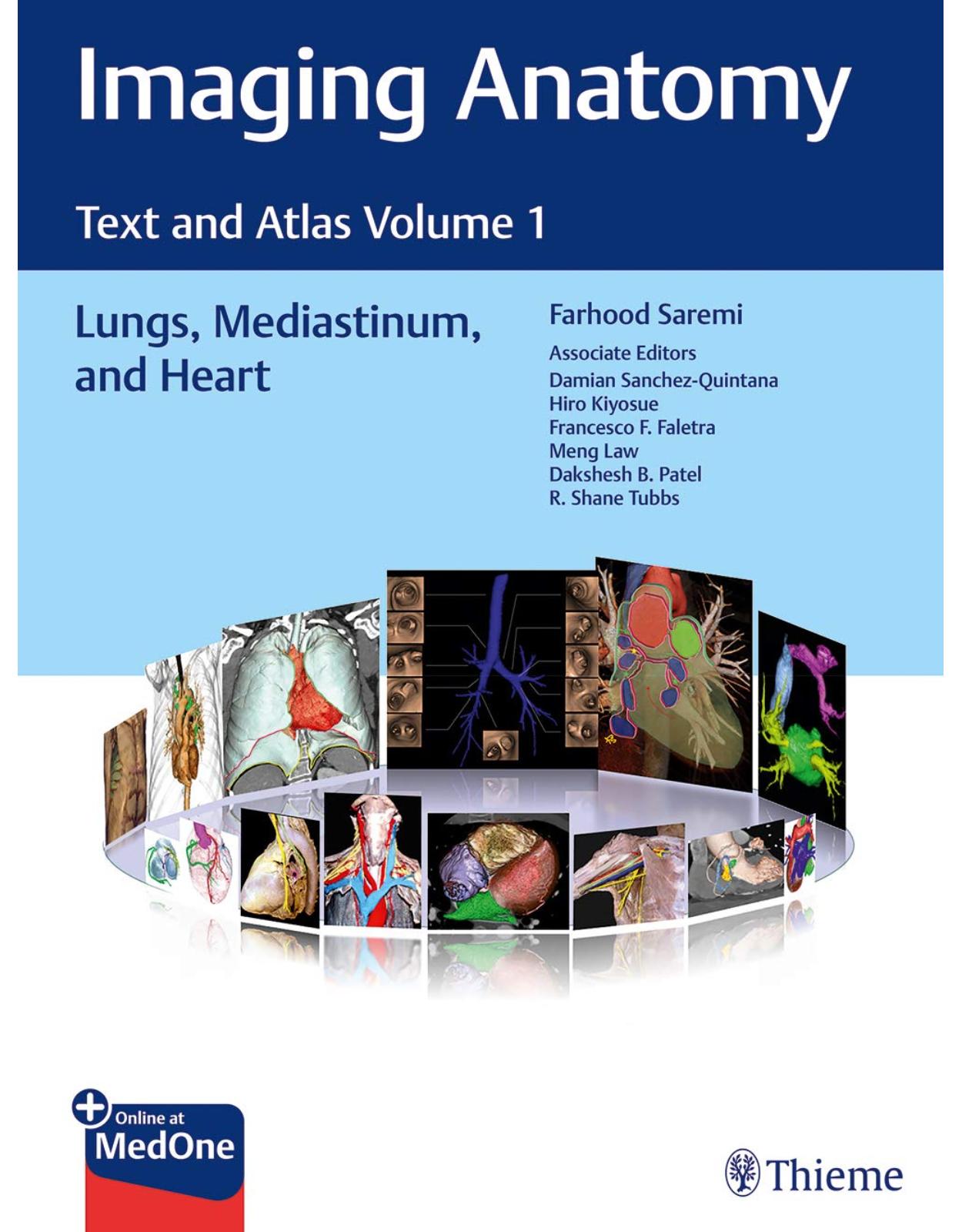
Imaging Anatomy: Text and Atlas Volume 1, Lungs, Mediastinum, and Heart (Atlas of Imaging Anatomy)
Livrare gratis la comenzi peste 500 RON. Pentru celelalte comenzi livrarea este 20 RON.
Disponibilitate: La comanda in aproximativ 4-6 saptamani
Autor: Farhood Saremi
Editura: Thieme
Limba: Engleza
Nr. pagini: 1196
Coperta: Hardcover
Dimensiuni: 30.48 x 22.86 cm
An aparitie: 15 Mar. 2020
First volume in state-of-the-art radiologic text-atlas series details anatomy of the lungs, mediastinum, and heart
Normal imaging anatomy and variants, including both diagnostic and surgical anatomy, are the cornerstones of radiologic knowledge. Imaging Anatomy: Text and Atlas Volume 1, Lungs, Mediastinum, and Heart is the first in a series of four richly illustrated radiologic references edited by distinguished radiologist Farhood Saremi and coedited by Damian Sanchez-Quintana, Hiro Kiyosue, Francesco F. Faletra, Meng Law, Dakshesh Patel, and Shane Tubbs, with contributions from an impressive cadre of international authors.
The exquisitely crafted atlas provides high-quality multiplanar and volumetric color-coded imaging techniques utilizing CT, MRI, or angiography, supplemented by cadaveric presentations and color drawings that best elucidate each specific anatomic region. Twenty-one chapters with concise text encompass thoracic wall, mediastinum, lung, vascular, and cardiac anatomy, providing readers with a virtual dissection experience. Many anatomical variants along with pathological examples are presented.
Key Highlights
More than 600 illustrations enhance understanding of impacted regions
Lung anatomy including the pleura, pulmonary arteries, pulmonary veins, and lymphatics
Discussion of the tracheobronchial system, mediastinum and thymus, thoracic aorta and major branches, systemic veins, lymphatics and nerves of the thorax, diaphragm, and breast
Heart anatomy including the atrioventricular septal region; aortic, pulmonary, mitral and tricuspid valves; coronary arteries and myocardial perfusion; coronary veins; and pericardium
This superb resource is essential reading for medical students, radiology residents and veteran radiologists, cardiologists, as well as cardiovascular and thoracic surgeons. It provides an excellent desk reference and practical guide for differentiating normal versus pathologic anatomy.
This book includes complimentary access to a digital copy on https://medone.thieme.com.
Table of Contents
1. Thoracic Wall
2. Tracheobronchial System
3. Mediastinum and Thymus
4. Lungs
5. The Pleura
6. Pulmonary Artery and Vein
7. Pulmonary and Systemic Veins
8. Thoracic Aorta and Major Branches
9. Lymphatics and Nerves of the Thorax
10. Diaphragm
11. Breast Anatomy
12. General Anatomy of the Heart
13. Atrioventricular Septal Region
14. The Aortic Valve
15. Pulmonary Valve
16. The Mitral Valve
17. The Tricuspid Valve Apparatus
18. Coronary Arteries and Myocardial Perfusion
19. The Coronary Veins
20. The Pericardium
21. Appendix
| An aparitie | 15 Mar. 2020 |
| Autor | Farhood Saremi |
| Dimensiuni | 30.48 x 22.86 cm |
| Editura | Thieme |
| Format | Hardcover |
| ISBN | 9781626239883 |
| Limba | Engleza |
| Nr pag | 1196 |


Clientii ebookshop.ro nu au adaugat inca opinii pentru acest produs. Fii primul care adauga o parere, folosind formularul de mai jos.