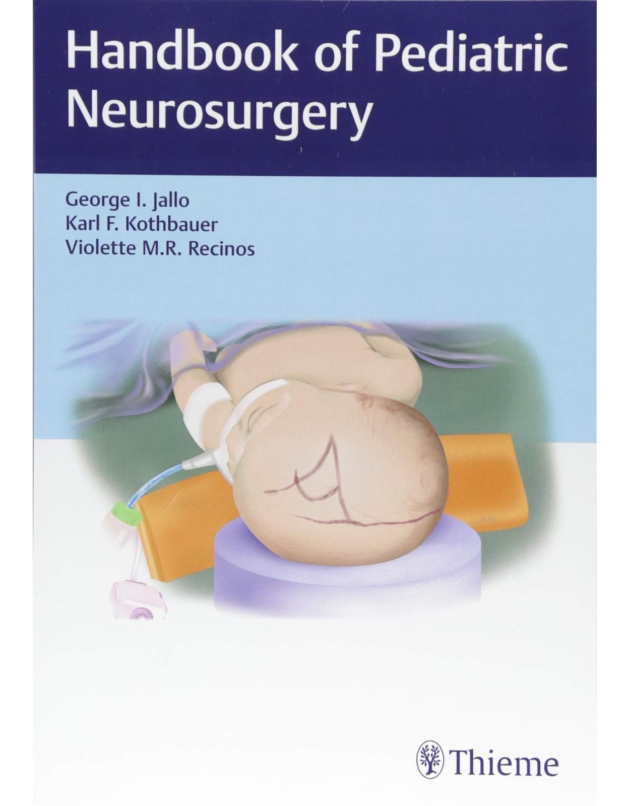
Handbook of Pediatric Neurosurgery
Livrare gratis la comenzi peste 500 RON. Pentru celelalte comenzi livrarea este 20 RON.
Disponibilitate: La comanda in aproximativ 4 saptamani
Editura: Thieme
Limba: Engleza
Nr. pagini: 1112
Coperta: Paperback
Dimensiuni: 25.4 x 17.78 cm
An aparitie: 30 April 2018
Description:
An essential backpack-size resource on the treatment of pediatric neurological conditions
Pediatric neurosurgery has witnessed considerable technological advances, resulting in more efficacious outcomes for young patients with hydrocephalus, epilepsy, brain tumors, spinal deformities, and a host of other conditions. The art of pediatric neurosurgery is a delicate balancing act—taking into account child and parents and emotional and disease challenges. As such, the management of serious neurological conditions in pediatric patients must encompass the big picture in addition to treating underlying pathologies.
Handbook of Pediatric Neurosurgery by George Jallo, Karl Kothbauer, and Violette Recinos covers the full depth and breadth of this uniquely rewarding subspecialty including congenital, developmental, and acquired disorders. The latest information is provided on anatomy, radiological imaging, and principles guiding the surgical and nonsurgical management of a full spectrum of neurological pathologies impacting infants and children. The book is divided into 11 sections and 56 chapters with state-of-the-art procedures, best practices, and clinical pearls from top pediatric neurosurgeons.
Key Features
Cranial disorders including Chiari malformations, encephaloceles, Dandy-Walker malformation, and craniosynostosis
Benign and malignant tumors—from the hypothalamus and optic pathways to the brainstem and spinal column
Spinal abnormalities such as spina bifida, tethered cord, and scoliosis
Clinical questions and answers at the end of chapters—ideal for self-testing and exam prep
Comprehensive and compact, this is the perfect backpack reference for neurosurgery residents and pediatric neurosurgery fellows to carry on rounds. It is also a must-have resource for seasoned pediatric neurosurgeons and all practitioners entrusted with the neurological care of pediatric patients.
Table of Contents:
Section I General and Critical Care
1 ICP Management
1.1 Introduction
1.2 Pathophysiology
1.3 ICP Monitoring
1.4 Treatment of Raised ICP
1.5 Summary
1.6 Questions
References
2 Pain Management for Pediatric Neurosurgical Patients
2.1 Introduction
2.2 Pediatric Pain Assessment
2.3 Pediatric Pharmacokinetics
2.4 Impact of Preexisting Comorbidities on Analgesic Management
2.5 Analgesic Considerations for Specific Neurosurgical Procedures
2.6 Common Analgesics Used in Children
2.7 Review Questions (True or False)
References
Section II Neuroradiology
3 Conventional Radiography
3.1 Introduction
3.2 Skull Radiography
3.3 Sinus Radiography
3.4 Stenvers and Schüller Radiography
3.5 Shunt Survey
3.6 Cervical Spine and Craniocervical Junction
3.7 Thoracic and Lumbosacral Radiography
3.8 Total Spine Radiography
4 Ultrasonography
4.1 Introduction
4.2 Anatomical Ultrasonography of the Brain
4.3 Doppler Ultrasonography of the Brain
4.4 Ultrasonography of the Spine
4.5 Ultrasonography of the Orbits
4.6 Ultrasonography of the Head and Neck
Reference
5 Computed Tomography
5.1 Introduction
5.2 Hounsfield Units
5.3 Intravenous Contrast Media
5.4 CT Angiography, CT Venography, and CT Perfusion
5.5 Limitations of CT Contrast Agents
5.6 Radiation Exposure
5.7 Indications
References
6 Conventional Magnetic Resonance Imaging
6.1 Introduction
6.2 T1- and T2-weighted Sequences
6.3 Contrast-enhanced T1-weighted MRI
6.4 Fat-saturated T1- and T2-weighted Sequences
6.5 Fluid-attenuated Inversion Recovery
6.6 T2*-Gradient Echo and Susceptibility-weighted Imaging
6.7 Inversion Recovery Sequences
6.8 Heavily T2-weighted MRI
6.9 Ultrafast MRI
6.10 Contraindications for MRI
7 Advanced MRI Techniques
7.1 Introduction
7.2 Magnetic Resonance Angiography and Venography
7.3 Cerebrospinal Fluid Flow Imaging
7.4 Diffusion-weighted Imaging and Diffusion Tensor Imaging
7.5 Perfusion-weighted Imaging
7.6 1H-Magnetic Resonance Spectroscopy
7.7 Functional MRI
7.8 Digital Subtraction Angiography
References
Section III Neurology
8 The Neurological Examination of the Infant and Child
8.1 Introduction
8.2 History
8.3 Examination
9 Spinal Cord Tracts and Development
9.1 Introduction
9.2 Ascending Tracts
9.3 Descending Tracts
9.4 Development
References
10 Migration Disorders
10.1 Introduction
10.2 Malformations
References
11 Neurofibromatosis Type 1
11.1 Introduction
11.2 Genetics and Cellular Biology
11.3 Diagnosis and Clinical Presentation
11.4 Management
11.5 Imaging
11.6 Cutaneous Manifestations
11.7 Optic Pathway Gliomas
11.8 Other NF1 Brain Tumors
11.9 NF1 Brain Function
11.10 NF1 Skeletal Manifestations
References
12 Medical Management for Epilepsy
12.1 Introduction
12.2 Treatment of Status Epilepticus
12.3 Specific Anticonvulsant Uses
12.4 Brief Overview of the Most Commonly Used Anticonvulsant Medications
12.5 Medication Side Effects
12.6 Drug–Drug Interactions
12.7 Discontinuation of Anticonvulsant Therapy
12.8 Treatment in the Setting of Comorbidities
12.9 Dietary Therapy
12.10 Common Clinical Questions
12.11 Answers to Common Clinical Questions
References
Section IV Tumor
13 Pediatric Scalp and Skull Lesions
13.1 Introduction
13.2 Cephalohematoma
13.3 Epidermoids/Dermoids
13.4 Langerhans Cell Histiocytosis
13.5 Fibrous Dysplasia
13.6 Hemangiomas
13.7 Sinus Pericranii
13.8 Growing Skull Fracture
13.9 Aneurysmal Bone Cyst
13.10 Osteoma, Osteoblastoma, and Osteoid Osteoma
13.11 Sarcoma and Neuroblastoma
References
14 Supratentorial Tumors
14.1 Introduction
14.2 Clinical Presentation
14.3 Clinical Evaluation
14.4 Imaging
14.5 Preoperative Management
14.6 Perioperative Management
14.7 Postoperative Management
14.8 Complications
14.9 Low-Grade Gliomas (Diffuse, Juvenile Pilocytic Astrocytoma)
14.10 High-Grade Gliomas
14.11 Pleomorphic Xanthoastrocytomas
14.12 Primitive Neuroectodermal Tumors
14.13 Atypical Teratoid Rhabdoid Tumors
14.14 Desmoplastic Neuroepithelial Tumors
14.15 Gangliogliomas
14.16 Ependymomas
14.17 Meningiomas
14.18 Surgical Pearls
14.19 Common Clinical Questions
14.20 Answers to Clinical Questions
References
15 Hypothalamic and Optic Pathway Tumors
15.1 Introduction/Epidemiology
15.2 Clinical Presentation
15.3 Diagnostic Imaging
15.4 Treatment
15.5 Summary
References
16 Craniopharyngiomas and Other Sellar Tumors
16.1 Introduction
16.2 Craniopharyngiomas
16.3 Rathke’s Cleft Cysts
16.4 Pituitary Adenomas
16.5 Embryology and Pathology
16.6 Papillary Craniopharyngiomas
16.7 Clinical Presentation
16.8 Craniopharyngiomas
16.9 Rathke’s Cleft Cysts
16.10 Pituitary Adenomas
16.11 Examination and Diagnostic Workup
16.12 Neuroimaging
16.13 Ophthalmological Evaluation
16.14 Endocrine Workup
16.15 Classification Systems
16.16 Nonsurgical Management
16.17 Surgical Approaches
16.18 Surgical Technique
16.19 Postoperative Care
16.20 Adjuvant Treatment
16.21 Outcomes
16.22 Clinical Pearls
16.23 Common Clinical Questions
16.24 Answers to Common Clinical Questions
References
17 Germ Cell Tumors
17.1 Introduction
17.2 Epidemiology Factors
17.3 Predisposing Factors
17.4 Pathophysiology
17.5 Pathology
17.6 Molecular Biology
17.7 Radiographic Findings of Intracranial Germ Cell Tumors
17.8 Presenting Signs and Symptoms
17.9 Laboratory Evaluation
17.10 Nonsurgical Management
17.11 Surgical Indications
17.12 Surgical Technique
17.13 Postoperative Care
17.14 Surgical Complications
17.15 Radiation Therapy and Chemotherapy
17.16 Prognosis
17.17 Surgical Pearls
17.18 Common Clinical Questions
17.19 Answers to Common Clinical Questions
References
18 Pineal Region Tumors
18.1 Introduction
18.2 Anatomy and Physiology
18.3 Classification of Tumors
18.4 Clinical Presentation
18.5 Investigations
18.6 Management
18.7 Treatment and Outcomes
18.8 Endoscopic and Microsurgical Approaches
18.9 Complications
18.10 Common Clinical Questions
18.11 Answers to Common Clinical Questions
References
19 Intraventricular Tumors in Children
19.1 Introduction
19.2 Ependymoma
19.3 Choroid Plexus Papilloma and Carcinoma
19.4 Subependymoma
19.5 Subependymal Giant Cell Astrocytoma
19.6 Central Neurocytoma
19.7 Clinical Presentation
19.8 Surgical Approach and Techniques
19.9 Postoperative Management and Complications
19.10 Common Clinical Questions
19.11 Answers to Common Clinical Questions
References
20 Posterior Fossa Tumors
20.1 Introduction
20.2 Neurosurgical Management of Pediatric Posterior Fossa Tumors
20.3 Surgical Considerations
20.4 Specific Pediatric Posterior Fossa Tumors
20.5 Common Clinical Questions
20.6 Answers to Common Clinical Questions
References
21 Brainstem Gliomas
21.1 Introduction
21.2 Anatomy
21.3 Examination
21.4 Nonsurgical Management
21.5 Surgical Indications
21.6 Surgical Technique
21.7 Complications
21.8 Postoperative Care
21.9 Outcomes
21.10 Surgical Pearls
21.11 Common Clinical Questions
21.12 Answers to Common Clinical Questions
References
22 Intramedullary Spinal Cord Tumors
22.1 Introduction
22.2 Neurosurgical Management of Intramedullary Spinal Cord Tumors
22.3 Specific Types of Intramedullary Spinal Cord Tumors
22.4 Summary
22.5 Common Clinical Questions
22.6 Answers to Common Clinical Questions
References
23 Tumors of the Spinal Column
23.1 Introduction
23.2 Clinical Presentation
23.3 Investigations
23.4 Tumor Types
23.5 Treatment Considerations
23.6 Complications and Their Prevention
23.7 Surgical Pearls
23.8 Common Clinical Questions
23.9 Answers to Common Clinical Questions
References
Section V Cerebrovascular Disorders
24 Aneurysms
24.1 Introduction and Anatomy
24.2 Epidemiology and Pathophysiology
24.3 Presentation and Evaluation
24.4 Treatment and Surgical Indications
24.5 Surgical Technique
24.6 Complications
24.7 Postoperative Care and Follow-up
24.8 Outcomes
24.9 Surgical Pearls
24.10 Common Clinical Questions
24.11 Answers to Common Clinical Questions
References
25 Arteriovenous Malformations
25.1 Introduction and Anatomy
25.2 Epidemiology and Pathophysiology
25.3 Presentation and Evaluation
25.4 Treatment and Surgical Indications
25.5 Surgical Technique
25.6 Complications
25.7 Postoperative Care and Follow-up
25.8 Outcomes
25.9 Surgical Pearls
25.10 Common Clinical Questions
25.11 Answers to Common Clinical Questions
References
26 Pediatric Cavernous Malformations
26.1 Introduction/Epidemiology
26.2 Pathology
26.3 Diagnostic Imaging
26.4 Clinical Presentation
26.5 Natural History
26.6 Surgical Indications, Techniques, and Outcomes
26.7 Radiosurgery
26.8 Common Clinical Questions
26.9 Answers to Common Clinical Questions
References
Section VI Developmental and Congenital Cranial Disorders
27 Chiari Malformations
27.1 Introduction
27.2 Anatomy
27.3 Clinical Findings
27.4 Diagnostic Evaluation
27.5 Nonsurgical Management
27.6 Surgical Indications
27.7 Surgical Technique
27.8 Complications
27.9 Postoperative Care
27.10 Outcomes
27.11 Surgical Pearls
27.12 Common Clinical Questions
27.13 Answers to Common Clinical Questions
References
28 Encephaloceles
28.1 Introduction
28.2 Classification
28.3 Pathogenesis
28.4 Epidemiology
28.5 Clinical Findings and Diagnosis
28.6 Surgical Management
28.7 Posterior Encephaloceles
28.8 Anterior Encephaloceles
28.9 Surgical Pearls
28.10 Common Clinical Questions
28.11 Answers to Common Clinical Questions
References
29 Congenital Arachnoid Cysts
29.1 Introduction
29.2 Pathogenesis
29.3 Epidemiology
29.4 Sylvian Fissure Cysts
29.5 Sellar Region Cysts
29.6 Cerebral Convexity Cysts
29.7 Interhemispheric Fissure Cysts
29.8 Quadrigeminal Plate Region Cysts
29.9 Infratentorial Arachnoid Cysts
29.10 Common Clinical Questions
29.11 Answers to Common Clinical Questions
References
30 Neuroenteric Cysts
30.1 Introduction
30.2 Radiographic Features
30.3 Surgical Management and Outcome
30.4 Common Clinical Questions
30.5 Answers to Common Clinical Questions
References
31 Intracranial Lipomas
31.1 Introduction
31.2 Anatomy
31.3 Presentation
31.4 Radiographic Imaging and Differential Diagnosis
31.5 Surgical Considerations
31.6 Surgical Pearls
31.7 Common Clinical Questions
31.8 Answers to Common Clinical Questions
References
32 The Dandy–Walker Malformation: Disorders of Cerebellar Development and Their Management
32.1 Introduction: Incidence, Genetics, and Evaluation
32.2 Differential Diagnosis
32.3 Management and Outcomes
References
33 Craniosynostosis
33.1 Introduction
33.2 Classification
33.3 Epidemiology
33.4 Etiology and Genetic Factors
33.5 Craniosynostoses and Their Treatment
33.6 Surgical Treatment
33.7 Conclusion
33.8 Common Clinical Questions
33.9 Answers to Common Clinical Questions
References
34 Management of Craniofacial Syndromes
34.1 Introduction
34.2 Embryology/Development
34.3 Genetics
34.4 Surgical Approaches
34.5 Summary
34.6 Common Clinical Questions
34.7 Answers to Common Clinical Questions
Reference
35 Germinal Matrix Hemorrhage in Premature Infants
35.1 Introduction
35.2 Pathophysiology of Germinal Matrix
35.3 Epidemiology
35.4 Etiology of Posthemorrhagic Ventricular Dilation
35.5 Clinical Presentation
35.6 Diagnosis
35.7 Pathology Associated with IVHp
35.8 Treatment
35.9 There is no Data to Prove Superiority of Either VSG Shunt or Reservoir Placement
35.10 Prognosis
35.11 Conclusion
References
36 Surgical Management of Hydrocephalus
36.1 Introduction
36.2 Surgical Indications
36.3 Shunt Implantation
36.4 Endoscopic Third Ventriculostomy
36.5 Common Clinical Questions
36.6 Answers to Common Clinical Questions
References
37 Treatment of Hydrocephalus in the Developing World
37.1 Introduction
37.2 Acquired
37.3 Treatment
37.4 Surgical Indications and Contraindications
37.5 Surgical Technique
37.6 Complications
37.7 Postoperative
37.8 Common Clinical Questions
37.9 Answers to Common Clinical Questions
References
Section VII Developmental and Congential Spinal Disorders
38 Abnormalities of the Craniocervical Junction and Cervical Spine
38.1 Introduction
38.2 Anatomy
38.3 Examination
38.4 Nonsurgical Management
38.5 Surgical Indications
38.6 Surgical Technique
38.7 Complications
38.8 Postoperative Care
38.9 Outcomes
38.10 Surgical Pearls
38.11 Common Clinical Questions
38.12 Answers to Common Clinical Questions
References
39 Spina Bifida: Classification and Management
39.1 Introduction
39.2 Spina Bifida Occulta
39.3 Spina Bifida Aperta
39.4 Embryology
39.5 Clinical Presentation
39.6 Prenatal Assessment
39.7 Postnatal Assessment
39.8 Common Clinical Questions
39.9 Answers to Common Clinical Questions
References
40 Pathophysiology and Management of Tethered Cord (Including Myelomeningocele)
40.1 Definitions
40.2 Pathophysiology of TCS
40.3 Pathophysiology of Recurrent Tethered Cord
40.4 Assessment of TCS
40.5 Imaging of TC
40.6 Treatment of Tethered Cord
40.7 Intraoperative Neuromonitoring in TCS
40.8 Outcome in TCS
40.9 Complications
40.10 Surgical Pearls
40.11 Common Clinical Questions
40.12 Answers to Common Clinical Questions
References
41 Intraspinal Lipomas
41.1 Introduction
41.2 Classification
41.3 Clinical Presentation
41.4 Imaging
41.5 Surgical Treatment
42 Scoliosis
42.1 Introduction
42.2 Nonsurgical Treatment
42.3 Surgical Treatment
42.4 Complications
42.5 Conclusion
42.6 Common Clinical Questions
42.7 Answers to Common Clinical Questions
References
43 Achondroplasia
43.1 Introduction
43.2 Cervicomedullary Compression
43.3 Spinal Stenosis
43.4 Hydrocephalus
References
Section VIII Functional Disorders
44 The Evaluation and Classification of Epilepsy
44.1 Introduction
44.2 Definitions
44.3 Classification of Epilepsy
44.4 Evaluation of Epilepsy
44.5 Common Clinical Questions
44.6 Answers to Common Clinical Questions
References
45 Surgical Management of Childhood Epilepsy: Temporal and Extratemporal Pathologies
45.1 Introduction
45.2 Epilepsy Disorders in Childhood Epilepsy
45.3 Presurgical Evaluation
45.4 Neuroimaging
45.5 EEG Investigations
45.6 Surgical Options
45.7 Summary
References
46 Surgical Management of Childhood Epilepsy: Corpus Callosotomy and Vagal Nerve Stimulation
46.1 Introduction
46.2 Corpus Callosotomy
46.3 Vagal Nerve Stimulation
46.4 Conclusion
46.5 Common Clinical Questions
46.6 Answers to Common Clinical Questions
References
47 Surgical Management of Pediatric Spasticity
47.1 Introduction
47.2 Epidemiology and Pathophysiology
47.3 Diagnosis and Examination
47.4 Nonsurgical Management
47.5 Surgical Indications
References
Section IX Trauma
48 Neonatal Brachial Plexus Injury
48.1 Introduction
48.2 Anatomy
48.3 Examination
48.4 Nonsurgical Management
48.5 Surgical Indications
48.6 Surgical Technique
48.7 Complications
48.8 Postoperative Care
48.9 Outcomes
48.10 Surgical Pearls
48.11 Common Clinical Questions
48.12 Answers to Common Clinical Questions
References
49 Pediatric Spine Trauma
49.1 Introduction
49.2 Epidemiology
49.3 Pathophysiology
49.4 Anatomy
49.5 Specific Injuries
49.6 Examination
49.7 Prehospital Management
49.8 Nonsurgical Management
49.9 Surgical Indications
49.10 Surgical Technique
49.11 Complications
49.12 Outcomes
49.13 Postoperative Care and Rehabilitation of Spinal Cord Injury
49.14 Surgical Pearls
49.15 Common Clinical Questions
49.16 Answers to Common Clinical Questions
References
50 Pediatric Peripheral Nervous System Trauma
50.1 Introduction
50.2 Peripheral Nerve Anatomy
50.3 Mechanism of Neuronal Injury
50.4 Sunderland’s Classification
50.5 Examination
50.6 Neurophysiology
50.7 Imaging
50.8 Nonsurgical Management
50.9 Surgical Repair
50.10 Pearls
50.11 Postoperative Care/Follow-up
50.12 Brachial Plexus
50.13 Extremities
50.14 Upper Extremity
50.15 Lower Extremity
50.16 Common Clinical Questions
50.17 Answers to Common Clinical Questions
References
51 Management of Pediatric Head Trauma
51.1 Introduction
51.2 Initial Emergency Room Evaluation
51.3 Management of Mild TBI
51.4 Management of Severe TBI and Polytrauma
51.5 Surgical Pearls
51.6 Common Clinical Questions
51.7 Answers to Common Clinical Questions
References
52 Abusive Head Trauma
52.1 Definitions and Nomenclature
52.2 Clinical Presentation
52.3 Differential Diagnosis of AHT
52.4 Work-up of AHT
52.5 Treatment
52.6 Outcome
52.7 Other Considerations
References
Section X Infections
53 Evaluation and Management of Pediatric Intracranial Infections
53.1 Introduction
53.2 Bacterial Meningitis
53.3 Viral Meningitis
53.4 Encephalitis
53.5 Brain Abscess, Subdural Empyema, and Extradural Empyema
53.6 Common Clinical Questions
53.7 Answers to Common Clinical Questions
References
54 Evaluation and Management of Pediatric Spinal Infections
54.1 Introduction
54.2 Anatomy
54.3 Examination
54.4 Nonsurgical Management
54.5 Surgical Indications
54.6 Surgical Technique
54.7 Complications
54.8 Postoperative Care
54.9 Outcomes
54.10 Surgical Pearls
54.11 Common Clinical Questions
54.12 Answers to Common Clinical Questions
References
55 Surgical Considerations in Tuberculosis, Fungal, and Parasitic Infections of CNS in Children
55.1 Introduction
55.2 Pathogenesis
55.3 Tuberculous Meningitis
References
Section XI Operating Room Basics
56 Neuronavigation in Pediatric Neurosurgery
56.1 Introduction
56.2 Terminology
56.3 Technique
56.4 Indications
56.5 Common Clinical Questions
56.6 Answers to Common Clinical Questions
References
Appendices
Index
| An aparitie | 30 April 2018 |
| Autor | George I. Jallo , Karl Kothbauer , Violette Recinos |
| Dimensiuni | 25.4 x 17.78 cm |
| Editura | Thieme |
| Format | Paperback |
| ISBN | 9781604068795 |
| Limba | Engleza |
| Nr pag | 1112 |




Clientii ebookshop.ro nu au adaugat inca opinii pentru acest produs. Fii primul care adauga o parere, folosind formularul de mai jos.