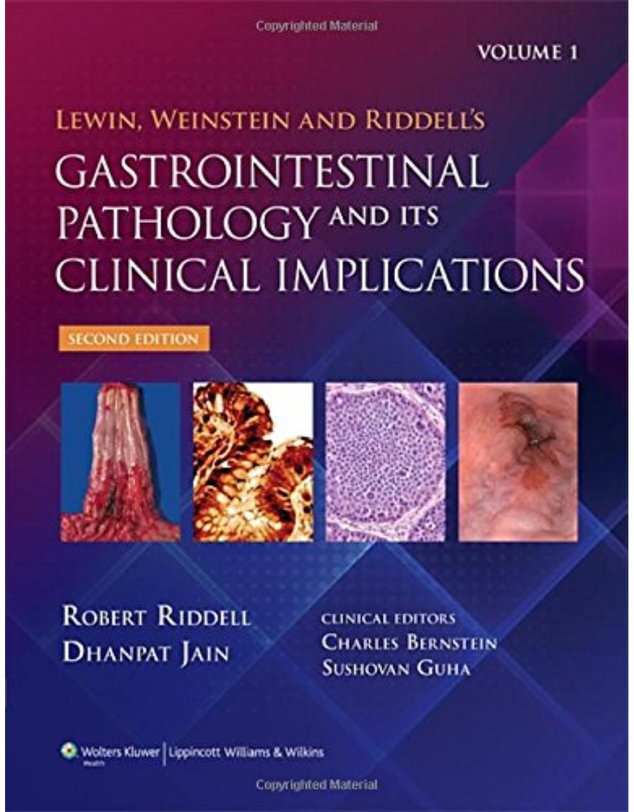
Gastrointestinal Pathology
Livrare gratis la comenzi peste 500 RON. Pentru celelalte comenzi livrarea este 20 RON.
Disponibilitate: La comanda in aproximativ 4-6 saptamani
Editura: LWW
Limba: Engleza
Nr. pagini: 1488
Coperta: Hardcover
Dimensiuni: 21.29 x 27.64 cm
An aparitie: 2014
Description:
Lewin Gastrointestinal Pathology and Its Clinical Implications, Second Edition This comprehensive, two-volume resource highlights the practical aspects of the pathology of biopsies and gross specimens, the clinical/pathological correlation, and differential diagnoses, and the ways in which these affect the management of patients with gastrointestinal disorders. The authors provide valuable insights on many important areas of gastrointestinal pathology, and openly address controversies within the specialty. This all-inclusive work stands alone in its illustrative quality and in its emphasis on the clinical implications of patient management as related to pathologic findings. The Second Edition has been completely revised to reflect two decades of advances in the field. The book's focus on clinical/pathological correlations and differential diagnoses emphasizes their affect on patient management. Major revisions of the chapters on colitis and gastritis feature new approaches to treatment. Over 2100 full-color illustrations highlight pathologic features to sharpen diagnostic skills and guide treatment choices. NEW to the Second Edition… • Completely revised content reflects two decades of advances in the field. • Focus on clinical/pathological correlations and differential diagnoses emphasizes their affect on patient management • Major revisions of the chapters on colitis and gastritis feature new approaches to treatment. • Over 2100 full-color illustrations highlight pathologic features to sharpen diagnostic skills and guide treatment choices. Now with the print edition, enjoy the bundled interactive digital edition, offering tablet, smartphone, or online access to: • Complete content with enhanced navigation • A powerful search that pulls results from content in the book, your notes, and even the web • Cross-linked pages, references, and more for easy navigation • Highlighting tool for easier reference of key content throughout the text • Ability to take and share notes with friends and colleagues • Quick reference tabbing to save your favorite content for future use Pick up your copy today!
Table of Contents:
Volume I
Chapter 1: Dialogue, Biopsies–Taking and Handling; Resected Specimens; Protocols
Chapter 1 Introduction
Mucosal Biopsy
Table 1-1: Recipe to Improve Biopsy Quality and Interpretation Dramatically
Usual Endoscopic Pinch Biopsies
Hot Biopsy Forceps
Cold Biopsies
Figure 1-1
Electrocautery Snare Biopsy
Snare Polypectomy
Snare Polypectomy after Submucosal Injection (“Lift-and-Cut” Technique)
Shave Biopsy
Endoscopic Mucosal Resection and Endoscopic Submucosal Dissection
Endoscopic mucosal resection
Endoscopic submucosal dissection
Submucosal lesions
Figure 1-2
Figure 1-4
Figure 1-3
Ancillary Techniques Used at Endoscopy
Diagnosis of Infections—Smears, Brushings, Aspiration, and Culture
Cytology
Direct-vision brush cytology
Balloon mesh cytology
Fine-needle aspirates
Chromoendoscopy
Barrett’s esophagus
Inflammatory bowel disease
Screening and surveillance colonoscopy for adenomas in otherwise healthy individuals
Virtual Histology
Biopsy specimen handling and processing
Figure 1-5
Figure 1-6
Figure 1-7
Figure 1-8
Figure 1-9
Figure 1-10
Table 1-2: Guide to Sites for Taking Biopsies
Handling of the Biopsy Specimen Prior to Immersion in Fixatives
Handling Polyps
Routine Fixation
Tissue Processing, Embedding, and Cutting
Figure 1-11
Figure 1-12
Figure 1-13
Figure 1-14
Figure 1-15
Description of Endoscopic Findings
Table 1-3: Suggested Descriptive Terms for Benign-Appearing Lesions at Endoscopy
Biopsy Specimen Location
Table 1-4: Recommended Approach to Describe the Locations of Endoscopic Lesions and Biopsy Sites
Number and Size of Biopsy Specimens
The History and the Question for the Pathologist
Approach to the Microscopic Examination
A Systematic Approach to Biopsy Specimen Interpretation
Table 1-5: Example of a Systematic Approach to the Examination of Gastrointestinal Biopsy Specimens
Table 1-6: Some Infections That May Be Found in Exudate or as Attachments to the Surface Epithelium
Technical Problems in Interpretation
Mucosal Hemorrhage and Edema
Pseudoerosions
Other Artifacts
Figure 1-16
Figure 1-17
Figure 1-18
Figure 1-19
Figure 1-20
Figure 1-21
The Pathologist’s Interpretation
Mild Nonspecific Chronic Inflammation
Special Fixatives, Stains, or Storage Conditions
Immunohistochemical Applications in Gastrointestinal Disorders
Interpretation of Immunohistochemical Stains
Infections
Tuberculosis and Mycobacterium avium-intracellulare
Table 1-7: Disorders That May Require Special Fixatives, Stains, or Storage Conditions
Surgically Resected Specimens
Examination of the Specimen
Frozen Sections
Photography
Opening the Specimen
Fixation
Insufflation with Fixative
Injection studies for vascular diseases
Examination and dissection of the fixed specimen
Reexamination of the Fixed Specimen
Dissection
Dissections of Tumors
Lymph Node Dissections
Depth of Tumor Penetration
Venous Invasion by Tumor
Sections of Resected Margins
Incidental Findings
Chapter 2: Vascular Disorders and Related Diseases
Chapter 2 Introduction
Vascularization of the Digestive Tract—Overview
Vascularization of the Specific Segment of the Digestive Tract
Esophagus
Stomach, Small Intestine, and Large Intestine
Intramural circulation
Extramural (Splanchnic) circulation
Venous drainage
Collateral blood supply
Figure 2-1
Figure 2-2
Figure 2-3
Ischemia of the Gastrointestinal Tract
Pathophysiology
Etiology and Clinical Manifestations
Esophageal ischemia
Gastric ischemia
Acute mesenteric ischemia
Chronic mesenteric ischemia
Pathology
Gross features
Microscopy
Ischemic colitis
Ischemic proctitis
Table 2-1: Causes of Acute and Chronic Mesenteric Ischemia
Table 2-2: Cellular Effects of Ischemia
Figure 2-4
Figure 2-5
Table 2-3: Major Causes of Acute Mesenteric Ischemia
Figure 2-6
Figure 2-7
Figure 2-8
Figure 2-9
Figure 2-10
Figure 2-11
Figure 2-12
Table 2-4: Etiology of Ischemic Colitis in Young Adults (n = 42)
Figure 2-13
Figure 2-14
Figure 2-15
Figure 2-18
Figure 2-16
Figure 2-17
Figure 2-19
Figure 2-21
Figure 2-22
Figure 2-20
Inflammatory Vascular Disorders of the Gastrointestinal Tract (Vasculitides)
Introduction
Classification
Clinical Presentation
Different Types of Vasculitides
Large vessel vasculitides
Infectious vasculitides
Medium vessel vasculitides
Medium and small vessel vasculitis (ANCA-associated vasculitides)
Small vessel vasculitis
Miscellaneous conditions
Malignant-atrophic papulosis (Kohlmeier–Degos Syndrome)
Biopsy Diagnosis of Ischemic Colitis and Differential Diagnosis
Stercoral ulcers
Table 2-5: Classification of Vasculitis According to Histologic Pattern
Table 2-6: Classification of Vasculitides by Caliber of the Vessel
Table 2-7: Frequency of Intestinal Involvement in Different Vasculitides
Table 2-8: Laboratory Parameters Important for Patients Clinically Suspected of Vasculitis
Figure 2-23
Figure 2-24
Figure 2-25
Figure 2-26
Figure 2-27
Figure 2-28
Figure 2-29
Figure 2-30
Mechanical Obstruction and Ischemia of the Digestive Tract
Pathogenesis and Clinical Features
Adhesions
Hernias
Hernia of the anterior abdominal wall
Inguinal and femoral hernia
Umbilical hernia
Internal hernias
Volvulus
Intussusception
Figure 2-31
Iatrogenic Disorders of the Vascular System
Iatrogenic Intestinal Ischemia
Arterial obstruction or constriction
Drug-induced vascular lesions
Neutropenic colitis
Iatrogenic Gastrointestinal Bleeding
Radiation Injury
Pathophysiology
Acute radiation injury
Chronic (late) radiation injury
Figure 2-32
Figure 2-33
Figure 2-34
Figure 2-35
Table 2-9: Vascular Changes in Radiation-Induced Injury
Figure 2-36
Figure 2-37
Figure 2-38
Figure 2-39
Vascular Abnormalities of the Gastrointestinal Tract
Vascular Ectasia (Angiodysplasia)
Gastric Antral Vascular Ectasia
Dieulafoy Malformation (Caliber-Persistent Arteriole)
Telangiectasias
Hereditary hemorrhagic telangiectasia (Rendu–Osler–Weber Syndrome)
Arteriovenous Malformation
Phlebectasia
Diseases Affecting Blood Vessels
Disorders of Connective Tissue Affecting Blood Vessels
Pseudoxanthoma elasticum
Ehlers–Danlos syndrome
Table 2-10: Classification of Vascular Anomalies of the GI Tract
Figure 2-40
Figure 2-41
Figure 2-42
Figure 2-43
Figure 2-44
Handling of Specimens
Endoscopic Biopsies
Surgical Specimens
Ischemic disease
Vascular malformations
Figure 2-45
Chapter 3: Immunodeficiency Disorders
Chapter 3 Introduction
Intestinal Host Defences
Functional Anatomy of the GI Immune System
Normal Distribution of Gut-associated Lymphoid Tissue
Humoral immune system of the gut
Cellular immune system of the gut
Figure 3-1
Figure 3-2
Figure 3-3
Figure 3-4
Figure 3-5
Figure 3-6
Immunodeficiency Disorders of the Intestinal Tract
Clinical features
Histology
Table 3-1: Immunologic Tests for the Categorization of Primary Immunodeficiency Disease
Table 3-2: Pathology of the GI Tract in Primary Immunodeficiency Disorders
Figure 3-7
Figure 3-8
Table 3-3: Incidence of Neoplasia in Immunodeficiency Disorders
Primary Immunodeficiency Disorders
Predominant Antibody Defects
Common variable hypogammaglobulinemia (Late-onset acquired hypogammaglobulinemia, common variable immunodeficiency [CVID], Bruton’s X-linked agammaglobulinemia [XLAG])90,91,92
Selective IgA deficiency
Secretory component deficiency
Infantile X-linked agammaglobulinemia (Bruton’s agammaglobulinemia, congenital agammaglobulinemia)
Miscellaneous B-cell disorders
Predominant Cell-mediated Immunodeficiency
Severe combined immunodeficiency disease (Swiss-type agammaglobulinemia, hereditary thymic dysplasia)
IPEX syndrome
Chronic mucocutaneous candidiasis
Table 3-4: GI Manifestations of Primary Immunodeficiency Syndromes
Figure 3-9
Figure 3-10
Figure 3-11
Immunodeficiency Associated with Other Defects
DiGeorge’s Syndrome—Third and Fourth Pouch/Arch Syndrome (Thymic Hypoplasia, Cellular Immunodeficiency with Hypoparathyroidism)
Phagocytic and Other Cell Dysfunction
Chronic Granulomatous Disease
Systemic Mastocytosis
Secondary (Acquired) Immunodeficiency Disorders
Bone Marrow Transplantation
Transplantation regimen
Infection
Graft versus host disease (GVHD)
Chronic GVHD
Intestinal Transplantation
The Acquired Immunodeficiency Syndrome (AIDS)
Pathogenesis and clinical features
GI AIDS infections
GI neoplasms in AIDS
Table 3-5: Common GI Infections and Parasitic Infestations in Bone Marrow Transplantation
Figure 3-12
Table 3-6: Grading Scheme for Acute Cellular Rejection in Small Bowel Allografts
Figure 3-13
Table 3-7: Common Opportunistic GI Infections in AIDS
Figure 3-14
Figure 3-15
Figure 3-16
Workup of the Immunodeficient Patient
Chapter 4: Lymphoproliferative Disorders of the Gastrointestinal Tract
Chapter 4 Introduction
Overview
Introduction
Definition
Incidence
Pathogenesis of GI Lymphoma
Classification of GI Lymphomas
Clinical Presentation and Other Practical Diagnostic Issues
Role of Molecular Diagnosis in GI Tract Lymphomas
Workup of Lymphoproliferative Disorders of the GI Tract
Table 4-1: Tumor Site of Gastrointestinal Lymphomas
Table 4-2: Classification of GI Lymphomas
Table 4-3: Modified Ann-Arbor Staging System for Primary Gastrointestinal Lymphomas
Lymphoid Hyperplasia of the Gastrointestinal Tract
Localized Lymphoid Hyperplasia of the Stomach—Gastric Lymphoid Hyperplasia (Pseudolymphoma)
Pathology
Angiofollicular Hyperplasia (Lymphoid Hyperplasia with “Castleman-like” Features)
Immunohistochemical features and molecular genetics
Localized Lymphoid Hyperplasia of the Small Intestine
Localized (lymphoid) hyperplasia of the terminal ileum and appendix
Localized lymphoid hyperplasia of the duodenum and small intestine excluding the terminal ileum
Localized Lymphoid Hyperplasia of the Rectum
Diffuse Nodular Lymphoid Hyperplasia of the Intestine
Diffuse nodular lymphoid hyperplasia with hypogammaglobulinemia
Diffuse nodular lymphoid hyperplasia without hypogammaglobulinemia
Figure 4-1
Figure 4-2
Figure 4-3
Figure 4-4
Figure 4-5
Figure 4-6
Figure 4-7
Figure 4-8
Figure 4-9
Figure 4-10
Figure 4-11
Figure 4-13
Figure 4-12
Lymphoproliferative Disorders of the Esophagus
Lymphoproliferative Disorders of the Stomach
MALT Lymphoma (Extra Nodal Marginal Zone Lymphoma)
Pathogenesis
Clinical features
Pathology
Immunophenotype and molecular genetics
Treatment and prognosis
Reporting MALT lymphomas
Follow-up of patients with treated MALT lymphoma
Role of the pathologist
Differential diagnosis and practical approach to the difficult diagnosis
Diffuse Large B-cell Lymphoma of the Stomach
Primary Gastric T-cell Lymphoma
Pathology
Other Miscellaneous Lymphoproliferative Disorders
Figure 4-14
Figure 4-15
Figure 4-16
Figure 4-18
Figure 4-17
Figure 4-19
Figure 4-20
Lymphoproliferative Disorders of the Small Intestine
MALT Lymphoma (Extranodal Marginal Zone Lymphoma) and Diffuse Large B-cell Lymphoma of the Small Intestine (Western-type Lymphoma)
Immunoproliferative Small Intestinal Disease (Mediterranean Lymphoma, α-Chain Disease)
Etiopathogenesis
Clinical features
Pathology
Immunophenotype and molecular genetics
Diagnosis and differential diagnosis
Treatment and prognosis
Burkitt’s and Burkitt’s-like Lymphoma (Malignant Lymphoma of the Small, Noncleaved Type)
Clinical presentation
Pathology
Immunophenotype and molecular genetics
Prognosis and treatment
Enteropathy-type T-cell Lymphoma
Pathogenesis
Clinical presentation
Pathology
Immunophenotype and molecular genetics
Treatment and prognosis
CD4 Positive Small Intestinal T-cell Lymphoma
Other Miscellaneous Lymphoproliferative Disorders
Figure 4-21
Figure 4-22
Table 4-4: Galian Staging System for IPSID
Figure 4-23
Figure 4-24
Figure 4-25
Lymphoproliferative Disorders of the Appendix, Colon, and Anal Canal
MALT Lymphomas of the Colon
Mantle Cell Lymphoma
Multiple Lymphomatous Polyposis
Figure 4-26
Miscellaneous Lymphoproliferative Disorders of the GI Tract
Follicular Lymphomas (Follicular and Diffuse Types)
Clinical features
Pathology
Immunophenotype and molecular genetics
Differential diagnosis
Treatment and prognosis
Lymphoplasmacytic Lymphoma/Waldenström’s Macroglobulinemia
Immunodeficiency-Associated Lymphoproliferative Disorders
Primary immunodeficiency-associated lymphoproliferative disorders
Acquired immunodeficiency-associated lymphoproliferative disorders
Solitary Plasmacytomas of the Gastrointestinal Tract
Lymphomatoid Granulomatosis (Angiocentric Lymphoproliferative Lesion)
Mycosis Fungoides Involving the Gastrointestinal Tract
Anaplastic Large-cell Lymphoma (Ki-1 Lymphoma)
Extranasal NK Cell or NK-like T-cell Lymphoma
NK cell enteropathy
Hodgkin’s Lymphoma of the Gastrointestinal Tract
True Histiocytic Lymphomas (Histiocytic Sarcoma) of the GI Tract
Langerhans Cell Histiocytosis (LCH, Histiocytosis X) of the GI Tract
Other Miscellaneous Lymphomas of the GI Tract
Angioimmunoblastic Lymphadenopathy Involving GI Tract
The Gastrointestinal Tract in Leukemia and Granulocytic Sarcoma
Figure 4-27
Table 4-5: Immunohistochemical Differentiation of Small Lymphocytic Gastrointestinal Lymphomas
Figure 4-28
Table 4-6: Lymphomas Associated with HIV Infection
Figure 4-29
Figure 4-30
Figure 4-31
Figure 4-32
Figure 4-33
Chapter 5: Disorders of Endocrine Cells
Chapter 5 Introduction
Introduction and Historical Perspective
From the APUD System (Amine Precursor Uptake and Decarboxylation) and on
Figure 5-1
Figure 5-2
The Normal Endocrine Cells at Specific Gastrointestinal Sites: Where They Are and What They Do
The Diseases: Perspectives Based on Clinical Implications
Table 5-1: Classification of Endocrine Tumors in the Gastrointestinal Tract
Table 5-2: Endocrine Tumors in the Gastrointestinal Tract and Their Distribution
Genetics of Endocrine Tumors and Endocrine Syndromes Involving the Gut
Carcinoid Tumors (Well-Differentiated Endocrine Tumors)
General Information
Gross Examination
Gross appearance
Gross dissection recommendations
Staging and Grading
Figure 5-3
Figure 5-4
Figure 5-5
Carcinoid Tumors (NETs) in Specific Sites
Carcinoid Tumors (NETs) of the Esophagus
Carcinoid Tumors (NETs) of the Stomach
Type I carcinoid tumors
Type II carcinoid tumors
Type III carcinoid tumors
Potential type IV carcinoid tumors
Carcinoid Tumors of the Duodenum
Gastrinomas
Somatostatinomas
Gangliocytic paraganglioma
Carcinoid Tumors of the Jejunum and Ileum (Midgut Carcinoids, NETs)
Incidental finding of carcinoid in biopsies of the cecum or terminal ileum
Carcinoid Tumors of the Appendix
Carcinoid Tumors of the Abdominal Colon
Carcinoid Tumors of the Rectum
Table 5-3: Gastric Enterochromaffin-like Cell (ECL-Cell) Carcinoid Tumors Subtypes
Figure 5-6
Figure 5-7
Figure 5-8
Figure 5-9
Figure 5-10
Figure 5-11
Figure 5-12
Figure 5-13
Figure 5-14
Figure 5-15
Figure 5-16
Figure 5-17
Figure 5-18
Figure 5-19
Figure 5-20
Poorly Differentiated Endocrine Neoplasms (Neuroendocrine or Endocrine Carcinomas)
General Information
Esophageal Tumors
Colonic and Rectal Tumors
Figure 5-21
Figure 5-22
Figure 5-23
Figure 5-24
Figure 5-25
Adenomas and Adenocarcinomas with Both Epithelial and Endocrine Differentiation
Tumors in Which Endocrine and Columnar Cells are Mixed
Tumors with Separate Components (Composite Tumors)
Figure 5-26
Figure 5-27
Endocrine Tumors Associated with Ulcerative Colitis and Crohn’s Disease
Hyperplasias
Figure 5-28
Figure 5-29
Figure 5-30
Enteroendocrine Cell Dysgenesis
Metastatic Endocrine Tumors with an Unknown Primary
Figure 5-31
Figure 5-32
Practical Approaches to Gastrointestinal Endocrine Abnormalities
General Information
Tumors
Chapter 6: Motility Disorders
Chapter 6 Introduction
Diverticular Disease of the Small and Large Intestines
Definition
Terminology
Large Bowel Diverticular Disease (Diverticulosis of the Colon)
Definitions
Pathogenesis
Clinical Features
Endoscopy
Imaging
Gross Pathology
Prediverticular disease
Handling of specimens
Histology
Defining diverticulitis
Figure 6-1
Figure 6-2
Figure 6-3
Figure 6-4
Figure 6-5
Figure 6-6
Figure 6-7
Figure 6-8
Figure 6-9
Figure 6-10
Figure 6-11
Figure 6-12
Figure 6-13
Figure 6-14
Polyps and Neoplasms Associated with and within Diverticula
Diverticular Polyps
Inverted diverticulum
Other Polyps
Carcinoma in diverticular disease
Figure 6-15
Diverticular Disease and Endometriosis
Diverticular Disease and IBD
Differential Diagnosis and Clinical Implications
Figure 6-16
Diverticulosis of the Cecum and Proximal Colon (Right-Sided Diverticulosis)
Pathogenesis
Clinical Features
Pathology
Figure 6-17
Diverticulosis of the Duodenum and Small Intestine
Pathogenesis
Duodenal Diverticula (Single or Isolated)
Extraluminal diverticula
Intraluminal duodenal diverticulum (prolapsed diaphragm)
Jejunoileal Diverticula
Pathogenesis and clinical features
Gross appearance
Histology
Meckel’s Diverticulum
Figure 6-18
Gastrointestinal Motility Disorders
The Normal Motility Apparatus of the Gut
Musculature of the Gut
Demonstration of Smooth Muscle in Motility Disorders
Figure 6-19
Enteric Nervous System
Interstitial Cells of Cajal
Figure 6-20
Figure 6-21
Figure 6-22
Figure 6-23
Figure 6-24
Figure 6-25
Motility Disorders
Table 6-1: Classification of Neuromuscular Disorders with GI Manifestations
Table 6-2: London Classification of GI Neuromuscular Pathology
Esophageal Disorders
Achalasia (Cardiospasm)
Gross appearance
Histology
Secondary Achalasia
Figure 6-26
Other Motor Disorders of the Esophagus
Figure 6-27
Figure 6-28
Motor Disorders of the Stomach
Pyloric Stenosis
Infantile pyloric stenosis (congenital hypertrophic pyloric stenosis)
Adult pyloric stenosis
Gastroparesis
Intestinal Pseudoobstruction
Definition
Acute intestinal pseudoobstruction
Ogilvie’s syndrome
Pathogenesis and Clinical Presentation of Chronic Pseudoobstruction
Chronic intestinal pseudoobstruction
Table 6-3: Chronic Idiopathic Intestinal Pseudoobstruction
Chronic Idiopathic (Primary) Intestinal Pseudoobstruction
Examining Resections for CIIP
Familial or Sporadic Visceral Myopathy
Pathology
Gross appearance
Histology
α-Smooth Muscle Actin Deficiency
Familial Visceral Myopathy with α-SMA–Positive Inclusion Bodies
Differential diagnosis
Desmin Myopathy
African Visceral Myopathy
Issues Reporting Motility Disorders
Table 6-4: Major Secondary Causes of CIIP
Figure 6-29
Figure 6-30
Figure 6-32
Figure 6-34
Figure 6-36
Figure 6-37
Figure 6-31
Figure 6-33
Figure 6-35
Figure 6-38
Figure 6-39
Figure 6-40
Figure 6-41
Figure 6-42
Figure 6-43
Primary Visceral Neuropathies
Familial Visceral Neuropathy
Paraneoplastic Syndromes
Sporadic Visceral Neuropathy
Differential diagnosis
Abnormalities of Interstitial Cells of Cajal
Hyperplasia of ICCs
Prognosis and therapy of CIIP
Diffuse Lymphoid Infiltration
Diffuse Eosinophilic Infiltrate
Slow Transit Constipation: Severe Idiopathic Constipation
Figure 6-44
Figure 6-45
Figure 6-46
Figure 6-47
Figure 6-48
Figure 6-50
Figure 6-49
Hirschsprung’s Disease
Clinical Presentation and Features
Etiology and Pathogenesis
Gross Appearance
Histology
Diagnosis of Hirschsprung’s Disease
Types of biopsy
Intraoperative frozen section
Variants of Hirschsprung’s Disease
Figure 6-51
Figure 6-52
Figure 6-53
Figure 6-54
Figure 6-55
Figure 6-56
Figure 6-57
Table 6-5: Variants of Hirschsprung’s Disease and Approximate Frequency
Intestinal Neuronal Dysplasia
Internal Sphincter Achalasia (Ultrashort Hirschsprung’s Disease)
Megacystis Microcolon Intestinal Hypoperistalsis
Diagnosis
Immature Ganglia
Mural Eosinophils in Hirschsprung’s Disease
Idiopathic Megacolon
Severe Idiopathic Constipation
Acquired Visceral Neuropathies
Toxic or Drug-Induced Visceral Neuropathy
Inflammatory Visceral Neuropathy (Acquired Aganglionosis)
Chagas’ Disease (American Trypanosomiasis)
Gross appearance
Histology
Differential diagnosis
Paraneoplastic Neuropathy
Miscellaneous Visceral Neuropathies
GI Manifestations Secondary to Neurologic Disorders of the Brain and Spinal Cord
Chronic Intestinal Pseudoobstruction Associated with Generalized Disease and the Muscular Dystrophies
Myotonic Muscular Dystrophy
Histology
Progressive Muscular Dystrophy
Acquired Jejunal Diverticulosis
Solitary Rectal Ulcer Syndrome of the Rectum and Inflammatory Cloacogenic Polyp (Mucosal Prolapse Syndromes)
Gross and Endoscopic Appearances
Histology
Differential Diagnosis
Proctitis (localized colitis) cystica profunda (hamartomatous inverted polyp of the rectum)
Gross and endoscopic appearances
Pathogenesis and histology
Nuclear atypicality, dysplasia, and carcinoma
Inflammatory cloacogenic polyp
Irritable Bowel Syndrome
Pathology of irritable bowel syndrome
Mastocytic enterocolitis
Role of the pathologist
Figure 6-58
Figure 6-59
Figure 6-60
Chapter 7: Mesenchymal Tumors
Chapter 7 Introduction
Introduction
Table 7-1: Classification of Gastrointestinal Mesenchymal Tumors
Gastrointestinal Stromal Tumors
Histogenesis
Demography and Clinical Aspects
Diagnostic Procedures
Targeted Therapy with Imatinib
Surgery for GIST
Frozen Section Diagnosis
Gross Examination and Appearances
Microscopic Appearances
Unusual morphologic variants
Posttreatment changes
Immunohistochemistry
False-positive immunoreactivity
CD117-negative GISTs
CD117- and DOG1-Positive Tumors Other Than GISTs
Molecular Features and Mutational Analysis
Succinyl dehydrogenase subunit B (SDHB) expression, NF1 mutations, and BRAF mutations
SDHB-deficient GISTs
Chromosomal alterations
Predicting Behavior
GIST Syndromes
Familial GIST syndrome
Neurofibromatosis type 1 (von Recklinghausen’s disease)
Carney’s triad
GIST-paraganglioma syndrome (Carney–Stratakis syndrome)
Sporadic multiple GIST
Pediatric GIST
Small GIST
Differential Diagnosis
Reporting GIST
Figure 7-1
Figure 7-2
Figure 7-3
Figure 7-4
Figure 7-5
Figure 7-6
Figure 7-7
Figure 7-8
Table 7-2: Potential GIST Mimics that May Express CD117 and/or DOG1
Figure 7-9
Table 7-3: The NIH 2001 Consensus Classification Scheme for Risk Stratification in GIST
Table 7-4: Risk Stratification Scheme Based on Long-term Follow-up Data from >1,600 Patients from the Armed Forces Institute of Pathology Prior to the Era of Imatinib Therapy
Table 7-5: Molecular Classification of Gastrointestinal Stromal Tumors
Table 7-6: Differential Diagnosis Based on Morphologic Subgroup
Table 7-7: Key Anatomic, Morphologic, and Immunohistochemical Features of Lesions to Be Considered in the Differential Diagnosis of Spindled, Cytologically Bland GIST
Table 7-8: Example of GIST Synoptic Report
Smooth Muscle Tumors
Leiomyomas
Epstein–Barr Virus–associated Smooth Muscle Tumors
Leiomyomatosis
Smooth Muscle Hamartoma
Leiomyomatosis Peritonealis Disseminata
Glomus Tumor
Leiomyosarcoma
Figure 7-10
Figure 7-11
Figure 7-12
Neurogenic Tumors
Schwannoma
Neurofibroma
Ganglioneuroma
Polypoid ganglioneuromas
Ganglioneuromatous polyposis
Diffuse ganglioneuromatosis
Mucosal Neuroma/Schwann Cell “Hamartoma”
Perineurioma and Fibroblastic Polyp
Perineurioma
Fibroblastic polyps
Biopsy Diagnosis of Polypoid Neural Lesions
Paraganglioma
Granular Cell Tumor
Malignant Peripheral Nerve Sheath Tumor
Mixed Neuronal Glial Tumor
Figure 7-13
Figure 7-14
Figure 7-15
Figure 7-16
Figure 7-17
Figure 7-18
Figure 7-19
Figure 7-20
Figure 7-21
Fibroblastic/Myofibroblastic Tumors
Desmoid Tumor (Intraabdominal Fibromatosis)
Inflammatory Fibroid Polyp
Plexiform Fibromyxoma of the Gastric Antrum
Inflammatory Myofibroblastic Tumor
Solitary Fibrous Tumor/Hemangiopericytoma
Calcifying Fibrous Tumor
Elastofibroma
Figure 7-22
Figure 7-23
Figure 7-24
Figure 7-25
Figure 7-26
Figure 7-27
Figure 7-28
Adipocytic Lesions
Lipohyperplasia of the Ileocecal Valve
Submucosal Lipoma
Atypical Lipoma
Angiolipoma
Lipomatous Polyposis and Epiploic Lipomatosis
Liposarcoma
Differential Diagnosis of Fatty Tumors
Figure 7-29
Figure 7-30
Figure 7-31
Endothelial and Vascular Tumors
Hemangioma
Angiosarcoma
Kaposi’s Sarcoma
Lymphangioma
Intestinal Vascular Lesions Associated with Clinical Syndromes
Figure 7-32
Figure 7-33
Figure 7-34
Figure 7-35
Figure 7-36
Striated Muscle Tumors
Rhabdomyoma
Rhabdomyosarcoma
Figure 7-37
Miscellaneous Sarcomas
Clear Cell Sarcoma
Malignant Gastrointestinal Neuroectodermal Tumor (GINECT)
Endometrial Stromal Sarcoma
Undifferentiated High Grade Pleomorphic Sarcoma/Malignant Fibrous Histiocytoma
Undifferentiated Sarcoma
Figure 7-38
Figure 7-39
Figure 7-40
Perivascular Epithelioid Cell Tumors
PEComa
Angiomyolipoma
Figure 7-41
Biphasic Epithelial–Mesenchymal Lesions
Synovial Sarcoma
Spindle Cell Carcinoma/Carcinosarcoma
Gastroblastoma (Epithelial Biphasic Tumor of Young Adults)
Figure 7-42
Figure 7-43
Figure 7-44
Figure 7-45
Nonmesenchymal Tumors That May Mimic Mesenchymal Neoplasms
Melanoma
Follicular Dendritic Cell Sarcoma
Lymphoma
Sarcomatoid Adult Granulosa Cell Tumor
Figure 7-46
Figure 7-47
Fibrosing Lesions of the Mesentery, Peritoneum, and Retroperitoneum
Sclerosing Mesenteritis
Sclerosing Peritonitis
Idiopathic Retroperitoneal Fibrosis
Weber–Christian Disease
Figure 7-48
Figure 7-49
Figure 7-50
Nonneoplastic Lesions That May Mimic Mesenchymal Neoplasms
Reactive Nodular Fibrous Pseudotumor
Pseudosarcomatous Granulation Tissue
Heterotopic Mesenteric Ossification
Xanthogranulomatous Pseudotumor
Mycobacterial Spindle Cell Pseudotumor
Figure 7-51
Figure 7-52
Figure 7-53
Chapter 8: Gastrointestinal Manifestations of Extraintestinal Disorders and Systemic Disease
Chapter 8 Introduction
Introduction
Connective Tissue Disorders (Collagen Vascular Diseases)
Scleroderma (Progressive Systemic Sclerosis)
Pathogenesis and Clinical Features
Gross Pathology and Histology
Clinical Implications
Dermatomyositis and Polymyositis
Systemic Lupus Erythematosus
Mixed Connective Tissue Diseases and the Overlap Syndrome
Rheumatoid Arthritis
Miscellaneous Disorders
Figure 8-1
Figure 8-2
Figure 8-3
Figure 8-4
Figure 8-5
Figure 8-6
Figure 8-7
Gastrointestinal Manifestations in Endocrine Disorders
Thyroid Gland
Hyperthyroidism
Hypothyroidism
Autoimmune Thyroid Disease
Thyroid Neoplasms
Parathyroid Gland
Hyperparathyroidism
Hypoparathyroidism
Endocrine Pancreas
Diabetes
Hyperfunction of Islets of Langerhans
Gastrinoma
VIPoma Syndrome (Verner–Morrisons Syndrome)
Somatostatinoma
Other Islet Cell Tumors
Adrenal Gland
Gonads
Pregnancy
Hypothalamus and Pituitary
Hypopituitarism
Pituitary Adenoma
Autoimmune Polyendocrinopathy Syndrome Type 1
The IPEX syndrome
Figure 8-8
Table 8-1: Gastrointestinal Manifestations of Multiple Endocrine Neoplasia (MEN) Syndromes
Gastrointestinal Manifestations in Renal Disease
Acute Renal Failure
Chronic Renal Failure
Endoscopic and Histologic Appearances
Other Findings
Renal Transplantation
Role of the Pathologist and Clinical Implications
Table 8-2: Interrelationship of Gastrointestinal and Renal Diseases
Gastrointestinal Manifestations of Hepatic Disorders
Portal Hypertension
Primary Sclerosing Cholangitis and Autoimmune Hepatitis
Liver Transplantation
Gastrointestinal Manifestations of Skin Disorders
Bullous Disorders
Epidermolysis bullosa
Epidermolysis Bullosa Acquisita
Pemphigus Vulgaris
Cicatricial Pemphigoid (Benign Mucous Membrane Pemphigoid)
Stevens–Johnson Syndrome
Herpes Simplex Virus Infection
Hyperkeratotic Disorders
Lichen planus
Tylosis
Miscellaneous Disorders
Acrodermatitis enteropathica
Darier’s Disease
Dermatogenic Enteropathy
Malignant Disease of the Gastrointestinal Tract and Skin Disease
Acanthosis Nigricans
Cowden’s Disease
Dermatomyositis
Miscellaneous
Table 8-3: Primary Skin Disorders Associated with Gastrointestinal Disease
Gastrointestinal Manifestations of Cardiac Disease
Congestive Cardiac Failure
Infective Endocarditis
Open Heart Surgery, Extracorporeal Circulation, and Cardiac Transplantation
Hematologic Disorders
Dysproteinemias
Hemolytic Uremic Syndrome
Coagulation Disorders
Hemophilia
Other Coagulation Disorders
Mastocytosis
Rosai–Dorfman Disease (Sinus Histiocytosis with Massive Lymphadenopathy)
Miscellaneous Disorders
Figure 8-9
Figure 8-10
Gastrointestinal Amyloid Deposition
General Properties and Classification
Clinical Features
Histologic Features
Diagnosis and Clinical Implications
Figure 8-11
Figure 8-12
Figure 8-13
Figure 8-14
Figure 8-15
Disorders of Lipid Metabolism
Fabry’s Disease
Tangier Disease
Wolman’s Disease
Abetalipoproteinemia
Granulomatous Disorders
Sarcoidosis
Chronic Granulomatous Disease
Miscellaneous Disorders
Endometriosis
Pellagra
Familial Mediterranean Fever (Familial Paroxysmal Polyserositis)
Figure 8-16
Figure 8-17
Neoplastic Disease
Figure 8-18
Figure 8-19
Chapter 9: Esophagus: Normal Structures, Developmental Abnormalities, and Miscellaneous Disorders
Chapter 9 Introduction
Structure of the Esophagus
Anatomy
Histology
Mucosa
Submucosa and muscularis propria
Figure 9-1
Figure 9-2
Figure 9-3
Figure 9-4
Figure 9-5
Esophageal Function
Age-Dependent Changes
Embryology and Development of the Esophagus
Figure 9-6
Figure 9-7
Developmental and Congenital Anomalies
Esophageal Atresia and Tracheoesophageal Fistulas
Bronchoesophageal Fistula
Developmental and Congenital Cysts
Duplication and congenital cysts
Neurenteric cysts/remnants
Bronchogenic cysts
Other cysts
Heterotopias
Gastric heterotopia in the esophagus
Other heterotopias
Other Developmental Anomalies
Congenital esophageal stenosis or stricture
Short esophagus
Pulmonary sequestrations
Table 9-1: Concomitant Anomalies in Individuals with Esophageal Atresia
Figure 9-8
Table 9-2: The Different Types of Esophageal Atresia
Figure 9-9
Figure 9-10
Figure 9-11
Figure 9-12
Table 9-3: Esophageal Stenosis
Esophageal Perforation
Spontaneous Rupture (Boerhaave’s Syndrome)
Pathogenesis and clinical features
Pathology
Nonspontaneous Rupture and Penetration
Pathogenesis and clinical features
Pathology
Table 9-4: Causes of Esophageal Perforation
Figure 9-13
Esophageal Hemorrhage
Esophageal Tears (Mallory–Weiss Syndrome)
Pathogenesis and clinical features
Pathology
Esophageal Varices
Pathogenesis and clinical features
Pathology
Figure 9-14
Figure 9-15
Esophageal Fistula
Acquired Esophageal Stenosis or Stricture
Esophageal webs and rings
Figure 9-16
Figure 9-17
Figure 9-18
Figure 9-19
Figure 9-20
Diverticula and Pseudodiverticula
Upper Esophageal Diverticula (Zenker’s)
Pathogenesis and clinical features
Pathology
Mid- and Lower Esophageal Diverticula
Pathogenesis and clinical features
Pathology
Atypical Esophageal Diverticula in Scleroderma
Esophageal Intramural Pseudodiverticulosis/Retention Cysts
Pathogenesis and clinical features
Pathology
Figure 9-21
Figure 9-22
Figure 9-23
Figure 9-24
Other Miscellaneous Conditions
Mucosal Bridge
Glycogenic Acanthosis
Esophageal Xanthelasma
Figure 9-25
Chapter 10: Inflammatory Disorders of the Esophagus: Reflux and Nonreflux Types
Chapter 10 Introduction
Gastroesophageal Reflux Disease (Reflux Esophagitis–GERD/GORD)
Definition of GERD
Symptoms of GERD
Correlation between GERD, symptoms, and endoscopy
Therapy
Long-term therapy for GERD and prognosis
The Gastroesophageal Junction and Z-Line
Etiology and Pathogenesis of GERD
Endoscopic Grading of Reflux Disease in Squamous Mucosa
Los Angeles Grading System for Reflux Disease in Squamous Mucosa
Nonerosive Reflux Disease and Its Pathology
Gross/Endoscopic Appearances of GERD
Where to Biopsy for GERD and Criteria Used
Erosions
Traditional biopsy sites
Cardia biopsies
Histologic Diagnosis and Criteria for GERD in Biopsies
Evaluating reactive changes
Grading reactive epithelial changes
Table 10-1: Classification of Esophagitis by Etiology
Figure 10-1
Figure 10-2
Figure 10-3
Figure 10-4
Figure 10-5
Table 10-2: Reflux-independent Esophageal Lesions
Figure 10-6
Figure 10-7
Figure 10-8
Figure 10-9
Figure 10-10
Inflammatory Cells
Intraepithelial Mononuclear Cells
Minor changes
Inflammatory Polyps
Reactive changes versus neoplasia following erosions and ulcers
Atypical Cells in Squamous Mucosa
Carditis as a Manifestation of GERD
Progression of GERD
Complications of GERD
GERD-related strictures
Cameron’s Ulcer
Figure 10-11
Figure 10-12
Figure 10-13
Figure 10-14
Figure 10-15
Figure 10-16
Figure 10-17
Barrett’s Esophagus (BE)
Definition
Practical Aspects of Making the Diagnosis of BE
Irregular Z-line versus “ultrashort” BE
Definitions of BE: short- versus long-segment BE
When BE Does Not Have Goblet Cells, Does It Matter?
When are goblet cells not present?
Risk of carcinoma in patients with non–goblet cell BE
Conclusions regarding the definition and cancer risk in Barrett’s (columnar-lined) esophagus
Epidemiology and Pathophysiology of Barrett’s Esophagus
Morphologic Development of Barrett’s Esophagus
Endoscopic Grading of Barrett’s Mucosa
Histology
Double muscularis mucosae
Palisaded vessels
Handling of Patients and Biopsies Postablation or Postendoscopic Resection
Differential Diagnosis of BE and Other Mucosal Types Encountered
Barrett-associated Neoplasia
Diagnosing and Grading Dysplasia
Indefinite for Dysplasia
Low-grade Dysplasia (Low-grade Intraepithelial Neoplasia—LGNIN)
High-grade Dysplasia (High-grade Intraepithelial Neoplasia—HGIEN)
Implication
Intramucosal Carcinoma
Invasive Carcinoma (Submucosal invasion)
Interobserver Variability and Need for a Second Opinion for Dysplasia
Ancillary Techniques
Surveillance biopsies
Figure 10-18
Figure 10-19
Figure 10-20
Figure 10-21
Figure 10-22
Figure 10-23
Figure 10-24
Figure 10-25
Figure 10-26
Figure 10-27
Figure 10-28
Figure 10-29
Figure 10-30
Figure 10-31
Figure 10-32
Nonreflux Esophagitis
Infections
Viral Infections
Herpes virus infection
Cytomegalovirus esophagitis
Human papillomavirus
Acute HIV infection
Other viral infections
Fungal Infections
Clinic symptoms and prognosis
Other fungal infections
Bacterial Esophagitis
Luetic infection
Other bacteria
Figure 10-33
Figure 10-34
Figure 10-35
Figure 10-36
Figure 10-37
Figure 10-38
Descriptive Esophagitides
Lymphocytic Esophagitis
Exfoliative (Sloughing) Esophagitis (Esophagitis Dissecans Superficialis)
Acute Necrotizing Esophagitis (Black Esophagus)
Eosinophilic Esophagitis
Pathogenesis
Symptoms
Clinical
Biopsy sites
Pathology
Differential diagnosis
Corrosive Esophagitis
Drug-induced Esophagitis (Pill Esophagitis)
Figure 10-39
Figure 10-40
Figure 10-41
Figure 10-42
Figure 10-43
Systemic Diseases Involving the Esophagus
Skin Diseases and Esophageal Inflammation
Glycogenic acanthosis
Skin Diseases and Esophageal Inflammation
Pseudoepitheliomatous Hyperplasia
Leukoplakia
Graft versus Host Disease
Behçet’s Disease
Crohn’s Disease
Lymphocytic esophagitis in Crohn’s disease
Granulomatous Esophagitis and Sarcoid
Ulcerative Colitis
Brown Bowel Syndrome (Ceroid Lipofuscinosis)
Diabetes Mellitus
Parkinson’s Disease
Figure 10-44
Figure 10-45
Figure 10-46
Figure 10-47
Figure 10-48
Chapter 11: Polyps and Tumors of the Esophagus
Chapter 11 Introduction
Table 11-1: Classification of Esophageal Tumors
Role of the Pathologist
Squamous Cell Carcinoma
Pathogenesis and Clinical Features
Risk Factors
Geographic distribution: association with smoking and alcohol
Diet
Genetic factors
Human papillomavirus infection
Other risk or protective factors
Predisposing Conditions
Celiac sprue
Tylosis palmaris et plantaris
Other carcinomas
Prior irradiation
Premalignant Lesions: Dysplasia and Intraepithelial Neoplasia
Gross and Endoscopic Appearances
Microscopic Appearances
Spread of Tumor, Staging, and Prognosis
TNM classification
Local spread and prognostic factors
Effect of chemotherapy and irradiation
DNA ploidy
Molecular factors as prognosticator
Failure and causes of death
Figure 11-1
Figure 11-2
Figure 11-3
Figure 11-4
Figure 11-5
Unusual Variants of Squamous Cell Carcinoma
Superficial Esophageal Carcinoma
Endoscopic mucosal resection
Superficial spreading carcinoma
Verrucous Carcinoma
Spindle Cell Carcinoma
Gross appearances
Microscopic appearances
Histogenesis
Biopsy diagnosis
Small Cell Carcinoma
Basaloid Squamous Cell Carcinoma
Figure 11-6
Figure 11-7
Figure 11-8
Figure 11-9
Figure 11-10
Figure 11-11
Adenocarcinoma
Adenocarcinoma in the Columnar-lined Esophagus (Barrett’s Esophagus)
Risk and pathogenesis
Clinical Features
Gross and Endoscopic Appearances
Microscopic appearances
Endoscopic Ablation Techniques
Endoscopic mucosal resection
Prognosis
Figure 11-12
Figure 11-13
Figure 11-14
Figure 11-15
Unusual Variants of Adenocarcinoma
Adenoid Cystic Carcinoma
Mucoepidermoid Carcinoma
Adenocarcinoma in Heterotopic Gastric Mucosa
Adenocarcinoma in Submucosal Glands
Adenosquamous Carcinoma and Adenoacanthoma
Choriocarcinoma and Hepatoid Adenocarcinoma
Figure 11-16
Biopsy Diagnosis of Squamous and Adenocarcinoma and Associated Problems
Regenerative changes
Granulation tissue
Barrett’s esophagus
Other Polyps and Tumors
Squamous papilloma and papillomatosis
Inflammatory Polyps
Pseudosarcomatous changes in inflammatory polyps
Mucosal Tags
Fibrovascular Polyps
Hamartomas, Choristomas, Thyroid Rests, Parathyroid Rests
Malignant Melanoma
Secondary Tumors
Mesenchymal Tumors
Figure 11-17
Figure 11-18
Figure 11-19
Figure 11-20
Figure 11-21
Figure 11-22
Figure 11-23
Figure 11-24
Figure 11-25
Figure 11-26
Figure 11-27
Figure 11-28
Figure 11-29
Figure 11-30
Figure 11-31
Chapter 12: Stomach: Normal Structures and Developmental Abnormalities
Chapter 12 Introduction
Embryology
Stomach
Cardia
Duodenum
Figure 12-1
Normal Structure of the Stomach
Anatomy
Gastric surfaces (relations of the stomach)
Anatomic regions
Blood vessels and lymphatics
Nerve supply
Histology
Mucosa
Submucosa
Muscularis propria, ICCs, and serosa
Figure 12-2
Figure 12-3
Figure 12-4
Figure 12-5a
Table 12-1: Endocrine Cells of the Stomach
Figure 12-6
Figure 12-7
Figure 12-8
Figure 12-9
Figure 12-10
Figure 12-11
Figure 12-12
Figure 12-13
Figure 12-15
Figure 12-14
Figure 12-16
Figure 12-17
Figure 12-18
Table 12-2: Developmental Abnormalities of the Stomach
Figure 12-19
Developmental Abnormalities of the Stomach
Pathogenesis and Clinical Features
Figure 12-20
Chapter 13: Stomach and Proximal Duodenum: Inflammatory and Miscellaneous Disorders
Chapter 13 Introduction
Classification of Gastritis and Gastropathy
Current Classification of Gastritis
Table 13-1: ABC Classification of Gastritis
Table 13-2: Gastritis Classification “Historical Prospective”
Figure 13-1
Figure 13-2
Table 13-3: Gastritis Classification “Sydney System”
Gastritis
Distinctive (Specific) Types of Gastropathies
Reactive (Predominant Epithelial) Changes
Reactive gastropathy
Toxic gastropathy
Reactive Changes with Erosions in Helicobacter—One or Two Diseases?
Distinction of Reactive Changes from Dysplasia
Reactive changes in intestinal metaplasia
Alcoholic gastropathy
Caustic-induced injury
Graft versus host disease
Chemotherapy and radiation
Ischemia
Predominantly Vascular Changes
Gastric antral vascular ectasia
Portal hypertension (congestive gastropathy)
Hemorrhagic gastropathy (“gastritis”) and “Curling’s ulcer”
Distinctive (Specific) Types of Gastritis
Infections
A H. pylori infection
Histology of H. pylori–associated gastritis
H. pylori diagnosis
Noninvasive methods
Invasive methods
Atrophic gastritis and gastric atrophy
Staging gastric atrophy
Disorders associated with H. pylori gastritis
Gastroduodenal erosions and ulcers (“peptic ulcers”)
Pathogenetic factors
Epidemiology
Atypical clinical presentations
Endoscopic appearance of peptic erosions and ulcers
Table 13-4: Classification by Predominant Histologic Change
Figure 13-3
Figure 13-4
Figure 13-5
Figure 13-6
Figure 13-7
Figure 13-8
Figure 13-9
Figure 13-10
Figure 13-11
Figure 13-12
Figure 13-13
Figure 13-14
Figure 13-15
Figure 13-16
Figure 13-17
Table 13-5: A Helicobacter-associated Diseases
Figure 13-18
Figure 13-19
Figure 13-20
Figure 13-22
Figure 13-21
Figure 13-23
Figure 13-24
Table 13-6: Stains for Detection of Helicobacter pylori
Figure 13-25
Figure 13-26
Figure 13-27
Figure 13-28
Figure 13-29
Figure 13-30
Figure 13-31
Figure 13-32
Figure 13-33
Figure 13-34
Table 13-7: OLGA
Figure 13-35
Figure 13-36
Table 13-8: Factors That Damage or Protect the Gastric Mucosa
Figure 13-37
Table 13-9: Evidence Linking H. pylori to the Pathogenesis of Duodenal Ulcer836–838
Figure 13-38
Figure 13-39
Figure 13-40
Figure 13-41
Figure 13-42
Figure 13-43
Figure 13-44
Figure 13-45
Figure 13-46
Figure 13-47
“Peptic Diseases” of the Duodenal Bulb and the Proximal Duodenum
Pathogenesis and Clinical Features of Duodenitis
Duodenal ulcer
Clinical features
Pathology of Duodenitis and Duodenal Erosions and Ulcer
Gross pathology
Histology
Differential diagnosis of duodenitis
Healing and healed ulcers
Complications of gastroduodenal ulcers
Treatment
The role of the pathologist and clinical implications
Autoimmune Gastritis
Pathogenesis
Subtypes of autoimmune gastritis (AIG) and their etiology
Clinical features
Pathology
Table 13-10: Associations of Erosive and Nonerosive Duodenitis
Figure 13-48
Figure 13-49
Figure 13-50
Figure 13-51
Figure 13-52
Figure 13-53
Figure 13-54
Figure 13-55
Figure 13-56
Figure 13-57
Figure 13-58
Figure 13-59
Figure 13-60
Table 13-11: Oxyntic (Corpus and Fundus) Predominant Gastritis
Figure 13-61
Lymphocytic Gastritis
Morphologic Separation of Etiologies
Table 13-12: Lymphocytic Gastritis Associations
Figure 13-62
Figure 13-63
Granulomatous Gastritis
Figure 13-64
Table 13-13: Conditions Associated with Granulomatous Gastritis
Carditis
Figure 13-65
Non–H. pylori Bacterial Infections
Non–H. pylori Helicobacter Species (NHPH)/“Helicobacter heilmannii”
Gastric disease associated with non–H. pylori Helicobacter species/“Helicobacter heilmannii”
Diagnosis
Tuberculosis
Syphilis
Enterococcal Gastritis
Phlegmonous and Emphysematous Gastritis
Figure 13-66
Table 13-14: Non–Helicobacter pylori Detected in the Stomach of Humans
Figure 13-67
Viral Infections
Cytomegalovirus Infection (HHV-5)
Herpes Viruses (HHV-1,2)
Epstein–Barr Virus (EBV—HHV-4)
Other HHV Viruses
Other Viruses
Figure 13-68
Fungal Infections
Candida albicans
Histoplasmosis
Mucormycosis (Zygomycosis)
Aspergillosis
Figure 13-69
Parasites and Nematodes
Cryptosporidium
Anisakiasis
Other Parasites and Nematodes
Figure 13-70
Other Gastritides
Clinical features
Endoscopic features
Histologic features
Eosinophilic Gastritis
Eosinophilic gastritis as part of gastric involvement in eosinophilic gastroenteritis
Differential diagnosis
Collagenous Gastritis
Diffuse collagenous gastroenterocolitis
Gastric Malakoplakia
Drug- and Chemotherapy-induced Gastritis
Figure 13-71
Figure 13-72
Figure 13-73
Figure 13-74
Table 13-15: Differential Diagnosis of Eosinophilic Gastroenteritis
Table 13-16: Diagnostic Workup for Eosinophilic Gastroenteritis
Figure 13-75
Figure 13-76
Figure 13-77
Figure 13-78
Hypertrophic Gastropathies and Ménétrier’s Disease
Primary/Idiopathic Ménétrier’s Disease
Clinical Presentation
Pathology of Primary Ménétrier’s Disease
Carcinoma Complicating Ménétrier’s Disease
Secondary Ménétrier’s Disease
Table 13-17: Hypertrophic Gastropathies
Figure 13-79
Hypertrophic Gastropathy-associated with Protein Loss
Cytomegalovirus-associated Hypertrophic Gastropathy
Hypertrophic Lymphocytic Gastritis
Helicobacter pylori–associated Hypertrophic Gastritis
HIV-associated Hypertrophic Gastritis
Large Gastric Folds Associated with Other Conditions
Other Types of Large Gastric Folds
Figure 13-80
Figure 13-81
Distinctive Endoscopic Entities
Miscellaneous Disorders of the Stomach
Gastric Calcinosis
Gastric Glandular Siderosis
Approach to the Interpretation of Gastric Biopsies
Surface epithelium
Figure 13-82
Table 13-18: The Differential Diagnosis in Gastric Pathology
Histology
Chapter 14: Gastric Epithelial Polyps and Tumors
Chapter 14 Introduction
Adenoma/Dysplasia/Intraepithelial Neoplasia
Definitions and Terminology
Special Stains for Characterizing Gastrointestinal Epithelial cells
Classification of Gastric Adenomas
Intestinal-type adenomas
Gastric-type adenomas
Pyloric gland adenoma
Foveolar adenoma
Fundic gland adenoma
Grading of Adenoma/Dysplasia/Intraepithelial Neoplasia
Low-grade adenoma/dysplasia/intraepithelial neoplasia
High-grade adenoma/dysplasia/intraepithelial neoplasia
Differential Diagnosis
Distinction of regenerative changes from adenoma (dysplasia/intraepithelial neoplasia)
Distinction of high-grade dysplasia/adenoma/intraepithelial neoplasia) from invasive carcinoma
Figure 14-1
Figure 14-2
Figure 14-3
Figure 14-4
Figure 14-5
Figure 14-5
Figure 14-5
Figure 14-6
Figure 14-7
Figure 14-8
Figure 14-9
Figure 14-10
Gastric Carcinoma
Introduction
Gastric Cardia Carcinoma
Carcinoma of the Gastric Antrum and Body
Pathogenesis
Risk Factors
Helicobacter pylori infection
Epstein–Barr virus, CIMP-H, and K-ras
JC virus
Dietary factors
Smoking
Genetic factors
Hereditary gastric cancer predisposition syndromes
Other genetic abnormalities predisposing to gastric cancer
Polymorphism in genes
Predisposing Conditions
H. pylori–related chronic atrophic gastritis
Postgastrectomy
Immunodeficiency disorders
Menetriér’s disease
Premalignant Lesions of the Stomach
Adenoma/dysplasia/intraepithelial neoplasm
Gastric polyps
Gross Features of Gastric Carcinoma
Gross Features of Superficial Cancers and Their Subdivision
Classification and Natural History of Early and Late (Advanced) Gastric Carcinoma
Early gastric carcinoma/cancer
Advanced gastric cancer
Histologic Classification of Gastric Carcinomas
Lauren classification
Nakamura classification
Ming classification
WHO histologic classification and grading of gastric cancer
Figure 14-11
Figure 14-12
Figure 14-13
Figure 14-14
Figure 14-15
Figure 14-16
Figure 14-17
Figure 14-18
Figure 14-19
Figure 14-20
Histological Subtypes of Carcinoma
Tubular Adenocarcinoma
Well-differentiated Adenocarcinoma
Moderately Well-differentiated Adenocarcinoma
Poorly Differentiated Adenocarcinoma
Papillary Adenocarcinoma
Mucinous Adenocarcinoma (syn. Mucoid, Mucus, Colloid, and Muconodular Adenocarcinoma)
Signet-ring Cell Carcinoma
Endocrine Cell Tumors
Rare Variants of Gastric Carcinoma
Gastric carcinoma with lymphoid stroma (syn. medullary carcinoma, gastric lymphoepithelioma-like carcinoma)
α-Fetoprotein-producing gastric carcinoma and hepatoid adenocarcinoma
Adenosquamous and squamous cell carcinoma
Choriocarcinoma (syn. chorioepithelioma)
Yolk sack tumor (syn. endodermal sinus tumor)
Paneth cell carcinoma
Micropapillary carcinoma
Gastric adenocarcinoma of fundic gland type (fundic gland carcinoma, combined parietal, and chief cell carcinoma) including parietal cell carcinoma and chief cell carcinoma
Fundic gland adenoma
Extremely well-differentiated adenocarcinoma
Pyloric gland carcinoma
Composite adenocarcinoma and endocrine tumor with pancreatic differentiation and pancreatic type tumors
Undifferentiated carcinoma
Serrated dysplasia and carcinoma
Gastric teratoma
Carcinosarcoma
Gastroblastoma
Growth Pattern of Gastric Adenocarcinoma
Superficial (spreading) carcinoma
Organoid differentiation
Stromal reaction
Differential Diagnosis and Clinical Implications
Early gastric cancer
Distinction from histiocytic lesions
Lymphoma
Metastatic carcinoma
Neuroendocrine (NET—carcinoid) tumors
Granular cell tumors
Bizarre undifferentiated cells (pseudosarcomatous changes)
Radiation therapy and chemotherapy
Spread of Gastric Carcinoma and Prognostic Factors
Prognosis of Gastric Carcinoma
Early gastric cancer
Risk of nodal metastases in early gastric carcinoma
Gastric remnant carcinoma
Advanced gastric cancer
Neoadjuvant therapy
Her2 status
K-ras status
Biopsy Diagnosis of Gastric Carcinoma
Biopsy before surgical resection
Misreading of biopsies for carcinoma
Value of Cytology in the Diagnosis of Gastric Carcinoma
Handling of Gastric Specimens and Intraoperative Evaluation
Polypectomy specimens
Endoscopic mucosal or submucosal specimens
Resection specimens
Figure 14-21
Figure 14-21
Figure 14-21
Figure 14-22
Figure 14-23
Figure 14-24
Figure 14-25
Figure 14-26
Figure 14-27
Figure 14-28
Figure 14-29
Figure 14-30
Figure 14-31
Figure 14-32
Figure 14-33
Figure 14-34
Table 14-1: Lymph Node Metastasis in Patients with D2 Gastrectomy Surgery for Early Gastric Cancer
Table 14-2: TNM Classification and Staging of Gastric Carcinoma (Seventh Edition)
Table 14-3: Grading of the Effects of Neoadjuvant Therapy
Table 14-4: Causes of Errors in the Biopsy Diagnosis of Gastric Carcinoma
Table 14-5: One-Piece and Piecemeal EMR in Early Gastric Cancer
Figure 14-35
Table 14-6: Examination of Gastric Resection Specimens
Figure 14-36
Nonneoplastic Polyps
Classification
Biopsy and Excision of Gastric Polyps: Role of the Pathologist
Approach to the biopsy diagnosis of gastric polyps
Fundic Gland Polyps
Pathology
Clinical implications
Hyperplastic and Inflammatory Polyps
Pathology
Dysplasia/carcinoma in hyperplastic polyps
Clinical implications for inflammatory/hyperplastic polyps
Differential Diagnosis
Single polyps
Multiple gastric polyps
Juvenile Polyps
Pathology
Peutz–Jeghers Polyps
Pathology
PTEN Hamartoma Tumor Syndrome
Familiar Gastric Hyperplastic Polyposis
Cronkhite–Canada Syndrome–Associated Polyps
Pathology
Polypoid Gastritis
Gastric Xanthomas (Xanthelasmas, Lipid Islands)
Pathology
Mucosal Bumps and Nodules
Solitary Polypoid Hamartoma (Gastric Hamartoma with Myxoid Stroma, Hamartomatous Inverted Polyp, Heterotopic Inverted Polyp)
Pathology
Gastritis Cystica Polyposa (Gastric Stomal Polypoid Hyperplasia, Gastric Stomal Polypoid Hypertrophic Gastritis)
Pathology
Mucosal and Submucosal Cysts
Gastritis Cystica Profunda
Pathology
Differential Diagnosis
Heterotopic Pancreas (Ectopic Pancreas, Pancreatic Rests, Adenomyoma, Adenomyomatous Hamartoma, Myoglandular Hamartoma, Myoepithelial Hamartoma)
Pathology
Differential Diagnosis
Brunner Gland Heterotopia/Nodule
Heterotopic Antral Mucosa in Oxyntic Mucosa
Table 14-7: Gastric Polyps
Figure 14-37
Figure 14-38
Figure 14-39
Figure 14-40
Figure 14-41
Figure 14-42
Figure 14-43
Figure 14-44
Figure 14-45
Figure 14-46
Figure 14-47
Figure 14-48
Figure 14-49
Figure 14-50
Figure 14-51
Figure 14-52
Volume Ii
Chapter 15: Appendix
Chapter 15 Introduction
Introduction
Function of the Appendix
The Normal Appendix
Normal Anatomy, Development, and Gross Appearance
Histology
Figure 15-1
Figure 15-2
Figure 15-3
Figure 15-4
Figure 15-5
Figure 15-6
Figure 15-8
Figure 15-7
Routine Pathologic Examination of the Appendix
Gross Handling of Dilated Appendices
Congenital Structural Abnormalities
Positional Abnormalities
Agenesis
Duplication
Appendix helicus and horseshoe appendix
Congenital fistula
Heterotopic tissue
Table 15-1: Modified Wallbridge Classification of Appendiceal Duplication
Figure 15-9
Acquired Structural Abnormalities
Diverticula
Torsion and Volvulus
Intussusception
Figure 15-10
Iatrogenic Use of the Appendix
Acute Appendicitis
Clinical Features
Pathogenesis
Gross Appearance
Histology
Resolution of Acute Appendicitis
Crypt architectural distortion
Localized epithelial hyperplasia (reactive hyperplasia)
Submucosal fibrosis
Luminal obliteration (obliterative fibrosis) and neuroma of the appendix
Terminology in Acute Appendicitis
Unusual variants of appendicitis
Issues with Appendicitis and a Practical Approach
Complications of Acute Appendicitis
Perforation
Appendiceal epithelium growing along the external surface of the appendix
Periappendiceal abscesses and productive periappendiceal fibrosis
Formation of multiple lumina (lumens)
Escaped fecalith
Fistula
Suppuration and suppurative pyelophlebitis and hepatic abscess formation
The Appendiceal Stump
Recurrent stump appendicitis
Endoscopic excision of the inverted appendix stump
Abscess formation
Xanthogranulomatous appendicitis and malacoplakia
Hemorrhage
Intussusception
Polyps
Figure 15-11
Figure 15-12
Figure 15-13
Figure 15-14
Figure 15-15
Figure 15-16
Figure 15-17
Figure 15-18
Figure 15-19
Figure 15-20
Specific Infections of the Appendix
Viral Infections
Adenovirus
Cytomegalovirus
Measles
Infectious mononucleosis
Bacterial Infections
Acute enteritis
Spirochetosis
Yersinia
Tuberculosis and mycobacterial infection
Actinomycosis
Whipple’s disease
Parasites
Figure 15-21
Figure 15-22
Figure 15-23
Figure 15-24
Figure 15-25
Figure 15-26
Miscellaneous Inflammatory Conditions Affecting the Appendix
Granulomatous Appendicitis and Crohn’s Disease Isolated to the Appendix
Isolated appendiceal Crohn’s disease
Ulcerative colitis involving the appendix
Histiocytic diseases
Arteritis (localized polyarteritis nodosa)
Inherited disorders
Figure 15-27
Figure 15-28
Figure 15-29
Figure 15-30
Figure 15-31
Figure 15-32
Tumors of the Appendix
Appendiceal Tumors are Not the Same as Large Bowel Tumors
Polyps and Other Benign Lesions of the Appendix
Adenomas
Tubular and tubulovillous adenomas
Villous adenomas
Serrated hyperplasia and polyps
Localised and diffuse mucosal hyperplasia (Serrated hyperplasia)
Hyperplastic polyps
Sessile serrated polyps (“sessile serrated adenomas”)
Sessile serrated adenomas/polyps with dysplasia
Molecular pathology of appendiceal serrated lesions
Table 15-2: Classification of Epithelial Polyps and Tumors of the Appendix
Figure 15-33
Figure 15-34
Table 15-3: Classifications of Serrated Appendiceal Lesions
Figure 15-35
Figure 15-36
Figure 15-37
Issues with Polyps of the Appendix and Their Sequela—A Practical Approach
Terminology and Differential Diagnosis
Terminology Issues
Mucocele
Cystadenomas and cystadenocarcinomas
Hybrid lesions
Mucinous Neoplasms of the Appendix and Pseudomyxoma Peritonei
Mucinous Neoplasms of Appendix
Terminology
Incidence
Clinical features
Gross appearances
Histology
Molecular Pathology and Immunohistochemistry of Appendiceal Neoplasia
Issues with Mucinous Neoplasms—Practical Approach
Grading of dysplasia and distinction of LAMN from mucinous adenocarcinoma
Problems when perforation is present
Rupture and mucin extravasation
Unusual variations of appendiceal mucinous neoplasia
Follow-up and therapy
Pseudomyxoma Peritonei
Gross appearance
Microscopic appearance
Management of pseudomyxoma peritonei and pathology of cytoreduction
Adenocarcinomas of the Appendix
Large bowel-type carcinoma
Grading appendiceal carcinomas
TNM staging of appendiceal tumors and prognosis of appendiceal neoplasms
Goblet Cell Carcinoma (Goblet Cell Carcinoid, Microglandular Goblet Cell Carcinoma, Adenocarcinoid, Microglandular Mucinous Carcinoma, Crypt Cell Carcinoma, Mucinous Carcinoid)
Terminology and histogenesis
Clinical features
Gross pathology
Histology
Immunohistochemistry
High-Grade Carcinomas Ex Goblet Cell Carcinoma/Carcinoid
Prognosis and treatment of GCC and carcinomas ex GCC
Combined Tumors of the Appendix
Comparative Survival of Appendiceal Adenocarcinomas and Carcinoids
Other Rare Tumors and Lesions of the Appendix
Metastatic Disease Involving the Appendix
Table 15-4: Terminology for Mucinous Appendiceal Tumors and PMP
Table 15-5: Comparing the WHO 2010 and Our Preferred Terminology
Table 15-6: Classifications of Low-Grade Mucinous Neoplasia
Figure 15-38
Figure 15-39
Figure 15-40
Figure 15-41
Figure 15-42
Figure 15-43
Figure 15-44
Table 15-7: Comparison of classifications of pseudomyxoma peritonei
Figure 15-45
Figure 15-45
Figure 15-45
Figure 15-46
Figure 15-47
Figure 15-48
Figure 15-49
Figure 15-50
Figure 15-51
Figure 15-52
Figure 15-53
Figure 15-54
Figure 15-55
Figure 15-56
Chapter 16: Small and Large Bowel Structure: Developmental and Mechanical Disorders
Chapter 16 Introduction
Structure of the Small and Large Intestines
External Appearance
Small intestine
Large intestine
Internal Appearance
Small intestine
Large intestine
Histology
Small intestinal mucosa
Ileocecal valve
Large intestinal mucosa
Specialized cells in the small and larger bowel epithelium
Stem cells and kinetics
Pericrypt fibroblast sheath
Lamina propria and immune system
Muscularis mucosae
Submucosa
Muscularis propria
Nerve supply, myenteric plexi, and interstitial cells of Cajal
Serosa
Blood vessels
Lymphatics
Figure 16-1
Figure 16-2
Figure 16-3
Figure 16-4
Figure 16-5
Figure 16-6
Figure 16-7
Figure 16-8
Figure 16-9
Figure 16-10
Figure 16-11
Figure 16-13
Figure 16-12
Figure 16-14
Figure 16-16
Figure 16-15
Figure 16-17
Figure 16-18
Figure 16-19
Figure 16-20
Figure 16-21
Figure 16-22
Figure 16-23
Figure 16-24
Figure 16-25
Figure 16-26
Figure 16-27
Figure 16-28
Figure 16-29
Embryology and Development of Small and Large Intestine
Congenital Anomalies
Heterotopia and metaplasia
Malrotation, lack of fixation, and volvulus
Congenital hernias
Abnormalities of intestinal musculature
Atresia, Webs, Diaphragms, and Stenoses
Atresia
Webs, diaphragms, and stenoses
Enteric duplications
Simple duplications
Complications
Hemorrhage and infarction
Malignancy
Abdominothoracic dorsal enteric diverticula
Meckel’s diverticulum and anomalies of the vitelline duct
Tumors in Meckel’s diverticulum
Miscellaneous anomalies
Mechanical Disorders and Disorders of Propulsion
Obstruction
Neonatal necrotizing enterocolitis and related disorders
Figure 16-30
Figure 16-31
Figure 16-32
Figure 16-33
Figure 16-34
Figure 16-35
Figure 16-36
Figure 16-37
Figure 16-38
Table 16-1: Classification of Intestinal Atresias
Figure 16-39
Figure 16-40
Figure 16-41
Figure 16-42
Figure 16-43
Figure 16-44
Figure 16-45
Figure 16-46
Figure 16-47
Figure 16-48
Chapter 17: Small Bowel Mucosal Disease
Chapter 17 Introduction
Small Bowel Mucosal Disease
Normal Appearances and Artifacts
Normal Villous Architecture
Regional variations in mucosal biopsy appearances
Geographic variation
Tangential artifact
Traumatic artifact
Evaluation of a Small Bowel Biopsy Specimen
The Abnormal Small Bowel Biopsy Specimen
The mild lesion (Marsh type 1)
The moderate lesion (Marsh type 3a and 3b)
The severe flat lesion (Marsh type 3c)
Alternative terminology
Figure 17-2
Figure 17-1
Table 17-1: Overview Classification of Small Bowel Biopsy
Figure 17-3
Figure 17-4
Figure 17-5
Figure 17-6
Figure 17-7
Figure 17-8
Disorders Associated with Abnormal Small Bowel Biopsy Specimens
Table 17-2: Severe (Flat) Lesions: Nonspecific Histologya
Table 17-3: Severe Flat Lesions: Specific/Distinctive Histology
Table 17-4: Variably Severe Lesions: Nonspecific Histologya
Table 17-5: Variably Severe Lesions—Specific/Distinctive Histologya
Table 17-6: Normal Villous Architecture: Specific/Distinctive Histology
Disorders Associated with a Severe Flat Mucosa: Nonspecific and Specific/Distinctive Histology
Celiac Sprue (Syn. Celiac Disease, Gluten-sensitive Enteropathy, Nontropical Sprue)
Pathogenesis
Clinical features
Associations of celiac sprue
Endoscopic appearance
Pathology
Complications of celiac sprue
Dermatitis herpetiformis and celiac sprue
Relatives of patients with celiac sprue
Latent celiac sprue and preclinical disease
Celiac crisis
Rebiopsy in patients with celiac sprue
Refractory celiac sprue
Unclassified sprue
Collagenous sprue
Autoimmune Enteropathy
Pathogenesis
Clinical features
Pathology
Treatment and prognosis
Food Hypersensitivity
Definitions of gastrointestinal reactions to food
Morphologic reactions in food hypersensitivity
Tropical Sprue
Childhood Kwashiorkor (Protein-Calorie Malnutrition)
Microvillus Inclusion Disease/Familial Enteropathy (Hypoplastic Villous Syndrome, Congenital Microvillus Atrophy)
Congenital Tufting Enteropathy
Figure 17-9
Figure 17-10
Figure 17-11
Figure 17-12
Figure 17-13
Figure 17-14
Figure 17-15
Diseases Associated with Variably Severe Lesions with an Intraepithelial Lymphocytosis
Immunodeficiency Syndromes
Common variable immunodeficiency/hypogammaglobulinemia (late-onset immunodeficiency)
Selective IgA deficiency
Acquired immunodeficiency syndrome (AIDS)
Infectious Gastroenteritis
Bacterial Overgrowth Syndrome (Stasis Syndromes, Contaminated Bowel Syndrome)
Pathogenesis and clinical features
Pathology
Zollinger–Ellison Syndrome (Gastrinoma)/Acid Dumping
Acrodermatitis Enteropathica
Figure 17-16
Figure 17-17
Figure 17-18
Figure 17-19
Diseases Associated with Variably Severe Lesions: Diagnostic/Distinctive Histology
Whipple’s Disease
Pathogenesis and clinical features
Endoscopic findings
Pathology
Differential diagnosis
Eosinophilic Gastroenteritis
Pathogenesis and clinical features
Clinical diagnosis and differential diagnosis
Endoscopic findings and gross pathology
Pathology
Differential diagnosis
Crohn’s Disease
Nonspecific Ulcers
Primary Intestinal Lymphoma
Infections
Intestinal Lymphangiectasia
Waldenstrom’s Macroglobulinemia
Impaired Epithelial Cell Replacement
Severe vitamin B12 deficiency, folic acid deficiency, acute radiation reaction, and chemotherapy
Figure 17-20
Figure 17-21
Table 17-7: Comparison of Whipple’s Disease with Mycobacterium avium-intracellare (MAI)
Figure 17-22
Figure 17-23
Figure 17-24
Figure 17-25
Figure 17-26
Normal Villous Architecture: Specific/Distinctive Histology
Abetalipoproteinemia and Hypobetalipoproteinemia
Pathogenesis
Pathology
Amyloidosis
Lipid Storage Diseases
Chronic Granulomatous Disease
Figure 17-27
Figure 17-28
Miscellaneous Disorders
Enzyme Deficiency States
Disaccharidase deficiencies
Ornithine transcarbamylase deficiency
Brown Bowel Syndrome
Protein-losing Enteropathy
Figure 17-29
Case Examples of Biopsy Interpretation and Clinical Implications
Case 1: Refractory Sprue
Part A
Part B
Part C
Part D
Case 2: The Mild Mucosal Lesion
Part A
Part B
Part C
Part D
Chapter 18: Inflammatory Bowel Diseases
Chapter 18 Introduction
Table 18-1: Normal Colonoscopy and Abnormal Histology
Microscopic Colitis
Introduction
Epidemiology
Etiology and Pathogenesis
Clinical Features and Endoscopic Findings
Microscopic Pathology
Collagenous colitis
Lymphocytic colitis
Microscopic colitis and involvement of small intestine
Differential Diagnosis of Lymphocytic Colitis
Treatment and Prognosis
Microscopic Colitis Variants
Brainerd Diarrhea
Microscopic Colitis in Children
Microscopic Colitis and Idiopathic Inflammatory Bowel Diseases
Table 18-2: Medications Potentially Related to the Development of Lymphocytic Colitis
Table 18-3: Autoimmune Disorders Associated with Microscopic Colitis
Table 18-4: Comparison of Collagenous and Lymphocytic Colitis
Figure 18-1
Figure 18-2
Figure 18-3
Figure 18-4
Figure 18-5
Figure 18-6
Chronic Idiopathic Inflammatory Bowel Diseases
Introduction
Epidemiology
Etiology and Pathogenesis
Genetic factors
Environmental factors
Smoking and IBD
Appendectomy and IBD
The immune system
Ulcerative Colitis
Clinical features
Endoscopic features and gross pathology
Microscopic pathology
Focality/patchiness in ulcerative colitis
Appendiceal involvement
Rectal sparing
Ileal involvement in ulcerative colitis
Pan enteritis post colectomy for ulcerative colitis
Prestomal ileitis
Ulcerative colitis in children
Proctitis/ulcerative proctitis
Fulminant colitis and toxic megacolon
Rectum in ulcerative proctocolitis following ileorectal anastomosis
Effect of medical therapy on the pathology of ulcerative colitis
Ileal pouches and their complications including pouchitis
Other causes of pouch problems
When are pouch biopsies done?
Dysplasia and cancer in pouches
Other diseases affecting pouches
Crohn’s Disease
Introduction and definition
Clinical features and classification
Endoscopic features and gross pathology
Microscopic pathology
Anal and perianal disease
Management
Differentiation Between Ulcerative Colitis and Crohn’s Disease
Macroscopic differences
Microscopic differences
Indeterminate Colitis/IBD-Unclassified
Introduction
Pathology
Synchronous and Metachronous Crohn’s Disease and Ulcerative Colitis
Upper Gastrointestinal Tract Involvement in Inflammatory Bowel Disease
The role of upper gastrointestinal biopsies in differentiating Crohn’s disease from ulcerative colitis
Extragastrointestinal Manifestations of IBD
Hepatobiliary disorders
Treatment and Prognosis of IBD
Crohn’s Disease and Small Intestinal Transplantation
Colitis Associated with Primary Sclerosing Cholangitis
Table 18-5: Genomic Regions Significantly Associated with IBD in Genome-wide Association Studies of Patients of European Ancestry
Figure 18-7
Figure 18-8
Figure 18-9
Figure 18-10
Figure 18-11
Figure 18-12
Figure 18-13
Figure 18-14
Figure 18-15
Figure 18-16
Figure 18-17
Figure 18-18
Table 18-6: Scoring System for the Assessment of Severity in Ulcerative Colitis
Table 18-7a: Simplified Grading System for Chronicity and Activity in IBD (Option A)*
Table 18-7b: Simplified Grading System for Ulcerative Colitis (Option B)
Figure 18-19
Figure 18-20
Figure 18-21
Figure 18-22
Figure 18-23
Figure 18-24
Figure 18-25
Figure 18-26
Figure 18-27
Table 18-8: Causes of Proctitis
Table 18-9: Clinical Criteria for the Diagnosis of Fulminant Colitis
Figure 18-28
Figure 18-29
Figure 18-30
Figure 18-31
Figure 18-32
Figure 18-33
Table 18-10: Vienna and Montreal Classification for Crohn’s Disease
Figure 18-34
Figure 18-35
Figure 18-36
Figure 18-37
Figure 18-38
Figure 18-39
Figure 18-40
Figure 18-41
Table 18-11: Common Causes of Endoscopic Aphthoid Ulcers
Figure 18-42
Figure 18-43
Table 18-12: Granuloma-associated Diseases in the Gastrointestinal Tract
Figure 18-44
Figure 18-45
Figure 18-46
Figure 18-47
Figure 18-48
Figure 18-49
Figure 18-50
Figure 18-51
Figure 18-52
Figure 18-53
Figure 18-54
Figure 18-55
Table 18-13: Gross Features Which Can Help to Distinguish Between Ulcerative Colitis and Crohn’s Disease
Table 18-14: Microscopic Features for the Distinction Between Ulcerative Colitis, Crohn’s Disease, and Indeterminate Colitis in Fulminant Colitis
Figure 18-56
Figure 18-56
Figure 18-56
Figure 18-56
Figure 18-56
Figure 18-57
Figure 18-58
Figure 18-59
Figure 18-60
Table 18-15: Extragastrointestinal Complications Associated with Inflammatory Bowel Disease
IBD Mimics and Related Disorders
Diverticular Disease–associated Chronic Colitis (Diverticular Colitis)
Diversion Colitis
Focal Active Colitis
Incidental Terminal Ileitis and Cryptogenic Multifocal Ulcerous Stenosing Enteritis (CMUSE)
Reporting Ileitis
Neutrophil Function Defects and IBD
Hermansky-Pudlak Syndrome
Intestinal Behcet Disease
Epidemiology
Clinical features
Pathology
Cryptogenic Multifocal Ulcerous Stenosing Enteritis (CMUSE) (Chronic Non-specific Ulcer [CNSU] of the Small Bowel)
Obstructive Colitis
NK Cell Enteropathy
Eosinophilic Colitis
Figure 18-61
Figure 18-62
Figure 18-63
Figure 18-64
Figure 18-65
Figure 18-66
Figure 18-67
Figure 18-68
Figure 18-69
Table 18-16: Common Causes of Eosinophilic Colitis
Figure 18-70
Differential Diagnosis of Chronic Inflammatory Bowel Diseases
Infections
Infections in patients with IBD
Drug-induced Mucosal Injury
Ischemia
Miscellaneous
Table 18-18: Enteric Pathogens That Cause Inflammation Resembling IBD
Table 18-17: Differential Diagnosis Between Acute Self-limited or Infectious-type Colitis and IBD
Table 18-19: Some Causes of Right-sided Colitis
Figure 18-71
Figure 18-72
Malignancy in Inflammatory Bowel Disease
Introduction
Molecular Pathogenesis of Cancer in IBD
Dysplasia in IBD
Terminology of dysplastic lesions and their management
Gross pathology of dysplasia
Microscopic pathology of dysplasia
Distinction from reactive changes
The classification and grading of dysplasia
Grading dysplasia
Reproducibility of dysplasia diagnosis and grading
Summary of problematic issues related to dysplasia in IBD
Role of immunohistochemistry and other ancillary techniques
The clinical implications of dysplasia in ulcerative colitis
Cancer in IBD
Pathologic characteristics of colorectal cancer in colitis
Management of carcinoma complicating ulcerative colitis
Cancer of the small and large bowel in Crohn’s disease
Anal carcinoma in IBD
Carcinoma in Indeterminate colitis
Inflammatory bowel disease and other malignancies
Carcinoma after surgery
Table 18-20: Genetic Alterations in Colitis-associated and Sporadic CRC
Figure 18-73
Table 18-21: Macroscopic Appearance of IBD-associated Intraepithelial Neoplasia
Figure 18-74
Figure 18-75
Figure 18-76
Figure 18-77
Figure 18-78
Figure 18-79
Figure 18-80
Figure 18-81
Figure 18-82
Figure 18-83
Figure 18-84
Table 18-22: Biopsy Classification of Dysplasia in IBD: Comparison of the Two Common Systems in Use
Figure 18-85
Figure 18-86
Figure 18-87
Figure 18-88
Figure 18-89
Figure 18-90
Case Studies
Case 1
Case 2
Case 3
Case 4
Case 5
Case 6
Table 18-23: Systematic Approach to Biopsy Evaluation for Inflammatory Disorders of the Bowel
Figure 18-91
Figure 18-92
Figure 18-93
Figure 18-94
Figure 18-95
Figure 18-96
Drug-Associated, Iatrogenic and Miscellaneous Diseases of the Small and Large Intestines
Table 18-24: Effects of Some Drugs and Medications on the Intestines
Role of the Pathologist
Mucosal Changes
Antineoplastic Drugs and Drugs Causing Increased Apoptosis (“Apoptotic” Colopathy)
Acute toxic injury
Abnormal mitosis
Dysplasia mimics related to medication
Neutropenic enterocolitis (typhlitis, neutropenic appendicitis)
Figure 18-97
Figure 18-98
Figure 18-99
Figure 18-100
Figure 18-101
Figure 18-102
Idiopathic Small and Large Bowel Ulcers, Fibrosis, Strictures
Gross and Endoscopic Appearances
Histology
Nonsteroidal Anti-inflammatory Drugs
Clinical features
Pathogenesis
NSAIDs and small intestinal disease
NSAIDs and large intestinal disease
NSAIDs and diverticular disease
NSAIDs in idiopathic IBD
NSAIDs and left-sided colonic increase of eosinophils
Potassium Chloride
Table 18-25: Some Causes of Idiopathic Ulcers of the Intestine
Figure 18-103
Figure 18-104
Figure 18-105
Drug-Associated and Iatrogenic Vascular Injuries
Ischemia Following Angiography
Trauma to the vessel wall
Embolization
Anticoagulant-associated Hemorrhage and Hematoma
Retroperitoneal hematomas
Arterial or Venous Thrombosis
Drug-induced vasculitis
Estrogen and oral contraceptives
Oral contraceptive and IBD
Changes Secondary to Contrast Media
Perforation
Ischemic necrosis
Arterial Spasm
Vasopressin
Hypotensive and hypovolemic agents
Neonatal Perforation
Stevens-Johnson Syndrome
Figure 18-106
Figure 18-107
Iatrogenic and Drug-associated Inflammatory Changes
Pseudomembranes and Ischemic Colitis
Acute Infectious-type Colitis
Methyldopa
Iatrogenic and Drug-associated Granulomatous and Eosinophilic Enteritis/Colitis
Chronic Inflammation with/without Focal Active Colitis
Mycophenolate mofetil
Rituximab
Ipilimumab and anti PD-1
Anti-tumor necrosis factor (TNFa) drugs
Anti vascular endothelial growth factor receptor antibodies (bevacizumab)
Gold enteritis and colitis
5-Flucytosine
Penicillamine
Cimetidine
Isotretinoin
Figure 18-108
Figure 18-109
Figure 18-110
Changes due to Other Drugs and Chemicals
Changes Due to Enemas and Laxatives
Sodium phosphate (fleets enema) and bisacodyl
Hypertonic saline enemas
Chemical colitis mimicking ischemia induced by glutaraldehyde, hydrogen peroxide or peracetic acid
Soap
Polyethylene glycol
Changes Due to Laxatives
Cathartic colon
Myenteric plexus damage, pseudo-obstruction, and stercoral ulceration
Alcohol
Obstruction from Ingested Drugs
Pneumatosis Cystoides Intestinalis
Pathogenesis
Clinical features
Gross and endoscopic disease
Histology
Prognosis
Iatrogenic Gas Introduced at Endoscopy (Pseudolipomatosis)
Gross and endoscopic appearances
Histology
Figure 18-111
Figure 18-112
Figure 18-113
Figure 18-114
Figure 18-115
Figure 18-116
Other Miscellaneous Conditions, Disorders, and Diseases
Compensatory Changes
Sequelae of Instrumentation
Extrinsic Damage to the Bowel
Large-bowel Disease in Acute Pancreatitis
Colitis (and Enteritis) Cystica Profunda
Adaptation to Prolapse
Rectal Mucosal Prolapse from Other Causes
Enteritis and Colitis Cystica Superficialis
Malabsorption
Diarrhea
Chemotherapeutic agents
Mycophenolate mofetil
Proton pump inhibitors
Neoplasms (Following Immune Suppression and Radiation)
Figure 18-117
Table 18-26: Causes of Cystica Superficialis and Glandularis/Cystica Profunda Lesions
Benign Disorders Involving Histiocytes
Normal Histiocytes
Histiocytes Associated with Drug Use
Iron-containing macrophages
Pseudomelanosis coli (melanosis coli)
Gross and endoscopic appearances
Histology
Histiocytes Associated with Infections
Mycobacterium avium-intracellulare
Whipple’s disease
Malakoplakia and xanthogranulomatous inflammation
Histiocytes Associated with Storage Disease
Histiocytes Associated with Miscellaneous Disorder
Iron-containing macrophages
Muciphages
Xanthoma/xanthelasma
Black Pigments in the Terminal Ileum and Large Bowel
Aluminum silicate titanium-containing pigment in terminal ileum
Injected India ink as a tattoo
Table 18-27: Disorders Associated with an Excess of Abnormal Histiocytes/Macrophages
Figure 18-118
Figure 18-119
Figure 18-120
Figure 18-121
Table 18-28: The Involvement of Various Cell Types in a Range of Storage Diseases
Table 18-29: The Staining Patterns of the Neuronal Storage Material
Figure 18-122
Table 18-30: Iatrogenic Causes of Ischemia
Figure 18-123
Figure 18-124
Abnormal Deposits (Crystal Deposition)
Barium Granuloma
Oleogranulomas (Paraffinomas)
Radiologic Masses, Baroliths, and Acute Appendicitis
Mercury
Figure 18-125
Chapter 19: Enteric Infections and Associated Diseases
Chapter 19 Introduction
Introduction
Table 19-1: Important Enteric Infections Classified According to Major Classes or Organisms
Host Defense Mechanisms
Acidity of Gastric Juices
Intestinal Motility
Mucus
Normal Resident Bacterial Flora
Gut-Associated Immune System
Infection of the Normal Host
Mechanisms of Infection
Adherence, Initial Attachment, and Colonization
Tissue Invasion
Toxin Production
Enteroadherent and Other Mechanisms of Diarrhea
Persistence of Infection
Approach to Diagnosis of GI Infections
Introduction
Clinical Features
Endoscopic and Gross Findings
Stool Examination, Culture, and Other Studies
Biopsy Diagnosis
No change
Typical acute infectious (self-limited) colitis
Mild acute or chronic inflammation
Specific histologic pattern
Detection of specific organisms or their cytopathic changes
Special stains
Electron microscopy
Molecular tests
Gastrointestinal Infections in Specific Clinical Circumstances
Traveler’s diarrhea
Point outbreaks of infection
Health care–associated (“nosocomial”) infection
Oral–anal sexual practices
Table 19-2: Pathogens Causing Distinctive Cellular Changes or Tissue Reactions
Figure 19-1
Table 19-3: Infectious Agents for Which Monoclonal or Polyclonal Antibodies or Both Are Available for Immunofluorescence or Immunohistochemistry
Table 19-4: Infectious Agents for Which DNA Probes for In Situ Hybridization Assays Are Available
Table 19-5: Infectious Agents for Which PCR Assays Are Available
Table 19-6: Identification of Specific Enteropathogens Shown as Percentage of Total Cases in Studies of the Etiology of Traveler’s Diarrhea Carried Out in Latin America/Caribbean, Africa, and Southern Asia, 1973–2004
Table 19-7: Infections in Male Homosexuals and Sites of Involvement
Acute Infectious (Self-Limited) Colitis and Proctitis
Pathogenesis and Clinical Features
Exacerbations of Inflammatory Bowel Disease
Gross and Endoscopic Appearances
Histology
Diagnosis
Bacterial Infections
Vibrios
Epidemiology
Clinical features
Pathogenesis
Pathology
Diagnosis
Treatment and follow-up
Escherichia coli
Enterotoxigenic E. coli
Enteroinvasive E. coli
Enteropathogenic organisms
Enteroadherent E. coli
Enterohemorrhagic E. coli
Hemolytic uremic syndrome and thrombotic thrombocytopenic purpura
Verotoxin-producing E. coli in inflammatory bowel disease
Shigella
Pathogenesis
Clinical features
Gross and endoscopic appearances
Histology
Diagnosis, differential diagnosis, and treatment
Salmonella
Classification of Salmonella
Salmonella enterocolitis
Fulminant infections
Salmonella infection in inflammatory bowel disease
Typhoid fever
Campylobacter
Pathogenesis
Clinical and endoscopic features
Gross and endoscopic findings
Histology
Complications
Aeromonas hydrophila, Plesiomonas shigelloides, and Edwardsiella tarda
Pathology
Legionella
Yersinia Infection
Pathogenesis and clinical features
Gross pathology and endoscopic appearances
Histology
Yersinia pseudotuberculosis infection
Klebsiella Granulomatis (Granuloma Inuinale)
Differential diagnosis
Prognosis
Clostridial Infections
Clostridium difficile
Antibiotic-associated colitis
Clostridium Perfringens (Previously Called C. welchii)
Enteritis necroticans
Clostridium botulinum
Neutropenic enterocolitis
Phlegmonous enterocolitis
Mycobacterial Infections
Mycobacterium tuberculosis and bovis
Mycobacterium avium-intracellulare
Gonococcal Proctitis
Gross and endoscopic appearances
Histology
Spirochetes
Syphilis
Intestinal spirochetosis
Brucella
Actinomyces
Rhodococcus equi (formerly known as Corynebacterium equi)
Chlamydia
Lymphogranuloma venereum
Chlamydial proctitis
Rickettsial Disease
Rocky mountain spotted fever
Table 19-8: The Various Pathotypes and the Common Clinical Syndromes Associated with E. coli
Figure 19-2
Figure 19-3
Figure 19-4
Figure 19-5
Figure 19-6
Figure 19-7
Figure 19-8
Figure 19-9
Figure 19-10
Figure 19-11
Figure 19-12
Figure 19-13
Figure 19-14
Figure 19-15
Figure 19-16
Figure 19-17
Table 19-9: Pathologic Features Differentiating Tuberculosis and Crohn’s Disease
Figure 19-18
Figure 19-19
Figure 19-20
Figure 19-21
Figure 19-22
Figure 19-23
Figure 19-24
Figure 19-25
Viral Infections
Herpesvirus Infections
Human herpesviridae infections
Herpes simplex virus
Cytomegalovirus infection
Measles
Enteric Viruses
Nonpathogenic enteric viruses
Pathogenic enteric viruses causing diarrhea
Rotavirus infections
Norovirus (formerly called Norwalk virus)
Enteric adenoviruses
Other causes of viral diarrhea
Figure 19-26
Figure 19-27
Figure 19-28
Figure 19-29
Figure 19-30
Figure 19-31
Parasitic Infections
Protozoa
Giardia
Amoeba
Potentially pathogenic protozoa
Dientamoeba fragilis
Balantidium coli
Blastocystis hominis
Coccidiosis
Nematodes
Ancylostomiasis
Angiostrongylus costaricensis
Anisakiasis
Ascariasis (round worm)
Capillariasis
Enterobiasis
Esophagostomiasis
Strongyloidiasis
Trichinosis
Trichuriasis
Trematodes
Schistosomiasis
Fasciolopsis buski
Cestodes
Diphyllobothrium latum (fish tapeworm)
Taenia saginata (beef tapeworm)
Taenia solium (pork tapeworm)
Hymenolepis nana (dwarf tapeworm)
Figure 19-32
Figure 19-33
Figure 19-34
Figure 19-35
Figure 19-36
Figure 19-37
Figure 19-38
Figure 19-39
Figure 19-40
Figure 19-41
Figure 19-42
Figure 19-43
Figure 19-44
Figure 19-45
Fungal Infections
Candidiasis
Histoplasmosis
Cryptococcosis
Phycomycosis/Mucormycosis
Aspergillosis
South American Blastomycosis
Pneumocystis
Figure 19-46
Figure 19-47
Figure 19-48
Figure 19-49
Acknowledgment
Chapter 20: Small and Large Bowel Polyps and Tumors
Chapter 20 Introduction
Introduction
Small Bowel Polyps
Nondysplastic Small Bowel Mucosal Polyps
Brunner’s gland nodules and polyps (“hyperplasia”)
Brunner’s gland adenoma
Heterotopic gastric mucosa
Heterotopic pancreatic tissue (myoepithelial hamartoma, ectopic pancreas, and adenomyoma)
Lymphoid nodules and tumors
Other duodenal mucosal bumps
Dysplastic Mucosal Polyps (Adenomas)
Clinical features
Gross and endoscopic appearances
Histology
Management
Serrated Polyps of the Small Bowel
Figure 20-1
Figure 20-2
Figure 20-3
Figure 20-4
Figure 20-5
Figure 20-6
Figure 20-7
Figure 20-8
Figure 20-9
Figure 20-10
Small Bowel Carcinoma
Introduction
Pathogenesis
Clinical features
Gross and endoscopic appearances
Histology
Immunophenotype and molecular pathology
Problems in diagnosis and management of early invasive carcinoma
Treatment
Staging and prognosis
Figure 20-11
Figure 20-12
Figure 20-13
Figure 20-14
Figure 20-15
Figure 20-16
Table 20-1: TNM Staging of Nonampullary Small Bowel Tumors
Table 20-2: Staging of Ampullary Tumors (Excluding Well-differentiated Endocrine Tumors)
Secondary Tumors of the Small Bowel
Miscellaneous Other Tumors and Tumor-Like Conditions
Large Bowel Polyps
Serrated/Hyperplastic Polyps
Introduction
Nomenclature and classification
Hyperplastic polyps
Sessile serrated polyps (synonym sessile serrated adenoma) (SSP, SSA, SSP/A)
Serrated polyps with dysplasia
Traditional serrated adenomas
Unclassified serrated polyps
Immunophenotype and molecular pathology of serrated polyps
Interobserver variability
Differential diagnosis and approach to diagnostic problems
Serrated polyp and associated mesenchymal tumors
Treatment and follow-up
Large Bowel Adenomas
Definition
Age, sex, and distribution
Invasive Carcinoma in Endoscopically Resected Polyps (Malignant Polyps)
Role of the pathologist
Gross and endoscopic appearances
Pathology
Histology
Problematic issues and approach to diagnosis
Histologic risk assessment of malignant polyps
Prognosis and management
Suggested follow-up for malignant polyps
Role of the pathologist in malignant polyps
Other Nondysplastic Polyps
Inflammatory polyps
Colonic mucosal–submucosal elongated polyp (mucosubmucosal polyp)
Polypoid prolapsing mucosal fold
Colonic elastosis and elastofibroma
Miscellaneous and other unclassifiable nonneoplastic polyps (benign unclassifiable mucosal bumps [BUMPs])
Other Distinct Polyps and Associated Polyposis Syndromes Involving the Small and Large Bowel
Peutz-Jeghers polyps and polyposis
Solitary Peutz-Jehjers polyps
Juvenile polyps and juvenile polyposis syndrome
Cowden’s syndrome
Cronkhite-Canada syndrome
Serrated/hyperplastic polyposis syndrome
Familial adenomatous polyposis (familial adenomatosis, familial polyposis coli) and its variants
Variants of FAP
MUTYH-associated polyposis (MYH-associated polyposis)
Lynch syndrome (HNPCC)
Multiple adenomas
Lymphoid polyposis with reactive lymphoid hyperplasia
Table 20-3: Classification of Serrated Polyps
Figure 20-18
Figure 20-17
Figure 20-20
Figure 20-19
Figure 20-21
Figure 20-22
Figure 20-23
Figure 20-24
Figure 20-25
Figure 20-26
Figure 20-27
Figure 20-28
Figure 20-29
Table 20-4: Serrated Polyps with Key Molecular Alterations
Figure 20-30
Table 20-5: Comparison of Microvesicular Hyperplastic Polyps with Sessile Serrated Polyps
Table 20-6: Comparison of Key Histologic Features Between Various Types of Serrated Polyps
Figure 20-31
Figure 20-32
Table 20-7: Guidelines for Surveillance Colonoscopy
Table 20-8: Gross Appearance of Polyps and Early Cancer-Paris Classification
Figure 20-33
Figure 20-34
Figure 20-35
Figure 20-36
Figure 20-37
Figure 20-38
Figure 20-39
Figure 20-40
Figure 20-41
Figure 20-42
Figure 20-43
Figure 20-44
Figure 20-45
Figure 20-47
Figure 20-46
Figure 20-48
Figure 20-49
Figure 20-50
Table 20-9: High-risk Histologic Features in Malignant Polyps That Require Colectomy
Figure 20-51
Figure 20-52
Figure 20-53
Figure 20-54
Figure 20-55
Figure 20-56
Table 20-10: Various Polyposis Syndromes of the GI Tract, Genes Involved, and Associated Cancer Risk
Figure 20-57
Figure 20-58
Figure 20-59
Figure 20-60
Figure 20-61
Figure 20-62
Figure 20-62
Figure 20-62
Figure 20-62
Figure 20-63
Figure 20-64
Figure 20-65
Figure 20-66
Figure 20-67
Figure 20-68
Table 20-11: Genotype–Phenotype Correlations in FAP
Figure 20-69
Table 20-12: Overview of Clinical Criteria for Lynch syndrome: Amsterdam I and II and Revised Bethesda Criteria
Figure 20-70
Figure 20-71
Colorectal Carcinoma
Introduction and Terminology
Incidence and Demographics
Predisposing Factors
Predisposing Conditions
Polyposis syndromes and family history of CRC
Prior colorectal neoplasia
IBD and chronic infection
Prior local irradiation
Prior cholecystectomy
Prior ureterosigmoidostomy
Other possible associations
Premalignant Lesions
Molecular Genetics and Pathogenesis
Chromosomal instability pathway
Microsatellite instability pathway
Methylation pathways
Other pathways
Major genes and signaling pathways involved in colorectal carcinogenesis
Cytogenetic abnormalities
Genotype–phenotype correlations in CRC
Growth and Development of Adenomas
The adenoma–carcinoma (dysplasia–carcinoma) sequence
Aberrant crypt foci
Growth and kinetics of colorectal adenomas and carcinomas
Spontaneous disappearance of colorectal adenomas and carcinomas
“De novo” carcinoma
Clinical Features
Distribution within the Large Bowel
Gross and Endoscopic Appearances
Handling the resected carcinoma specimen
Histology
Usual/conventional-type colorectal carcinoma
MSI-H Carcinoma
MSS CRCs with CpG island methylation
Post–Chemotherapy and Radiation Changes
Other Histologic Variants of Colon Carcinoma
Mucinous (colloid) carcinoma
Diffuse-type signet ring carcinomas
Goblet cell carcinoma (adenocarcinoid, goblet cell carcinoid, crypt cell carcinoma—see chapter 15, appendix)
Micropapillary colon carcinoma
Medullary carcinoma (lymphoepithelioma-like carcinoma)
Serrated adenocarcinoma
Clear cell carcinoma
Adenosquamous carcinoma
Squamous carcinoma and nonanal basaloid (cloacogenic) carcinoma
Carcinosarcoma (spindle cell, sarcomatoid or metaplastic carcinoma)
Choriocarcinoma-like carcinoma (“carcinoma with trophoblastic differentiation”)
Small cell carcinoma and composite adenoma/carcinoma–carcinoid tumors
Melanotic adenocarcinoma and malignant melanoma
Cribriform or comedocarcinoma
Rhabdoid tumors
Intramucosal carcinoma
Colon Cancer in the Young
Prognostic Factors for Colorectal Carcinoma
Clinical factors
Pathologic features influencing prognosis
Treatment and Prognosis
Figure 20-72
Figure 20-73
Table 20-13: Emerging Molecular Classification of CRC Based on Genotype–Phenotype Correlations
Figure 20-74
Figure 20-75
Figure 20-76
Figure 20-77
Figure 20-78
Figure 20-79
Figure 20-80
Figure 20-81
Figure 20-82
Figure 20-83
Figure 20-84
Figure 20-85
Figure 20-86
Figure 20-87
Figure 20-88
Figure 20-89
Figure 20-90
Figure 20-91
Figure 20-92
Figure 20-93
Figure 20-94
Figure 20-95
Figure 20-96
Figure 20-97
Figure 20-98
Figure 20-99
Figure 20-100
Figure 20-101
Figure 20-102
Figure 20-103
Figure 20-104
Table 20-14: Pathologic Staging for Colorectal Cancer
Table 20-15: Colon Cancer: Expanded Changes in AJCC Substaging for Stages II and III Based on Expanded SEER Data
Table 20-16: Rectal Cancer: Expanded Changes in AJCC Substaging for Stages II and III Based on Expanded SEER Data
Tumors in Urinary Conduits
Tumors in Ileal Conduits
Tumors at Ureterosigmoidostomy Sites
Polyps
Carcinomas
Management
Nephrogenic adenoma of the large intestine
Secondary Tumors of the Large Bowel
Figure 20-105
Miscellaneous Conditions Mimicking Carcinoma
Endometriosis
Figure 20-106
Figure 20-107
Chapter 21: The Anal Canal
Chapter 21 Introduction
Anatomic and Histologic Characteristics
Terminology Used for the Anus, Distal Rectum, and Perianal Skin and Problems with These Terms
Surface Anatomy
Musculature
Blood Supply
Lymphatic Supply
Microscopic Anatomy
Columnar zone
Anal transition zone
Squamous zone distal to the dentate line
Perianal skin
Anal ducts and glands
Nerves and Biopsy Implications for Hirschsprung’s Disease
Figure 21-1
Figure 21-2
Figure 21-3
Figure 21-4
Figure 21-5
Figure 21-6
Developmental Abnormalities
Table 21-1: Classification of Anorectal Anomalies
Hemorrhoids
Pathogenesis
Clinical Features
Histologic Features
Role of the Pathologist in Dealing with Hemorrhoidectomy Specimens
Figure 21-7
Figure 21-8
Perianal Hematoma (Clotted Venous Saccule)
Inflammatory Lesions
Fissures and Fistulae
Infections
Viral infections—herpes and papillomavirus
Syphilis
Lymphogranuloma venereum
Granuloma inguinale
Figure 21-9
Figure 21-10
Prolapse and Inflammatory Cloacogenic Polyps
Figure 21-11
Anal Tags (Fibroepithelial Polyps)
Figure 21-12
Tumors of the Anus
Epithelial Hyperplasia Without Dysplasia (Pseudoepitheliomatous Hyperplasia)
Condyloma Acuminatum
Verrucous Carcinoma (Giant Condyloma of Bushke and Lowenstein)
Anal Intraepithelial Neoplasia, Dysplasias, Including Bowen’s Disease of the Perianal Skin, and Their Relation to Human Papillomavirus infection
Use of biomarkers in AIN
Unsuspected AIN Found in Hemorrhoidectomy Specimens or in Tags or Other Polyps (Referred to as “Minor Surgical Specimens”)
Bowen’s Disease (Squamous Carcinoma In Situ) of Perianal Skin
Invasive Squamous Cell Carcinoma
Risk factors for anal squamous cell carcinoma
Premalignant lesions
Clinical features
Gross appearances
Histologic features
Biopsy of irradiated tumors
Spread, staging, treatment, and prognosis
Minimally invasive/Superficial carcinoma of the anal and perianal region
Treatment with chemotherapy, radiotherapy, or both
Role of the pathologist in anal squamous neoplasia
Adenomas and Adenocarcinoma
Anal gland hidrocystoma and adenoma
Surface colorectal-type adenomas
Adenocarcinoma
Anal gland carcinoma
Other unusual adenoma-like columnar dysplasias and carcinomas
Mucoepidermoid carcinoma
Malignant Melanoma
Mesenchymal Tumors
Figure 21-13
Figure 21-14
Figure 21-15
Figure 21-16
Figure 21-17
Figure 21-18
Figure 21-19
Figure 21-20
Figure 21-21
Figure 21-22
Figure 21-23
Figure 21-24
Figure 21-25
Figure 21-26
Figure 21-27
Lesions of the Perianal Skin
Keratoacanthoma
Bowen’s Disease (Squamous Cell Carcinoma In Situ) and Bowenoid Papulosis
Invasive Squamous Cell Carcinoma of the Perianal Skin
Basal Cell Carcinoma
Paget’s Disease
Lichen Sclerosus
Hidradenitis Suppurativa
Figure 21-28
Appendix
Remarks
| An aparitie | 2014 |
| Autor | Robert Riddell MD, Dhanpat Jain MD |
| Dimensiuni | 21.29 x 27.64 cm |
| Editura | LWW |
| Format | Hardcover |
| ISBN | 9780781722162 |
| Limba | Engleza |
| Nr pag | 1488 |
| Versiune digitala | DA |






Clientii ebookshop.ro nu au adaugat inca opinii pentru acest produs. Fii primul care adauga o parere, folosind formularul de mai jos.