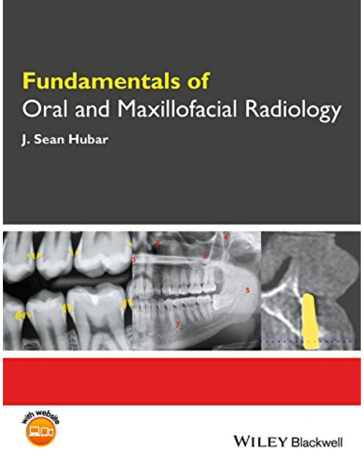
Fundamentals of Oral and Maxillofacial Radiology
Livrare gratis la comenzi peste 500 RON. Pentru celelalte comenzi livrarea este 20 RON.
Disponibilitate: La comanda in aproximativ 4-6 saptamani
Autor: J. Sean Hubar
Editura: Wiley
Limba: Engleza
Nr. pagini: 264
Coperta: Paperback
Dimensiuni: 18.29 x 1.27 x 24.13 cm
An aparitie: 4 July 2017
DESCRIPTION:
Fundamentals of Oral and Maxillofacial Radiology provides a concise overview of the principles of dental radiology, emphasizing their application to clinical practice.
Distills foundational knowledge on oral radiology in an accessible guide
Uses a succinct, easy-to-follow approach
Focuses on practical applications for radiology information and techniques
Presents summaries of the most common osseous pathologic lesions and dental anomalies
Includes companion website with figures from the book in PowerPoint and x-ray puzzles
TABLE OF CONTENTS:
Acknowledgments ix
About the Companion Website x
Part One: Fundamentals 1
A. Introduction 3
What is dental radiology? 3
What are x rays? 3
What’s the big deal about x‐ray images? 5
B. History 6
Discovery of x rays 6
Who took the world’s first “dental” radiograph? 8
Dr. C. E. Kells, Jr., a New Orleans dentist and the early days of dental radiography 8
C. Generation of X Rays 11
D. Exposure Controls 13
Voltage (V) 13
Amperage (A) 13
Exposure timer 14
E. Radiation Dosimetry 15
Exposure 15
Absorbed dose 15
Equivalent dose 15
Effective dose 16
F. Radiation Biology 17
What happens to the dental x‐ray photons that are directed at a patient? 18
Determinants of biologic damage from x‐radiation exposure 19
G. Radiation Protection 22
1. Radiation protection: Patient 22
Protective apron 23
Collimation 24
Filtration 25
Digital versus analog 26
Exposure settings 26
Operator technique 26
2. Radiation protection: Office personnel 27
How much occupational radiation exposure is permitted? 29
H. Patient Selection Criteria 30
I. Film versus Digital Imaging 32
Film 32
Digital imaging 33
Imaging software 36
J. What do Dental X‐ray Images Reveal? 38
Alterations to the dentition 38
Periodontal disease 39
Growth and development 39
Alterations to periapical tissues 40
Osseous pathology 40
Temporomandibular joint disorder 40
Implant assessment (pre‐ and post‐placement) 40
Identification of a foreign body 40
K. Intraoral Imaging Techniques 41
1. Paralleling technique 42
Maxillary incisors paralleling projection 45
Maxillary cuspid paralleling projection 45
Maxillary bicuspid paralleling projection 46
Maxillary molar paralleling projection 46
Mandibular incisor paralleling projection 47
Mandibular cuspid paralleling projection 48
Mandibular bicuspid paralleling projection 48
Mandibular molar paralleling projection 49
2. Bisecting angle technique 50
Maxillary incisor bisecting angle projection 51
Maxillary cuspid bisecting angle projection 51
Maxillary bicuspid bisecting angle projection 52
Maxillary molar bisecting angle projection 52
Mandibular incisor bisecting angle projection 53
Mandibular cuspid bisecting angle projection 53
Mandibular bicuspid bisecting angle projection 54
Mandibular molar bisecting angle projection 54
3. Bitewing technique 55
Bicuspid bitewing 56
Molar bitewing 56
Anterior bitewing projection 56
4. Distal oblique technique 57
5. Occlusal imaging technique 58
Maxillary occlusal projection 59
Mandibular occlusal projection 60
L. Intraoral Technique Errors 61
Cone‐cut 61
Apex missing 62
Elongation 63
Foreshortening 63
Overlapped contacts 64
Missing contacts 64
Overexposure and underexposure 65
Motion artifact 66
Foreign object 66
M. Extraoral Imaging Techniques 68
1. Panoramic imaging 68
Positioning the patient 69
Exposure settings 71
Advantages and disadvantages 71
Technique errors 74
Anatomic landmarks 84
2. Lateral cephalograph imaging 85
3. Cone beam computed tomography 86
Introduction 86
Anatomic landmarks 89
N. Quality Assurance 96
O. Infection Control 97
Excerpt from “CDC
Guidelines for Infection Control in Dental Health‐Care Settings” 97
General instructions for cleaning and disinfecting a solid‐state receptor (courtesy of Sirona™) 98
P. Occupational Radiation Exposure Monitoring 100
Q. Hand‐held X‐ray Systems 102
Dental radiographic examinations: recommendations for patient selection and limiting radiation exposure 102
Commentary 102
Part Two: Interpretation 105
R. Localization of Objects (SLOB Rule) 107
S. Recommendations for Interpreting Images 111
T. X‐ray Puzzles: Spot the Differences 113
U. Radiographic Anatomy 124
1. Dental anatomy 124
2. Anatomic landmarks of the maxillary region 126
Radiopaque landmarks 126
Radiolucent landmarks 129
3. Anatomic landmarks of the mandibular region 133
Radiopaque landmarks 133
Radiolucent landmarks 136
V. Dental Caries 141
Limitations to visualizing caries on x‐ray images 141
Classification of caries 143
W. Dental Anomalies 149
Number 149
Size 149
Shape 151
Developmental factors 157
Environmental factors 161
X. Osseous Pathology (Alphabetic) 170
Y. Lagniappe (Miscellaneous Oddities) 188
Part Three: Appendices 195
Appendix 1: FDA Recommendations for Prescribing Dental X‐ray Images 197
Appendix 2: X‐radiation Concerns of Patients: Question and Answer Format 200
1. How often should I get x rays taken? 200
2. How much radiation am I receiving from dental x rays? 200
3. Can I get cancer from dental x rays? 201
4. Why do I need to wear a protective apron for dental x rays and why does the assistant leave the room before taking my x rays, if dental x rays are so safe? 201
5. Your protective apron does not have a thyroid collar, why not? 201
6. I am pregnant, should I get dental x rays taken? 201
7. When should my child first get dental x rays taken? 201
8. Will I glow in the dark after all of the x rays that I received at the dental office? 202
9. What are 3‐D x rays? 202
10. Why does the dentist require additional 3‐D x rays before placing my dental implant? 202
Appendix 3: Helpful Tips for Difficult Patients 203
1. Hypersensitive gag reflex 203
2. Small mouth/shallow palate/ constricted arch/torus 204
3. Large frenulum 205
4. Trismus 205
5. Cuspid superimposition 205
6. Rubber dam 206
7. Third molar imaging 206
Appendix 4: Deficiencies of X‐ray Imaging Terminology 207
Survey results 207
Appendix 5: Tools for Differential Diagnosis 210
1. Number 210
2. Location 210
3. Density 211
4. Shape 211
5. Size 211
6. Borders 212
7. Changes to surrounding anatomic structures 212
Appendix 6: Table of Radiation Units 213
Appendix 7: Table of Anatomic Landmarks 214
Tooth 214
Tooth‐related structures 214
Landmarks associated with the maxilla 214
Landmarks associated with the mandible 214
Appendix 8: Table of Dental Anomalies 216
Number 216
Size 216
Shape 216
Developmental defects 216
Environmental effects 216
Appendix 9: Table of Osseous Pathology 217
Radiolucent anomalies in the maxilla and mandible 217
Radiopaque anomalies in the maxilla and mandible 217
Mixed (radiolucent–radiopaque) anomalies in the maxilla and mandible 218
Appendix 10: Common Abbreviations and Acronyms 219
Appendix 11: Glossary of Terms 221
Suggested Reading 238
Index 251
| An aparitie | 4 July 2017 |
| Autor | J. Sean Hubar |
| Dimensiuni | 18.29 x 1.27 x 24.13 cm |
| Editura | Wiley |
| Format | Paperback |
| ISBN | 9781119122210 |
| Limba | Engleza |
| Nr pag | 264 |
-
1,37400 lei 1,30600 lei






Clientii ebookshop.ro nu au adaugat inca opinii pentru acest produs. Fii primul care adauga o parere, folosind formularul de mai jos.