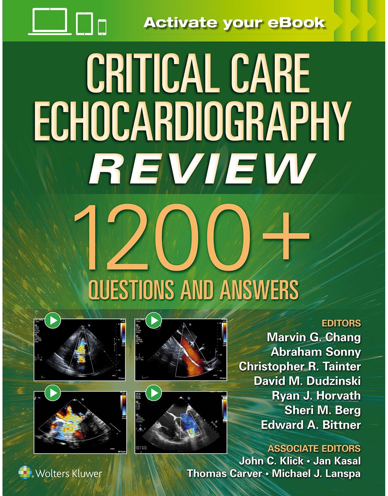
Critical Care Echocardiography Review: 1200+ Questions and Answers: Print + Digital Version with Multimedia
Livrare gratis la comenzi peste 500 RON. Pentru celelalte comenzi livrarea este 20 RON.
Disponibilitate: La comanda in aproximativ 4-6 saptamani
Editura: LWW
Limba: Engleza
Nr. pagini: 1088
Coperta: Paperback
Dimensiuni: 213 x 276 mm
An aparitie: 25/01/2022
Description:
Prepare for success on the Examination of Special Competence in Critical Care Echocardiography (CCEeXAM)! Critical Care Echocardiography Review is a first-of-its-kind, review textbook containing over 1,200 questions and answers. Helmed by Drs. Marvin G. Chang, Abraham Sonny, David Dudzinski, Christopher R. Tainter, Ryan J. Horvath, Sheri M. Berg, Edward A. Bittner as well as a team of associated editors and authors from institutions across the nation , this highly visual resource covers every aspect of the use of ultrasound for clinical diagnosis and management in the critical care setting, providing a thorough, effective review and helping you identify areas of mastery and those needing further study.
Enrich Your Digital Reading Experience
- Read directly on your preferred device(s), such as computer, tablet, or smartphone.
- Easily convert to audiobook, powering your content with natural language text-to-speech.
,
Table of Contents:
Interactive Presentation
1. Basic Ultrasound Wave Properties
2. Pulsed-Wave vs. Continuous-Wave Doppler
3. Ultrasound Propagation Through Tissue
4. Ultrasound Transducer and System
5. Ultrasound Modes
6. Longitudinal, Lateral, and Temporal Resolution
7. Doppler Effect and Principles
8. Physics of Ultrasound Bioeffects
9. Patient and Ultrasound Machine Positioning, Probe Selection and Orientation, Proper Ultrasound Care
10. Optimizing Probe Position and Knobology for Image Acquisition
11. Knobology, Probe Positioning, and Concepts of Image Acquisition
12. Transthoracic Echocardiography (TTE) and Transesophageal Echocardiography (TEE) Views: TTE Versus TEE
13. Three-Dimensional Echocardiography
14. Physics of Artifacts
15. Types of Artifacts
16. Ultrasound Artifacts Versus Pathological and Normal Anatomical Variants
17. Doppler Shift Principles
18. Pulsed Wave Doppler, Continuous Wave Doppler, and Tissue Doppler Imaging
19. Hemodynamic Calculations
20. Quantification Calculations
21. Quantification of Diastolic Function
22. Left Ventricular Systolic Function
23. Right Ventricular Function and Pulmonary Hypertension
24. Aortic Valvular Disease
25. Mitral Valvular Disease
26. Tricuspid Valvular Disease
27. Pulmonary Valve Diseases
28. Mechanical and Bioprosthetic Valves
29. Endocarditis and Other Pathologic and Normal Anatomic Variants
30. Left Atrial and Right Atrial Size, Function, and Pathology
31. Pericardial Disease
32. Aortic and Other Great Vessel Diseases
33. Ischemic/Nonischemic Cardiomyopathies and Congenital Heart Disease
34. Myocardial Ischemia, Infarction, and Wall Motion Abnormalities
35. Diastology
36. Mechanical Circulatory Support
37. Intracardiac Shunts
38. Intracardiac Masses, Abnormal Structures, Normal Anatomic Variants, and Artifacts
39. Transthoracic Echocardiogram Versus Transesophageal Echocardiogram
40. Obstructive Shock
41. Hypovolemic shock
42. Distributive, Vasodilatory, Neurogenic, and Septic
43. Cardiogenic Shock
44. Various Protocols: FATE, FEEL, FEER, FAST
45. Cardiac Arrest and Peri-Arrest
46. E-FAST
47. Hypoxemia
48. Mechanical Circulatory Support and Cannulation Strategies
49. Predicting and Measuring Fluid Responsiveness
50. Clinical Applications of Diastology
51. Echocardiography in Assessment and Management of Acute and Chronic Right Heart Failure
52. Pneumothorax
53. Cardiogenic Pulmonary Edema, Noncardiogenic Pulmonary Edema (ARDS), Diffuse Parenchymal Lung Disease (Interstitial Pneumonitis)
54. Pulmonary Consolidations
55. Pleural Effusions
56. Differentiating Pulmonary Edema, Pneumonia, COPD/Asthma, Bronchiolitis, ARDS, Pulmonary Embolism
57. Esophageal Versus Endotracheal Intubation
58. Optimization and Facilitation of Weaning from the Ventilator
59. Other Causes of Hypoxemia
60. Trauma Ultrasound and E-FAST Examination
61. Liver and Gallbladder
62. Kidney
63. Detection of Free Fluid and Air
64. Small Bowel Obstruction, Perforation, and Other Bowel Pathology
65. Bladder Ultrasound
66. Obstetrics and Gynecology
67. Abdominal Vascular Ultrasound
68. Gastric Ultrasound
69. Abdominal Emergencies
70. Deep Vein Thrombosis
71. Pseudoaneurysm
72. Aneurysm
73. Aortic Dissection
74. Invasive Line Placement: Central Line, Arterial Line, and Peripheral IV Placement
75. Thoracentesis and Chest Tube Placement
76. Paracentesis
77. Pericardiocentesis
78. Endotracheal Intubation
79. Abscess Evaluation and Drainage
80. Lumbar Puncture
81. Transcranial Doppler
Static Presentation
I. Physics of Ultrasound
1. Basic Ultrasound Wave Properties
2. Pulsed-Wave vs. Continuous-Wave Doppler
3. Ultrasound Propagation Through Tissue
4. Ultrasound Transducer and System
5. Ultrasound Modes
6. Longitudinal, Lateral, and Temporal Resolution
7. Doppler Effect and Principles
8. Physics of Ultrasound Bioeffects
II. Image Acquisition, Optimization, Knobology, and Ultrasound Care
9. Patient and Ultrasound Machine Positioning, Probe Selection and Orientation, Proper Ultrasound Care
10. Optimizing Probe Position and Knobology for Image Acquisition
11. Knobology, Probe Positioning, and Concepts of Image Acquisition
12. Transthoracic Echocardiography (TTE) and Transesophageal Echocardiography (TEE) Views: TTE Versus TEE
13. Three-Dimensional Echocardiography
III. Artifacts
14. Physics of Artifacts
15. Types of Artifacts
16. Ultrasound Artifacts Versus Pathological and Normal Anatomical Variants
IV. Quantification and Hemodynamic Calculations
17. Doppler Shift Principles
18. Pulsed Wave Doppler, Continuous Wave Doppler, and Tissue Doppler Imaging
19. Hemodynamic Calculations
20. Quantification Calculations
21. Quantification of Diastolic Function
V. Cardiac
22. Left Ventricular Systolic Function
23. Right Ventricular Function and Pulmonary Hypertension
24. Aortic Valvular Disease
25. Mitral Valvular Disease
26. Tricuspid Valvular Disease
27. Pulmonary Valve Diseases
28. Mechanical and Bioprosthetic Valves
29. Endocarditis and Other Pathologic and Normal Anatomic Variants
30. Left Atrial and Right Atrial Size, Function, and Pathology
31. Pericardial Disease
32. Aortic and Other Great Vessel Diseases
33. Ischemic/Nonischemic Cardiomyopathies and Congenital Heart Disease
34. Myocardial Ischemia, Infarction, and Wall Motion Abnormalities
35. Diastology
36. Mechanical Circulatory Support
37. Intracardiac Shunts
38. Intracardiac Masses, Abnormal Structures, Normal Anatomic Variants, and Artifacts
39. Transthoracic Echocardiogram Versus Transesophageal Echocardiogram
VI. Differentiating Shock
40. Obstructive Shock
41. Hypovolemic shock
42. Distributive, Vasodilatory, Neurogenic, and Septic
43. Cardiogenic Shock
44. Various Protocols: FATE, FEEL, FEER, FAST
VII. Rescue Echo
45. Cardiac Arrest and Peri-Arrest
46. E-FAST
47. Hypoxemia
48. Mechanical Circulatory Support and Cannulation Strategies
VIII. Predicting and Measuring Fluid Responsiveness
49. Predicting and Measuring Fluid Responsiveness
IX. Clinical Applications of Diastology
50. Clinical Applications of Diastology
X. Pulmonary Hypertension and Cor Pulmonale
51. Echocardiography in Assessment and Management of Acute and Chronic Right Heart Failure
XI. Lung and Pleural Ultrasound
52. Pneumothorax
53. Cardiogenic Pulmonary Edema, Noncardiogenic Pulmonary Edema (ARDS), Diffuse Parenchymal Lung Disease (Interstitial Pneumonitis)
54. Pulmonary Consolidations
55. Pleural Effusions
56. Differentiating Pulmonary Edema, Pneumonia, COPD/Asthma, Bronchiolitis, ARDS, Pulmonary Embolism
57. Esophageal Versus Endotracheal Intubation
58. Optimization and Facilitation of Weaning from the Ventilator
59. Other Causes of Hypoxemia
XII. Trauma Ultrasound and E-FAST Exam
60. Trauma Ultrasound and E-FAST Examination
XIII. Abdominal Ultrasound
61. Liver and Gallbladder
62. Kidney
63. Detection of Free Fluid and Air
64. Small Bowel Obstruction, Perforation, and Other Bowel Pathology
65. Bladder Ultrasound
66. Obstetrics and Gynecology
67. Abdominal Vascular Ultrasound
68. Gastric Ultrasound
69. Abdominal Emergencies
XIV. Vascular Ultrasound
70. Deep Vein Thrombosis
71. Pseudoaneurysm
72. Aneurysm
73. Aortic Dissection
XV. Procedures
74. Invasive Line Placement: Central Line, Arterial Line, and Peripheral IV Placement
75. Thoracentesis and Chest Tube Placement
76. Paracentesis
77. Pericardiocentesis
78. Endotracheal Intubation
79. Abscess Evaluation and Drainage
80. Lumbar Puncture
81. Transcranial Doppler
Appendix
Index
| An aparitie | 25/01/2022 |
| Autor | Marvin G. Chang, Abraham Sonny MD, FASE, David Dudzinski, Christopher R. Tainter, Ryan J. Horvath MD, PhD, Sheri M. Berg MD, Edward A Bittner |
| Dimensiuni | 213 x 276 mm |
| Editura | LWW |
| Format | Paperback |
| ISBN | 9781975144135 |
| Limba | Engleza |
| Nr pag | 1088 |
| Versiune digitala | DA |
-
1,06300 lei 93000 lei
-
1,18700 lei 98300 lei


Clientii ebookshop.ro nu au adaugat inca opinii pentru acest produs. Fii primul care adauga o parere, folosind formularul de mai jos.