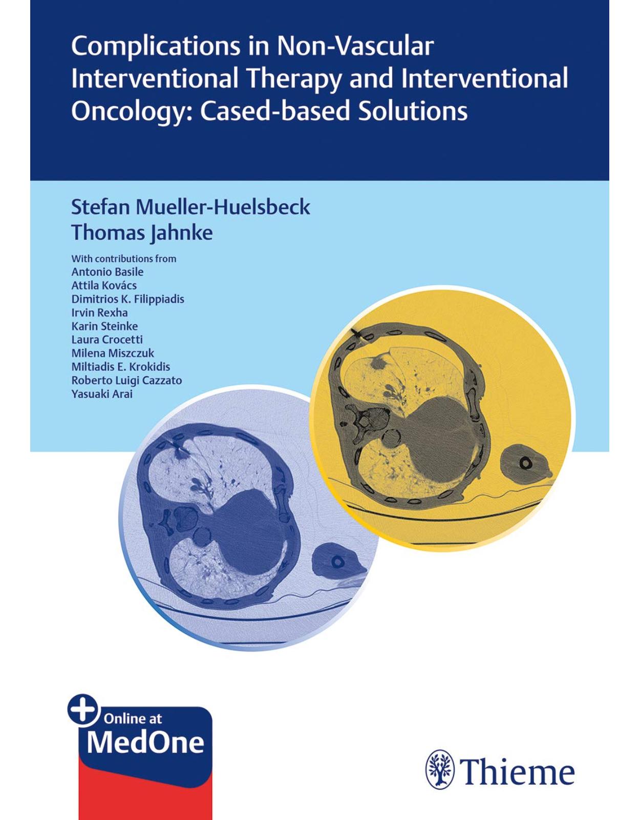
Complications in Non-vascular Interventional Therapy and Interventional Oncology: Case-based Solutions
Livrare gratis la comenzi peste 500 RON. Pentru celelalte comenzi livrarea este 20 RON.
Disponibilitate: La comanda in aproximativ 4-6 saptamani
Autor: Mueller-Huelsbeck; Jahnke
Editura: Thieme
Limba: Engleza
Nr. pagini: 150
Coperta: Hardcover
Dimensiuni: 17.53 x 1.52 x 24.38 cm
An aparitie: 2019
Description:
Learn to avoid and manage complications from non-vascular interventional and interventional oncological procedures
The range of non-vascular procedures that can be performed in interventional imaging is vast and includes management of a wide range of conditions, such as treatment of kidney stones, obtaining diagnostic biopsies in suspected cancers, bile duct occlusions, compression fractures, drainage of abscesses, collection of fluids, etc. In particular, various cancers often lend themselves well to local tumor destruction with interventional techniques, while holding morbidity and mortality to a minimum.
This compendium presents 45 cases in detail to provide a thorough review of potential complications that may occur during non-vascular interventional radiology and interventional oncological procedures. Each case also includes a list of take-home messages discussing vital prevention strategies for each problem.
Key Features:
Content presented in case-based format to help the reader benefit from the real-life experiences of the authors and motivate them to take part in identifying the problem and finding a solution to a specific situation
Solid coverage of characteristic complications of special technologies, such as thermal ablation and percutaneous CT-guided interstitial high-dose brachytherapy
A wealth of information and advice for optimizing patient safety before, during, and after interventional therapy
Take-home messages at the end of each case providing vital prevention strategies
Complications in Non-vascular Interventional Therapy and Interventional Oncology: Case-based Solutions is an invaluable sourcebook for radiology residents and fellows, experienced interventional radiologists, and all physicians actively performing non-vascular and oncological interventions. This book discusses methods to both avoid and manage complications, thus having the potential to directly enhance patient care.
Table of Contents:
1 Introduction
2 Minor and Major Complications
2.1 Definition and Reporting System of Complications
2.2 Avoiding Complications
2.2.1 Patient Safety
2.2.2 Patient Safety Checklist
2.2.3 Periprocedural Documentation
2.3 General Complications Related to Non-vascular and Oncologic Procedures
2.3.1 Impaired Renal Function
2.4 Known Allergic Reactions to Contrast Material
2.5 Radiation Exposure
2.6 Infection
2.7 Management of Complications
2.7.1 Arterial Hemorrhage
2.7.2 Preventing Arterial Hemorrhage
2.7.3 Device Malfunction
2.7.4 Preventing Device Malfunction
3 Case-Based Procedure-Related Complications
3.1 Bleeding
3.1.1 Bleeding after Percutaneous Biopsy of Liver Tumor
3.1.2 Hemothorax during Electroporation for Hepatocellular Carcinoma Treatment
3.1.3 Cervical Hematoma after Thyroid Fine Needle Aspiration Biopsy
3.1.4 Hepatic Intraparenchymal Hemorrhage after CT-Guided Liver Biopsy
3.1.5 Hemodynamic Instability, Presumed to be Related to Worsening Retroperitoneal Hemorrhage during and after Cryoablation for Renal Tumor Treatment
3.1.6 Mediastinal Hemorrhage and Hemothorax after Anterior Mediastinal Puncture
3.1.7 Hemoptysis after Percutaneous Lung Biopsy
3.1.8 Delayed Bleeding after Biliary Drainage
3.1.9 Bleeding during Diagnostic CT-Guided Liver Puncture
3.1.10 Bleeding after Radiofrequency Ablation for Hepatocellular Carcinoma Treatment
3.1.11 Massive Pleural Hemorrhage after Lung Radiofrequency Ablation
3.1.12 Delayed Bleeding after Microwave Ablation for a Recurrent Colorectal Liver Metastasis
3.2 Cement Extravasation
3.2.1 Pulmonary Cement Embolization after Vertebroplasty for Lumbar Fracture Treatment
3.2.2 Endplate Cement Extravasation after Balloon Kyphoplasty for Treatment of Osteoporotic Fracture
3.2.3 Intra-articular Cement Leakage after Bone Augmentation in the Peripheral Skeleton
3.3 Device Failure
3.3.1 Two Cases of Short Antenna during Microwave Ablation for Treatment of Lung Nodules
3.3.2 Antenna Fracture during Microwave Ablation for Treatment of Non-Small-Cell Lung Cancer
3.3.3 Thermal Ablation: Cutting Off the Leg of a Radiofrequency Ablation Device during a Simultaneous Biopsy
3.4 Infection
3.4.1 Hepatic Abscess after Transarterial Chemoembolization for Hepatocellular Carcinoma Treatment
3.4.2 Liver Abscess after Transarterial Chemoembolization for Hepatocellular Carcinoma Treatment
3.4.3 Injury of the Liver and Biliary System after Drug Eluting Beads Transcatheter Intra-arterial Chemoembolization
3.5 Non-vascular Miscellaneous Cases
3.5.1 Renal Defect after Cryoablation of Renal Tumor
3.5.2 A Bronchial Fistula Following Percutaneous Lung Microwave Ablation
3.5.3 Lethal Hepatocellular Tumor Rupture after Incomplete Chemoembolization: What Went Wrong?
3.5.4 Interstitial Pneumonitis after Microwave Ablation for Metastatic Lesion Treatment
3.5.5 Insufficiency Fracture Following Bone Cryoablation
3.5.6 Postablation Biloma with Further Sequelae after Percutaneous Microwave Ablation for Treatment of Recurrent Metastasis
3.5.7 Hip Joint Destruction following Radiofrequency Ablation and Cementoplasty of an Adjacent Bone Metastasis
3.5.8 Calyceal Leakage after Renal Biopsy
3.5.9 Hemopericardium Following Malplacement of a Radiofrequency Ablation Electrode during Thermal Ablation of the Liver—A Potentially Fatal Complication
3.5.10 Infection of the Ablation Cave after Electrochemotherapy of a Colorectal Liver Metastasis
3.6 Vascular Miscellaneous Cases
3.6.1 Arterioportal Fistula Following Microwave Ablation of Subcentimeter Liver Metastasis from Sigmoid Adenocarcinoma
3.6.2 Arteriovenous Fistula after Diagnostic Renal Puncture
3.6.3 Laceration of the Left Hepatic Artery during Biliary Drainage
3.6.4 Renal Artery Pseudoaneurysm Post Percutaneous Kidney Biopsy
3.7 Neurologic Event
3.7.1 Complete Motor Deficit of the Lower Limbs during Celiac Plexus Neurolysis
3.7.2 Nontarget Embolization during Transarterial Chemoembolization for Hepatocellular Carcinoma Treatment: Be Aware of the Arteria Radicularis Magna
3.8 Pneumothorax
3.8.1 Delayed Pneumothorax after Lung Biopsy
3.8.2 Pneumothorax during Microwave Ablation for Treatment of a Single Pulmonary Metastasis
3.8.3 Pneumothorax during Diagnostic CT-Guided Lung Puncture
3.8.4 Intraprocedural Pneumothorax after Biopsy before Microwave Ablation
3.8.5 Pneumothorax after Percutaneous Lung Interstitial Brachytherapy in Solitary Colorectal Adenocarcinoma Metastasis
3.9 Skin Burn
3.9.1 Skin Burn after Radiofrequency Ablation of Lung Nodule
3.9.2 Burned Skin after Radiofrequency Ablation for Osteoid Osteoma Treatment
3.9.3 Skin Burn after Dislocation of a Microwave Ablation Electrode during Ablation of Liver Metastases in Coaxial Technique
Index
| An aparitie | 2019 |
| Autor | Mueller-Huelsbeck; Jahnke |
| Dimensiuni | 17.53 x 1.52 x 24.38 cm |
| Editura | Thieme |
| Format | Hardcover |
| ISBN | 9783132412873 |
| Limba | Engleza |
| Nr pag | 150 |








Clientii ebookshop.ro nu au adaugat inca opinii pentru acest produs. Fii primul care adauga o parere, folosind formularul de mai jos.