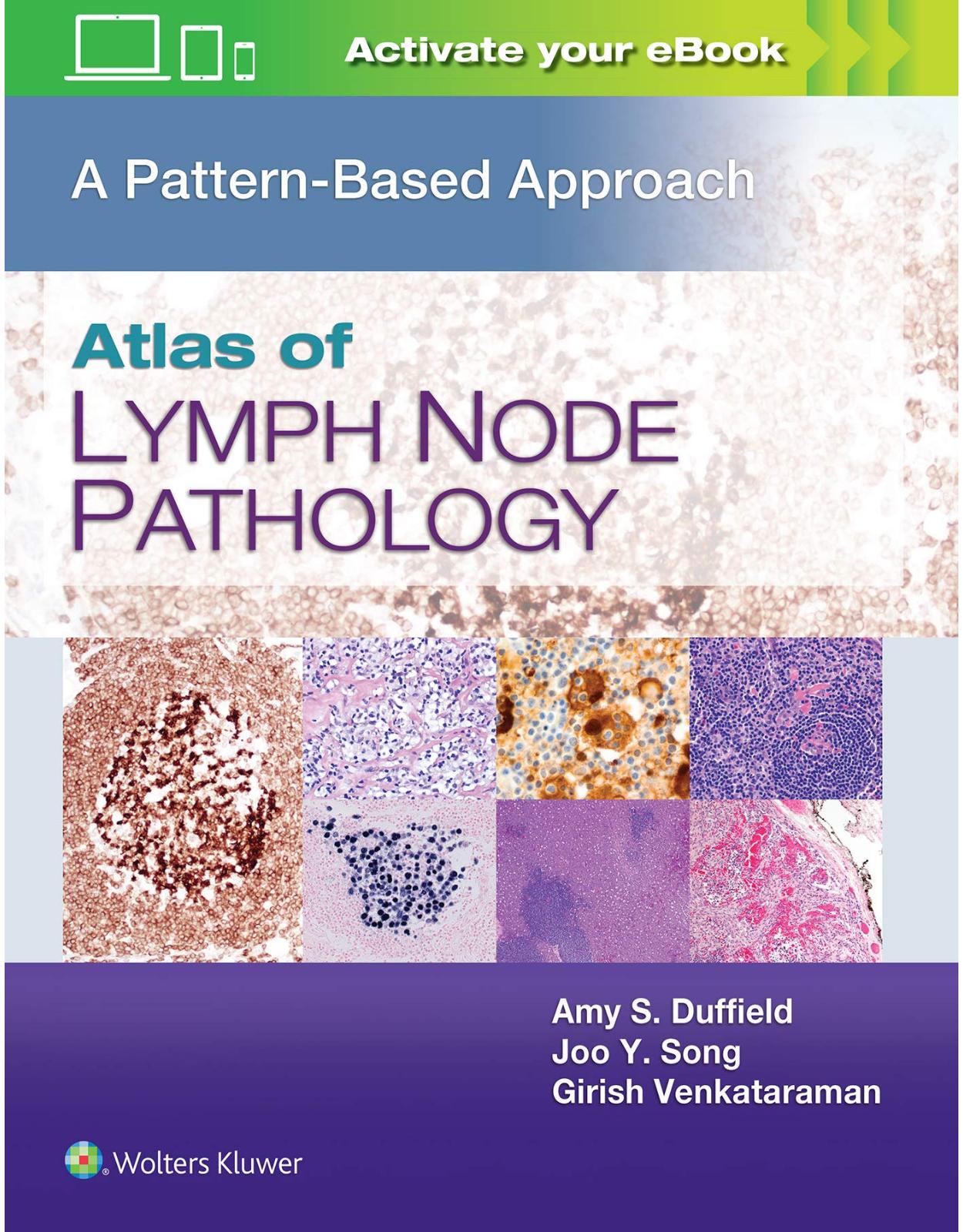
Atlas of Lymph Node Pathology: A Pattern Based Approach
Livrare gratis la comenzi peste 500 RON. Pentru celelalte comenzi livrarea este 20 RON.
Disponibilitate: La comanda in aproximativ 4-6 saptamani
Editura: LWW
Limba: Engleza
Nr. pagini: 320
Coperta: Hardcover
Dimensiuni: 21.59 x 1.78 x 28.19 cm
An aparitie: 1 Jan. 2021
Description:
Closely mirroring the daily sign-out process, Atlas of Lymph Node Pathology: A Pattern Based Approach is a highly illustrated, efficient guide to accurate diagnosis. This practical reference uses a proven, pattern-based approach to clearly explain how to interpret challenging cases by highlighting red flags in the clinical chart and locating hidden clues in the slides. Useful as a daily “scope-side guide,” it features numerous clinical and educational features that help you find pertinent information, reach a correct diagnosis, and assemble a thorough and streamlined pathology report.
Table of Contents:
1. INTRODUCTION TO THE LYMPH NODE
Lymph Node Structure
Capsule
Sinuses
Cortex
Paracortex
Medulla
Specimen Preparation
Fixation
Frozen Sections
Limited Specimens
Clinical Context
Anatomic Site
Patient Demographics
Clinical Information
Ancillary Studies
Flow Cytometry
Cytogenetic/FISH Studies
Molecular Studies
Near Misses
IgG4-Reactive Lymphadenopathy
Nodal Marginal Zone B-Cell Lymphoma With Colonization of Reactive Follicles
Intrafollicular Neoplasia
Interfollicular Classical Hodgkin Lymphoma
Early Angioimmunoblastic T-cell Lymphoma
2. THE LYMPH NODE CAPSULE
Normal Lymph Node Capsule
Absent Capsule
Lymph Node Versus Lymphoid Tissue
Capsular Inclusions
Disrupted Capsule
Thickened Capsule
Reactive Conditions
Neoplastic Conditions
Near Misses
Angioimmunoblastic T-cell Lymphoma
Kaposi Sarcoma
Tumor-Infiltrating Lymphocytes Versus Metastatic Disease
Capsular Nevi Versus Metastatic Melanoma
3. THE LYMPHATIC SINUSES
Introduction
Conditions With Dilated Prominent Sinuses
Sinus Histiocytosis and Dermatopathic Lymphadenopathy
Sinus Histiocytosis With Massive Lymphadenopathy (SHML)
Langerhans Cell Histiocytosis (LCH)
Lymphoplasmacytic Lymphoma
Node Draining Suppurative Area
Lipid-Associated Lymphadenopathy (Lymphangiogram, Prosthesis, or Storage Diseases)
Vascular Transformation of Sinuses
Near Miss
Sinusoidal Involvement by Kaposi Sarcoma Mimicking Vascular Transformation of Sinuses
Sinusoidal Lymphomatous Infiltrates
Near Miss
ALCL Mimicking Metastatic Carcinoma
Metastatic Cancer
Near Miss
Benign Mesothelial Cells
Leukemic Infiltrates
Other Perisinusoidal Cellular Clusters
Conditions With Inconspicuous/Obliterated Sinuses
Follicular Lymphoma
Castleman Disease
Angioimmunoblastic T-cell Lymphoma
4. CORTEX
Introduction
Clinical Correlation
Site-Specific Variations in Follicles
Abnormal Follicles
Reactive Follicular Hyperplasia
Giant Follicular Hyperplasia
Reactive Follicular Hyperplasia In Altered Immune States
Intrafollicular Plasmacytosis-IgG4 Disease
Other Clonal Conditions With Reactive Hyperplasia
Abnormal Expanded Mantle Zones
Castleman-Like Proliferations
Progressive Transformation of Germinal Centers
Kimura Disease
Attenuated Mantle Zones
Other Infectious Processes
Toxoplasma
5. PARACORTEX
Paracortical Hyperplasia
Dermatopathic Lymphadenopathy
Infection
Drug-Associated Lymphadenopathy
Autoimmune Conditions
Systemic Lupus Erythematosus Lymphadenopathy
Atypical Paracortical Hyperplasia
Histiocytic Proliferations
Singly/Small Clusters
Granulomas
Extensive/Diffuse
Vascular Changes
Vascular Proliferation
Abnormalities of Vessel Walls
Expansion/Infiltration of the Paracortex by Unexpected Cells
Atypical Lymphocytes/Lymphomas
Plasma Cells
Spindled Cells
Necrosis
Near Misses
Hamazaki-Wesenberg Bodies in Sarcoidosis Lymphadenopathy
Indolent T-Lymphoblastic Proliferation
Lymph Node Involvement by Myeloid/Lymphoid Neoplasms With PDGFRA Rearrangement
6. OBLITERATED NODULAR PATTERN
Introduction
List of Entities to Consider in Cases With Nodular Pattern
Follicular lymphoma
Some Histologic Variations That Represent Pitfalls in the Diagnosis of Follicular Lymphoma
Grading and Follicular Lymphoma
Unconventional Wisdom Relating to FL Grading
Immunostains and Approach in Nodular Proliferations
Scenarios and Clinical Details That Determine How You Look at and Sign Out Some Follicular Lymphoma Cases
Pitfall Cases of Nodular Proliferations
Pitfall Case 1: Nodal Marginal Zone lymphoma with Follicular Colonization
Pitfall Case 2: Germinotropic lymphoproliferative disorder, EBV+/ HHV8+
Pitfall Case 3: Follicular variant of peripheral T-cell lymphoma (Figures 6.66-6.70)
7. OBLITERATED NODAL ARCHITECTURE
B-Cell Lymphomas
Small Lymphocytic Lymphoma and Richter Syndrome
Mantle Cell Lymphoma
Diffuse Follicular Lymphoma
Follicular Lymphoma With Transformation to DLBCL
Plasmacytoma/Plasma Cell Myeloma
Diffuse Large B-Cell Lymphoma, Not Otherwise Specified
T-Cell–/Histiocyte-Rich Large B-Cell Lymphoma
Plasmablastic Lymphoma
Primary Mediastinal Large B-Cell Lymphoma
B-Cell Lymphoma, Unclassifiable, With Features Intermediate Between Diffuse Large B-Cell Lymphoma and Classical Hodgkin Lymphoma (Gray Zone Lymphoma)
Burkitt Lymphoma
High-Grade B-Cell Lymphoma
T-Cell Lymphomas
Anaplastic Large-Cell Lymphoma
Peripheral T-Cell Lymphoma
Lymphoepithelioid Variant of PTCL (Lennert Lymphoma)
Precursor Lesion
Myeloid Sarcoma
B-Lymphoblastic Lymphoma
T-Lymphoblastic Lymphoma
Near Misses
Blastoid Variant of Mantle Cell Lymphoma
Nodal Involvement by CD30-Positive T-Cell Lymphoproliferative Disorder
Peripheral T-Cell Lymphoma With Hodgkin-Like Cells
Anaplastic Large-Cell Lymphoma With Aberrant Expression of PAX5
8. NECROSIS
Introduction
Lymphoma/Aggressive Lymphoproliferative Neoplasms
B-Cell Lymphoma
B Lymphoblastic Lymphoma
Diffuse Large B-Cell Lymphoma, Not Otherwise Specified
EBV-Positive Diffuse Large B-Cell Lymphoma, Not Otherwise Specified
Post-Transplant Lymphoproliferative Disorder
Plasmablastic Lymphoma
Classical Hodgkin Lymphoma
T-Cell and NK Cell Lymphoma
Peripheral T-Cell Lymphoma, Not Otherwise Specified
NK/T-Cell Lymphoma
Anaplastic Large Cell Lymphoma
Infectious Etiologies
Mycobacterial Lymphadenitis
Cytomegalovirus Infection
Herpes Simplex Lymphadenitis
Cat Scratch Disease
Fungal Infection/Lymphadenitis
Benign Reactive Lymphoproliferative Disorders
Systemic Lupus Erythematosus
Near Misses
Epstein-Barr Virus: Infectious Mononucleosis
Kikuchi-Fujimoto Lymphadenitis
9. IMMUNOHISTOCHEMISTRY
Introduction
Key Features Related to Technical Points in Immunohistochemistry
List of Common Antibodies in Lymphoma Immunohistochemistry
Near Miss
CD20-Negative Follicular Lymphoma
Incidental Mantle Cell Lymphoma in Reactive Looking Node
Peripheral T-cell Lymphoma With Hodgkin-Like Cells of B-Cell Derivation
Stains Related to Microorganisms
Extracavitary Primary Effusion Lymphoma
Exercise in Interpretation, Sample Write-Ups in One Single Case, and Best Practices in Write-Ups
SELF-ASSESSMENT QUESTIONS
INDEX
| An aparitie | 1 Jan. 2021 |
| Autor | Amy S. Duffield MD, Joo Y. Song MD, Girish Venkataraman MD |
| Dimensiuni | 21.59 x 1.78 x 28.19 cm |
| Editura | LWW |
| Format | Hardcover |
| ISBN | 9781496375544 |
| Limba | Engleza |
| Nr pag | 320 |
-
1,07600 lei 92500 lei











Clientii ebookshop.ro nu au adaugat inca opinii pentru acest produs. Fii primul care adauga o parere, folosind formularul de mai jos.