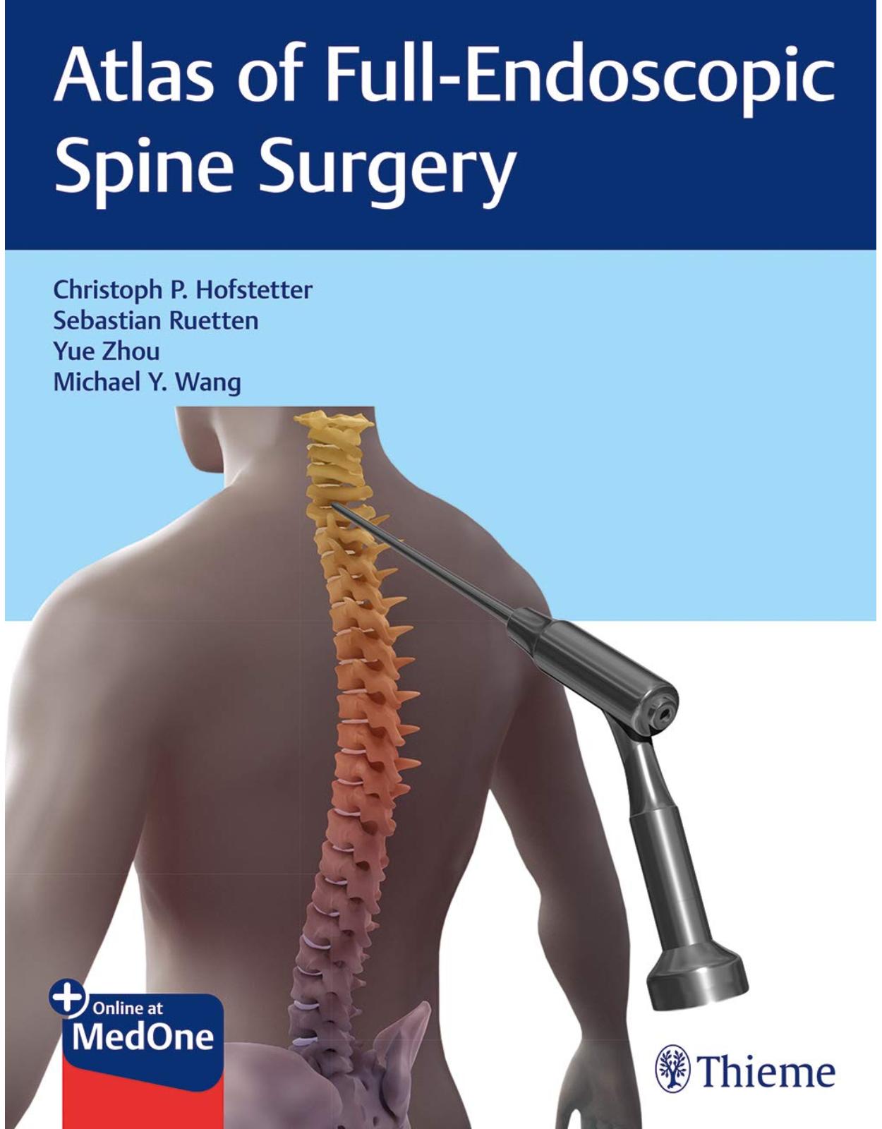
Atlas of Full-Endoscopic Spine Surgery
Livrare gratis la comenzi peste 500 RON. Pentru celelalte comenzi livrarea este 20 RON.
Disponibilitate: La comanda in aproximativ 4-6 saptamani
Editura: Thieme
Limba: Engleza
Nr. pagini: 320
Coperta: Paperback
Dimensiuni: 27.94 x 21.59 cm
An aparitie: 13 Dec. 2019
Description:
Endoscopic spine surgery essentials from expert spine surgeons
Atlas of Full-Endoscopic Spine Surgery by internationally renowned spine surgeons Christoph Hofstetter, Sebastian Ruetten, Yue Zhou, and Michael Wang provides concise, step-by-step guidance on the latest full endoscopic spine procedures. The book is targeted at practicing spine surgeons, fellows, and residents currently not trained in endoscopic spine surgery who have the desire to learn and incorporate these techniques into clinical practice. It is also an excellent curriculum resource for cadaveric training courses taught at the national and international level.
The book lays a solid foundation with opening chapters on anesthesia, OR setup and endoscopic tools, applied anatomy, basic endoscopic surgical tasks, and preoperative diagnostics. Additional sections include step-by-step descriptions of the full spectrum of cervical, thoracic, and lumbar endoscopic approaches. The last section provides invaluable pearls on overcoming challenges, avoiding pitfalls, and optimizing postoperative care.
Key Features:
Transforaminal endoscopic lumbar and thoracic discectomy approaches
Trans-SAP endoscopic approach for foraminal and lateral recess decompression
Interlaminar endoscopic lumbar discectomy
Cervical/thoracic and lumbar unilateral laminotomy for bilateral decompression
Special topics including endoscopic management of challenging cases, endoscopic revision surgery, and management of complications.
Neurosurgery residents, fellows, young practicing neurosurgeons, and all healthcare practitioners involved in the care of endoscopic spine surgery patients will gain invaluable insights from this book.
Table of Contents:
Part 1: Introduction
1 Anesthesia and Rapid Recovery
1.1 Introduction
1.2 General Principles of Rapid Recovery
1.3 Key Components
1.4 Anesthesia Protocol
1.5 Summary
2 Radiation Safety in Full-Endoscopic Spine Surgery
2.1 Introduction
2.2 Occupational Radiation Guidelines
2.3 Radiation Safety
2.4 ALARA (As Low As Reasonably Achievable) Principle
2.4.1 Restrict the Area and Duration of Exposure
2.4.2 Get as Much Distance to the Radiation Source as Achievable
2.4.3 Protect Yourself against Radiation Exposure
2.5 Emerging Technologies
2.6 Conclusion
3 Essential Imaging in Full-Endoscopic Spine Surgery
3.1 Introduction
3.2 Features of Intraoperative Fluoroscopy
3.2.1 Magnification
3.2.2 Distortion
3.2.3 Parallax
3.3 Fundamental Fluoroscopic Images for Full-Endoscopic Spine Surgery
3.3.1 Lumbar Spine
3.3.2 Interlaminar Technique
3.3.3 Transforaminal Technique
3.4 Thoracic Spine
3.5 Cervical Spine
3.6 Conclusion
4 Intraoperative Navigation for Full-Endoscopic Spine Surgery
4.1 Case Example
4.2 Background
4.3 Indications for Intraoperative Navigation
4.4 Preoperative Planning and Positioning
4.5 Surgical Technique
4.6 Conclusion
4.7 Pearls and Pitfalls
5 Endoscopic Instruments
5.1 Introduction
5.2 Working-Channel Endoscope
5.2.1 Optical System
5.2.2 Illumination
5.2.3 Irrigation Channels
5.2.4 Working Channel
5.3 Illumination
5.4 Video Equipment
5.5 Fluid Pump
5.6 Surgical Instruments
5.6.1 Instruments for Targeting and Creating an Approach Corridor
5.6.2 Instruments to Maintain the Artificial Working Space
5.6.3 Instruments for Dissection and Removing Tissue
5.6.4 Radiofrequency Instruments
5.6.5 Lasers
5.6.6 Motorized Instruments
5.6.7 Basic Surgical Instruments
6 Operating Room Setup: The Basics
6.1 Introduction
6.2 Operating Room Layout
6.3 Positioning
6.4 Equipment Setup
6.5 Operative Field
6.6 Staff
7 Applied Anatomy for Full-Endoscopic Spine Surgery
7.1 Cervical Spine Applied Anatomy
7.1.1 Posterior Fascia
7.1.2 Lamina and Ligamentum Flavum
7.1.3 Facet Joints and Intervertebral Foramen
7.2 Thoracic Spine Applied Anatomy
7.2.1 Lamina and Ligamentum Flavum
7.2.2 Facet Joints and Intervertebral Foramen
7.3 Lumbar Spine Applied Anatomy
7.3.1 Lamina and Ligamentum Flavum
7.3.2 Lateral Recess
7.3.3 Kambin’s Triangle
7.3.4 Intervertebral Foramen
7.3.5 Pedicle Morphology
8 Principles of Full-Endoscopic Surgical Technique
8.1 Progression of Full-Endoscopic Spine Surgery
8.1.1 Radiographic Identification of Target Area
8.1.2 Transition from Imaging to Palpation
8.1.3 Transition from Palpation to Direct Visualization
8.2 Holding the Working-Channel Endoscope
8.3 Utilizing the Tubular Retractor
8.4 Continuous Irrigation
8.5 Working with Tools via the Working Channel
8.6 Hemostasis
8.6.1 Areas of Frequent Bleeding
9 Essential Tasks of Full-Endoscopic Spine Surgery
9.1 Create a Working Space
9.2 Defining a Bony Edge
9.3 Optimizing Off-Axis Reach
9.4 Resection of Soft Tissue in Line with Endoscope
9.5 Off-Axis Resection of Soft Tissue
9.6 Retraction of Neural Elements
9.7 Using the Drill
10 Preoperative Diagnostic Workup
10.1 Introduction
10.2 History and Physical Examination
10.3 Imaging
10.4 Diagnostic Injections
Part 2: Lumbar
11 Interlaminar Endoscopic Lateral Recess Decompression
11.1 Case Example
11.2 Indications
11.3 Approach
11.4 Visualization of the Target Area
11.5 Hemilaminotomy
11.6 Enter Epidural Space
11.7 Identification of the Lateral Margin of Neural Elements
11.8 Lateral Recess Decompression
11.9 Retraction of Neural Elements
11.10 Ventral Decompression of Neural Elements
11.11 Pearls and Pitfalls
12 Interlaminar Endoscopic Lumbar Diskectomy
12.1 Case Example
12.2 Indications
12.3 Approach
12.3.1 Tissue Dilation and Placement of Working Tube
12.3.2 Visualization of the Target Area
12.3.3 Hemilaminotomy
12.4 Enter Epidural Space
12.4.1 Identify Lateral Margin of Neural Elements
12.4.2 Resection of Disk Sequester
12.4.3 Retraction of Neural Elements
12.4.4 Resection of Residual Disk Fragments and Exploration of the Annular Defect
12.5 Pearls and Pitfalls
13 Interlaminar Contralateral Endoscopic Lumbar Foraminotomy
13.1 Case Example
13.2 Indications
13.3 Approach
13.4 Identify Bony Landmarks
13.5 Laminotomy
13.6 Enter Epidural Space
13.7 Contralateral Recess Decompression
13.8 Contralateral Foraminotomy
13.9 Pearls and Pitfalls
14 Lumbar Endoscopic Unilateral Laminotomy for Bilateral Decompression
14.1 Case Example
14.2 Indications
14.3 Approach
14.4 Identification of Bony Landmarks
14.5 Laminotomy
14.6 Flavectomy
14.6.1 Lateral Recess Decompression
14.6.2 Mobilization of Neural Elements
14.7 Pearls and Pitfalls
15 Endoscopic Extraforaminal Lumbar Diskectomy
15.1 Case Example
15.2 Indications
15.3 Preoperative Planning
15.4 Approach
15.5 Identify Bony Landmarks
15.5.1 Foraminal Decompression
15.5.2 Extraforaminal Decompression
15.6 Pearls and Pitfalls
16 Transforaminal Endoscopic Lumbar Diskectomy
16.1 Case Example
16.2 Indications
16.3 Preoperative Planning
16.4 Approach
16.5 Identify Bony Landmarks
16.6 Identify the Traversing Nerve Root
16.7 Resect the Disk Fragment
16.8 Inspect the Annular Defect
16.9 Pearls and Pitfalls
17 Trans-Superior Articular Process Endoscopic Lumbar Approach
17.1 Case Example
17.2 Indications
17.3 Preoperative Planning
17.4 Approach
17.5 Identify Bony Landmarks
17.5.1 Decompression of the Intervertebral Foramen
17.5.2 Decompression of the Lateral Recess
17.6 Pearls and Pitfalls
18 Transforaminal Endoscopic Lumbar Interbody Fusion
18.1 Indications
18.2 Approach and Access
18.3 Diskectomy and Endplate Preparation
18.3.1 Interbody Cage Placement and Osteobiologics
18.3.2 Percutaneous Screw Fixation and Spondylolisthesis Correction
18.3.3 Postoperative Care
18.4 Pearls and Pitfalls
Part 3: Thoracic
19 Transforaminal Endoscopic Thoracic Diskectomy
19.1 Case Example
19.2 Indications
19.2.1 Operating Room Setup and Patient Position
19.2.2 Approach
19.3 Identify Bony Landmarks and Foraminoplasty
19.3.1 Delineate Dura–Disk Interface
19.3.2 Resect Disk Herniation
19.4 Pearls and Pitfalls
Part 4: Cervical
20 Anterior Endoscopic Cervical Diskectomy
20.1 Case Example
20.2 Indications
20.3 Approach
20.4 Tissue Dilation and Placement of Working Tube
20.5 Diskectomy
20.6 Foraminotomy
20.7 Pearls and Pitfalls
21 Posterior Endoscopic Cervical Foraminotomy
21.1 Case Example
21.2 Indications
21.3 Approach
21.4 Identification of Bony Landmarks
21.5 Hemilaminotomy
21.6 Identify the Index-Level Nerve Root
21.7 Foraminal Decompression
21.8 Optional Partial Caudal Pedicle Resection
21.9 Optional Resection of Disk-Osteophyte Complex
21.10 Pearls and Pitfalls
22 Cervical Endoscopic Unilateral Laminotomy for Bilateral Decompression
22.1 Case Example
22.2 Indications
22.3 Approach
22.4 Visualizing Target Area
22.5 Hemilaminotomy
22.6 Undercutting the Spinous Process
22.7 Contralateral Decompression
22.8 Pearls and Pitfalls
Part 5: Additional Topics
23 Adapting Full-Endoscopic Technique for Challenging Cases
23.1 Morbidly Obese Patients
23.1.1 Case Example
23.1.2 Pearls and Pitfalls
23.2 Adapting the Transforaminal Approach for Caudally Migrated Disk Herniations
23.2.1 Case Example
23.2.2 Pearls and Pitfalls
23.3 Adapting the Transforaminal Approach for Rostrally Migrated Disk Herniations
23.3.1 Case Example
23.3.2 Pearls and Pitfalls
23.4 Resection of Synovial Cysts
23.4.1 Case Example
23.4.2 Pearls and Pitfalls
23.5 Transforaminal Access to L5/S1 in Patients with a High Iliac Crest
23.5.1 Case Example
23.5.2 Pearls and Pitfalls
24 Endoscopic Revision Surgery
24.1 Principles
24.2 Indications
24.3 Illustrative Cases
24.3.1 Interlaminar Endoscopic Lumbar Diskectomy Revision
24.3.2 Transforaminal Endoscopic Lumbar Diskectomy Revision
24.3.3 Interlaminar Endoscopic Lateral Recess Decompression Revision
24.3.4 Transforaminal Endoscopic Lateral Recess Decompression Revision
24.3.5 Interlaminar Endoscopic Approach for Resection of Retropulsed Interbody Cage
24.3.6 Transforaminal Endoscopic Thoracic Diskectomy Revision
24.3.7 Interlaminar Endoscopic Approach for Resection of Sacroiliac Bolt
25 Complications Associated with Full-Endoscopic Spine Surgery
25.1 Types of Complications
25.2 Dural Lacerations
25.2.1 Dural Lacerations Encountered during the Transforaminal Approach
25.2.2 Case Examples
25.2.3 Dural Lacerations Encountered during the Interlaminar Approach
25.2.4 Case Examples
25.3 Dural Lacerations in Full-Endoscopic Spine Surgery
25.3.1 Clinical Symptoms
25.3.2 Management Algorithm
25.3.3 Surgical Technique
25.3.4 Pearls and Pitfalls
25.4 Hematomas
25.4.1 Case Examples
25.4.2 Pearls and Pitfalls
25.5 Paresthesias
25.5.1 Case Example
25.6 Incomplete Transforaminal Endoscopic Lumbar Diskectomy
25.6.1 Case Example
25.6.2 Pearls and Pitfalls
26 Perioperative Care in Full-Endoscopic Spine Surgery
26.1 Introduction
26.2 Infection Control
26.3 Pain Management
26.4 Wound Healing
26.5 Mobilization
26.6 Conclusion
Appendix: AOSpine Nomenclature System
Index
| An aparitie | 13 Dec. 2019 |
| Autor | Christoph Hofstetter, Sebastian Ruetten, Yue Zhou |
| Dimensiuni | 27.94 x 21.59 cm |
| Editura | Thieme |
| Format | Paperback |
| ISBN | 9781684200238 |
| Limba | Engleza |
| Nr pag | 320 |




Clientii ebookshop.ro nu au adaugat inca opinii pentru acest produs. Fii primul care adauga o parere, folosind formularul de mai jos.