
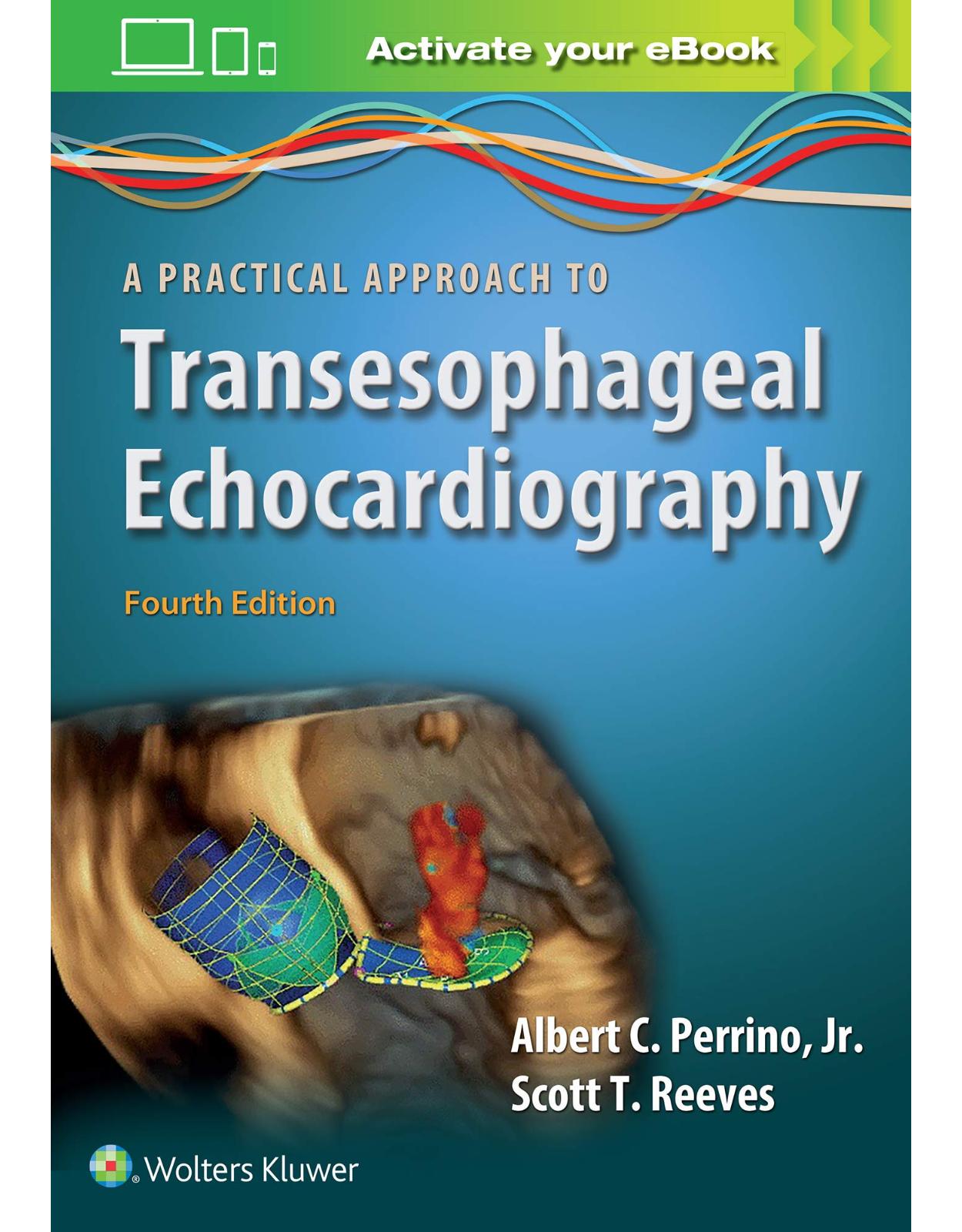
A Practical Approach to Transesophageal Echocardiography
Livrare gratis la comenzi peste 500 RON. Pentru celelalte comenzi livrarea este 20 RON.
Disponibilitate: La comanda in aproximativ 4 saptamani
Editura: LWW
Limba: Engleza
Nr. pagini: 592
Coperta: Paperback
Dimensiuni: 17.78 x 2.29 x 25.15 cm
An aparitie: 1 Aug. 2019
Description:
With updated content and new expert contributors, A Practical Approach to Transesophageal Echocardiography, Fourth Editionis every clinician’s go-to reference for TEE skill-building. The book is written concisely, so you can quickly digest and apply critical information in a real clinical setting. Numerous figures throughout each chapter and hundreds of video clips on the digital paint an important visual picture, illuminating the textual explanations and instructions.
Table of Contents:
SECTION I: ESSENTIALS OF 2D IMAGING
1. Principles and Technology of Two-Dimensional Echocardiography
INTRODUCTION
PHYSICAL PROPERTIES OF SOUND WAVES
Vibrations
Amplitude
Frequency and Wavelength
Propagation Velocity
WHAT IS SO SPECIAL ABOUT ULTRASOUND?
INTERACTIONS OF SOUND AND TISSUE
Reflection
Refraction
Attenuation
TRANSDUCER DESIGN AND BEAM FORMATION
Transducer Components
Formation of Ultrasound Waves: The Piezoelectric Crystal
The Three-Dimensional Ultrasound Beam
Resolution
Extraneous Sound Beams
SIGNAL RECEPTION AND PROCESSING
Cycling of Transducer Transmit and Receive Modes
Electrical Processing
DISPLAY FORMATS
The Golden Rule: Time Is Distance
Amplitude Mode
Brightness Mode
Motion Mode
2D Echocardiography
2D SCAN SYSTEMS
Linear Scanners
Sector Scanners
CREATING THE 2D IMAGE
Imaging a Sector
Image Quality and Dynamic Motion
Image Quality versus Dynamic Motion
ADVANCEMENTS
Harmonic Imaging
3D Imaging
TISSUE DOPPLER AND SPECKLE TRACKING
Tissue Doppler Imaging
Strain, Strain Rate, and Speckle Tracking
Speckle Tracking
SUMMARY
Review Questions
2. Two-Dimensional Examination
PROBE MANIPULATION
MULTIPLANE IMAGING ANGLE
PROBE INSERTION
GOALS OF THE EXAMINATION
THE COMPREHENSIVE EXAMINATION
Midesophageal Ascending Aortic Short-Axis View
Midesophageal Right Pulmonary Vein View
Midesophageal Ascending Aortic Long-Axis View
Midesophageal Aortic Valve Short-Axis View
Midesophageal Right Ventricular Inflow–Outflow
Midesophageal-Modified Bicaval Tricuspid Valve View
Midesophageal Aortic Valve Long-Axis View
Midesophageal Bicaval View
Midesophageal Right and Left Pulmonary Vein View
Midesophageal Five-Chamber View
Midesophageal Four-Chamber Views
Midesophageal Mitral Commissural View
Midesophageal Two-Chamber View
Midesophageal Left Atrial Appendage View
Midesophageal Left Pulmonary Vein View
Midesophageal Long-Axis View
Transgastric Basal Short-Axis View
Transgastric Midpapillary Short-Axis View
Transgastric Apical Short-Axis View
Transgastric Two-Chamber View
Transgastric Long-Axis View
Transgastric Right Ventricular Inflow View
Deep Transgastric Five-Chamber View
TG Right Ventricular Basal View
TG Right Ventricular Inflow–Outflow View
Descending Aorta Short-Axis View
Upper Esophageal Aortic Arch Long-Axis View
Upper Esophageal Aortic Arch Short-Axis View
Descending Aorta Long-Axis View
REAL-TIME BIPLANE VIEWS
THE ABBREVIATED CORE EXAMINATION
SUMMARY
Review Questions
3. Left Ventricular Systolic Performance and Pathology
MEASUREMENTS OF LV SIZE—WALL THICKNESS, CHAMBER SIZE, AND LV MASS
Left Ventricular Wall Thickness
Relative Wall Thickness
Chamber Size (LV Diameter)
Chamber Size (LV Volume)
Left Ventricular Mass
QUANTITATIVE MEASURES OF LEFT VENTRICULAR SYSTOLIC PERFORMANCE
OTHER MEASURES OF LEFT VENTRICULAR SYSTOLIC FUNCTION
Assessing Left Ventricular Systolic Function With Doppler Techniques
VENTRICULAR PATHOLOGY
Cardiomyopathies
Left Ventricular Hypertrophy
Left Ventricular True Aneurysm
Left Ventricular Pseudoaneurysm
CONCLUSION
Review Questions
4. Diagnosis of Myocardial Ischemia
ANATOMY AND PATHOPHYSIOLOGY OF THE MYOCARDIAL BLOOD SUPPLY
Coronary Anatomy
Variations in Coronary Anatomy and Main Distribution Types
Echocardiographic Assessment of Coronary Arteries
Detection of Coronary Artery Pathology
PHYSIOLOGIC BASIS FOR THE DETECTION OF ISCHEMIA
Clinical Syndromes of Myocardial Ischemia
ECHOCARDIOGRAPHIC ASSESSMENT OF ISCHEMIA
Anatomic Localization of Ischemia: The 17-Segment Model
Echocardiographic Imaging of the 17 Myocardial Segments
Coronary Perfusion of LV Segments
Assessment and Grading of Regional Wall Contraction Abnormalities
Important Methodologic Aspects of Wall Contraction Analysis and Potential Pitfalls
Limitations of Echocardiographic Detection of Myocardial Ischemia
Alternative Echocardiographic Methods for Detection of Ischemia
Right Ventricular Ischemia
COMPLICATIONS OF MYOCARDIAL ISCHEMIA
SUMMARY
Review Questions
SECTION II: ESSENTIALS OF DOPPLER ECHO
5. Doppler Technology and Technique
INTRODUCTION
DOPPLER FREQUENCY SHIFT
The Doppler Effect
Signal Frequency and Blood Flow
DOPPLER ANALYSIS
The Doppler Equation: Linking the Frequency Shift to Velocity
Implications of Beam Orientation
Clinical Caveats in Transesophageal Echocardiographic Doppler Examinations
Isolating the Doppler Frequency Shift
PRESENTATION OF DOPPLER DATA
Audible Broadcast
Spectral Display
DOPPLER TECHNIQUES
Pulsed-Wave Doppler
Continuous-Wave Doppler
Color Flow Mapping
SUMMARY
Review Questions
6. Quantitative Doppler and Hemodynamics
VOLUMETRIC FLOW CALCULATIONS
Doppler Measurements of Stroke Volume and Cardiac Output
Regurgitant Volume
Intracardiac Shunts
Valve Area: The Continuity Equation
INTRACARDIAC PRESSURES AND PRESSURE GRADIENTS: THE BERNOULLI EQUATION
Assessment of Valvular Disease
Measurement of Intracavitary Pressures
Vascular Resistance
Determination of Filling Pressures
HEART RHYTHM
SUMMARY
Review Questions
7. Echocardiographic Evaluation of Ventricular Diastolic Function
INTRODUCTION
BASICS OF DIASTOLIC PHYSIOLOGY
Intrinsic Myocardial Factors Affecting Diastolic Performance
Extrinsic Nonmyocardial Factors Affecting Diastolic Performance
PATHOPHYSIOLOGY OF DIASTOLIC DYSFUNCTION AND PROGRESSION OF DISEASE
ECHOCARDIOGRAPHIC EVALUATION OF LEFT VENTRICULAR DIASTOLIC FUNCTION
Two-Dimensional Echocardiography
Doppler Echocardiographic Evaluation of LV Filling
Influence of Physiologic Variables on Doppler Flow Profiles
Assessment of Diastolic Function in Special Clinical Scenarios
Right Ventricular Diastolic Function
Pericardial Diseases: Constrictive Pericarditis and Cardiac Tamponade
A PRACTICAL APPROACH TO DIASTOLIC FUNCTION ASSESSMENT IN THE PERIOPERATIVE PERIOD
SUMMARY
Review Questions
SECTION III: VALVULAR DISEASE
8. Mitral Regurgitation
INTRODUCTION
ANATOMY
NOMENCLATURE
ETIOLOGY AND MECHANISM OF MR
APPROACH TO THE TRANSESOPHAGEAL ECHOCARDIOGRAPHY EVALUATION OF MR
Where on the Mitral Valve Is the MR and What Is the Mechanism of MR?
How Severe Is the MR?
PITFALLS IN THE EVALUATION OF MR
SECONDARY (FUNCTIONAL) MR
CONCLUSION
Review Questions
9. Mitral Valve Stenosis
INTRODUCTION
MITRAL VALVE ANATOMY
ETIOLOGY OF MITRAL STENOSIS
TRANSESOPHAGEAL ECHOCARDIOGRAPHIC EVALUATION OF MITRAL STENOSIS
Two-Dimensional Echocardiography
Physiologic Assessments
Advanced Concepts That May Be Helpful in Difficult Cases
A PRACTICAL APPROACH TO THE EVALUATION OF MITRAL STENOSIS
Three-Dimensional Echocardiographic Evaluation of Mitral Stenosis
Use of Echocardiography for New Procedures
SUMMARY
Review Questions
10. Mitral Valve Repair
INDICATIONS
Primary versus Secondary Mitral Regurgitation
ECHOCARDIOGRAPHIC EVALUATION
The Prerepair Evaluation
Help in Planning the Surgical Procedure
MITRAL VALVE REPAIR TECHNIQUES
The Fundamental Goals of MV Repair for Type II Valve Dysfunction
7. Repair Technique of Type I Valve Dysfunction
8. Repair Technique of Type III Valve Dysfunction
Repair Technique for Carpentier Type I Valve Dysfunction
Repair Technique for Carpentier Type III Valve Dysfunction
Postrepair TEE Examination
Preweaning From Bypass TEE Examination
Postbypass TEE Examination
Identification of the Circumflex Coronary Artery Perfusion
Checking for Complete Deairing of the Left Ventricle
Residual Mitral Regurgitation
Postrepair Mitral Stenosis
Screening for Potential Complications
SUMMARY
Review Questions
11. Aortic Regurgitation
ETIOLOGY AND MECHANISMS OF AORTIC REGURGITATION
HEMODYNAMICS OF AORTIC REGURGITATION
ECHOCARDIOGRAPHIC EVALUATION OF THE AORTIC VALVE
RECOMMENDED VIEWS
APPROACHES TO THE QUANTITATIVE ASSESSMENT OF AORTIC REGURGITATION
Color Flow Doppler
Calculation of Regurgitant Volume, Fraction, and Orifice Area
Pulsed- and Continuous Wave Doppler
Special Clinical Situations
SUMMARY
REFERENCES
Review Questions
12. Aortic Stenosis
INTRODUCTION
PATHOPHYSIOLOGY
ECHOCARDIOGRAPHIC EVALUATION OF THE AORTIC VALVE
Quantitative Doppler Assessment of Aortic Stenosis
Planimetry of the Aortic Orifice
SPECIAL CONSIDERATIONS
Pressure Recovery
Systemic Hypertension and Arterial Compliance
Classical and “Paradoxical” Low-Flow, Low-Gradient Aortic Stenosis
Assessment of Aortic Stenosis Gradient in Aortic Regurgitation
Pre- and Postoperative Subaortic Obstruction
TECHNICAL CONSIDERATIONS
SUMMARY
Review Questions
13. Prosthetic Valves
INTRODUCTION
ROLE OF TRANSESOPHAGEAL ECHOCARDIOGRAPHY IN THE EVALUATION OF PROSTHETIC CARDIAC VALVES
TECHNICAL CONSIDERATIONS IN PERFORMING TEE EXAMINATIONS OF PROSTHETIC VALVES
ECHOCARDIOGRAPHIC CHARACTERISTICS OF THE INDIVIDUAL PROSTHETIC CARDIAC VALVE TYPES
Mechanical Heart Valves
Biologic Valves or Tissue Valves
CLINICAL CAVEATS FOR THE ECHOCARDIOGRAPHIC DIAGNOSIS OF PROSTHETIC VALVE DYSFUNCTION
CONCLUSION
Review Questions
14. Right Ventricle, Right Atrium, Tricuspid and Pulmonic Valves
INTRODUCTION
RIGHT VENTRICLE
Anatomy
Transesophageal Echocardiographic Views
Assessment of Right Ventricular Structure
Assessment of Right Ventricular Systolic Function
Assessment of Right Ventricular Diastolic Function
Assessment of Regional Right Ventricular Function
Hemodynamic and Extracardiac Manifestation of Right Ventricular Dysfunction
RIGHT ATRIUM
Anatomy
Transesophageal Echocardiographic Views
TRICUSPID VALVE
Anatomy
Transesophageal Echocardiographic Views
Tricuspid Regurgitation
Tricuspid Stenosis
Etiology of Tricuspid Valve Disease
PULMONIC VALVE
Anatomy
Transesophageal Echocardiographic Views
Pulmonic Regurgitation
Pulmonic Stenosis
REAL-TIME THREE-DIMENSIONAL TRANSESOPHAGEAL ECHOCARDIOGRAPHY
Review Questions
SECTION IV: CLINICAL CHALLENGES
15. Transesophageal Echocardiography for Coronary Revascularization
INTRODUCTION
SAFETY AND COMPLICATIONS
PREBYPASS EXAMINATION
Assessment of Ventricular Function
New Advances in Intraoperative Assessment of Global and Regional Cardiac Function
Monitoring of Ischemia Visualizing the Aortic Root, Coronary Arteries, and Coronary Blood Flow
Shortcomings of TEE
NEW RWMA AFTER CARDIOPULMONARY BYPASS
What Does it Signify?
What Should We Do?
ACUTE CARDIAC DYSFUNCTION: ASSESSMENT AND MANAGEMENT
Hypovolemia
Dynamic Mitral Regurgitation
Right Ventricular Dysfunction
Low Ejection Fraction
Unexpected Diagnosis of Coexisting Problems
Intracardiac Shunts; Patent Foramen Ovale or Atrial Septal Defect
Pleural Effusions
Previously Unknown Aortic Valve Pathology
Coexisting Mitral Valve Regurgitation—Combined CABG and MV Surgery
Use of 3D Echo in Coronary Artery Revascularization
TEE for Specific Surgical Situations
Role of TEE in Vascular Cannulations
Endoscopic Vein Harvesting (EVH)
Failure to Wean From CPB
Impella Endovascular Devices
Ventricular Assist Devices
SUMMARY
Review Questions
16. Intraprocedural Echocardiographic Guidance for Transcatheter Interventions in Aortic and Mitral Position
INTRODUCTION
TRANSCATHETER AORTIC VALVE REPLACEMENT
Current Devices
Procedural Performance (TAVR)
Key Points and Practical Approach
MITRAL VALVE INTERVENTIONS
Mitraclipø Intervention
TRANSAPICAL MITRAL VALVE REPLACEMENT
Steps for Transapical Mitral Valve Replacement
Echocardiographic Implications
TRANSAPICAL BEATING HEART MV REPAIR
Procedure and Guidance
Direct and Indirect Annuloplasty
CONCLUSION
Review Questions
17. Transesophageal Echocardiography of the Thoracic Aorta
ANATOMY AND TEE IMAGING
ACUTE AORTIC SYNDROMES
Acute Aortic Dissection
Intramural Hematoma
Penetrating Atherosclerotic Ulcer
Following Repair
Thoracic Aortic Aneurysms
AORTIC ATHEROMA
Intra-Aortic Balloon Pump Placement
CONCLUSION
Review Questions
18. Critical Care Echocardiography
INTRODUCTION
A PROPOSED APPROACH TO HEMODYNAMIC INSTABILITY
Step #1: Ultrasound Imaging of the Inferior Vena Cava and Abdominal Venous Flow
Step #2: CCUS in Patients With Reduced Mean Systemic Venous Pressure
Step #3: CCUS in Patients With Raised Right Atrial Pressure
Step #4: CCUS in Increased Resistance to Venous Return
HYPOXEMIA AND LUNG ULTRASOUND
LIMITATIONS
Caveats
CONCLUSION
Review Questions
19. Transesophageal Echocardiography for Congenital Heart Disease in the Adult
INTRODUCTION
INCIDENCE OF CONGENITAL HEART DISEASE AND PREVALENCE IN THE ADULT POPULATION
CLASSIFICATION OF CONGENITAL HEART DISEASE
Based on Severity of Disease
Based on the Presence or Absence of Cyanosis
Based on the Physiology of the Defect
GUIDELINES FOR TRANSESOPHAGEAL ECHOCARDIOGRAPHY IN CONGENITAL HEART DISEASE
INDICATIONS FOR TRANSESOPHAGEAL ECHOCARDIOGRAPHY IN THE ADULT WITH CONGENITAL HEART DISEASE
TEE for Diagnostic Applications
TEE During Cardiothoracic Surgery
TEE in the Cardiac Catheterization/Electrophysiology Laboratory
SELECTED CONGENITAL HEART DEFECTS
ATRIAL SEPTAL DEFECT
Anatomy
Pathophysiology
Management
Transesophageal Echocardiographic Evaluation
Goals of the Two-Dimensional Examination
Goals of the Doppler Examination
Goals of the Examination After Surgery
Goals of the Examination During Transcatheter Intervention
Applications of Three-Dimensional Imaging
VENTRICULAR SEPTAL DEFECT
Anatomy
Pathophysiology
Management
Transesophageal Echocardiographic Evaluation
Goals of the Two-Dimensional Examination
Goals of the Doppler Examination
Goals of the Examination After Surgery
Goals of the Examination During Transcatheter Intervention
Applications of Three-Dimensional Imaging
PATENT DUCTUS ARTERIOSUS
Anatomy
Pathophysiology
Management
Transesophageal Echocardiographic Evaluation
Goals of the Two-Dimensional Examination
Goals of the Doppler Examination
Goals of the Examination After Surgery
COARCTATION OF THE AORTA
Anatomy
Pathophysiology
Management
Transesophageal Echocardiographic Evaluation
Goals of the Two-Dimensional Examination
Goals of the Doppler Examination
Goals of the Examination After Surgery
AORTIC STENOSIS
Anatomy
Pathophysiology
Management
Transesophageal Echocardiographic Evaluation
Goals of the Two-Dimensional Evaluation
Goals of the Doppler Examination
Goals of the Examination After Surgery
Goals of the Examination During Transcatheter Intervention
Applications of Three-Dimensional Imaging
PULMONARY VALVE (PULMONIC) STENOSIS
Anatomy
Pathophysiology
Management
Transesophageal Echocardiographic Evaluation
Goals of the Two-Dimensional Evaluation
Goals of the Doppler Examination
Goals of the Examination After Surgery
Applications of Three-Dimensional Imaging
TETRALOGY OF FALLOT
Anatomy
Pathophysiology
Management
Transesophageal Echocardiographic Evaluation
Goals of the Two-Dimensional Examination
Goals of the Doppler Examination
Goals of the Examination After Surgery
Goals of the Examination During Transcatheter Intervention
Applications of Three-Dimensional Imaging
TRANSPOSITION OF THE GREAT ARTERIES
Anatomy
Pathophysiology
Management
Transesophageal Echocardiographic Evaluation
Goals of the Two-Dimensional Examination
Goals of the Doppler Examination
Goals of the Examination After Surgery
Goals of the Examination During Transcatheter Interventions
CONGENITALLY CORRECTED TRANSPOSITION OF THE GREAT ARTERIES
Anatomy
Pathophysiology
Management
Transesophageal Echocardiographic Evaluation
Goals of the Two-Dimensional Examination
Goals of the Doppler Examination
Goals of the Examination After Surgery
EBSTEIN ANOMALY
Anatomy
Pathophysiology
Management
Transesophageal Echocardiographic Evaluation
Goals of the Two-Dimensional Examination
Goals of the Doppler Examination
Goals of the Examination After Surgery
Goals of the Examination During Transcatheter Intervention
Applications of Three-Dimensional Imaging
SINGLE-VENTRICLE LESIONS OR UNIVENTRICULAR HEART
Anatomy
Pathophysiology
Management
Transesophageal Echocardiographic Evaluation
Goals of the Two-Dimensional Examination
Goals of the Doppler Examination
Goals of the Examination After Surgery
Goals of the Examination During Transcatheter Intervention
CONGENITAL CORONARY ARTERY ANOMALIES
Anatomy
Pathophysiology
Management
Transesophageal Echocardiographic Evaluation
Goals of the Two-Dimensional Examination
Goals of the Doppler Examination
Goals of the Examination After Surgery/Intervention
TRANSESOPHAGEAL ECHOCARDIOGRAPHY FOR NONCARDIAC SURGERY IN ADULTS WITH CONGENITAL HEART DISEASE
LIMITATIONS OF TRANSESOPHAGEAL ECHOCARDIOGRAPHY IN CONGENITAL HEART DISEASE
SUMMARY
Review Questions
20. Cardiac Masses and Embolic Sources
INTRODUCTION
CARDIAC TUMORS
Myxoma
Lipoma
Papillary Fibroelastoma
Rhabdomyoma
Fibroma
Cardiac Sarcomas
Metastatic Tumors
Embolization
INTRACARDIAC THROMBUS
Practical Considerations in the Evaluation of Cardiac Masses
TEE in the Assessment of Cardiac Sources of Emboli
Probable Sources of Emboli With High Embolic Potential
Possible Sources of Emboli With Low Embolic Potential
Review Questions
SECTION V: MAN AND MACHINE
21. 3D TEE Imaging
INTRODUCTION
3D TECHNOLOGY
TEE Probes
3D Imaging
3D IMAGE ACQUISITION
Imaging Modes
POSTPROCESSING
Image Optimization
Image Artifacts
Cropping and Orientation
Measurements
QUANTIFICATION ANALYSIS MODELS
CLINICAL APPLICATIONS
Mitral Valve
Left and Right Ventricles
Aortic Valve
Tricuspid Valve and Pulmonic Valve
Aorta
Congenital Lesions
Masses
Hypertrophic Obstructive Cardiomyopathy
Interventional Procedures
ACKNOWLEDGMENTS
Review Questions
22. Common Artifacts and Pitfalls of Clinical Echocardiography
INTRODUCTION
NORMAL ANATOMIC VARIANTS IN TWO-DIMENSIONAL IMAGING
Crista Terminalis
Eustachian Valve or Chiari Network
Lipomatous Hypertrophy of the Atrial Septum
Coumadin Ridge
Pericardial Sinuses
Lambl Excrescences
Moderator Band and False Tendon
Pleural Effusion
TWO-DIMENSIONAL ECHOCARDIOGRAPHIC IMAGING ARTIFACTS
Suboptimal Image Quality
Acoustic Shadowing
Lateral Resolution
Side Lobe and Beam Width
Reverberation and Mirroring
Electronic Noise
ARTIFACTS IN SPECTRAL AND COLOR FLOW DOPPLER
Aliasing
Acoustic Shadowing in Color Flow
Nonparallel Beam Angle
Mirroring
Color Flow Reverberation and Gain-Related Anomalies
Beam Width Flow Artifacts
Range Ambiguity With Pulsed-Wave Doppler
THREE-DIMENSIONAL ECHOCARDIOGRAPHIC IMAGING ARTIFACTS
Artifacts Unique to Three-Dimensional TEE
SUMMARY
Review Questions
23. Techniques and Tricks for Optimizing Transesophageal Images
INTRODUCTION
TWO-DIMENSIONAL CONTROLS
Preprocessing Versus Postprocessing Controls
Transmit Power
Gain
Time Gain Compensation
Lateral Gain Compensation
Depth
Focus
Frequency
Dynamic Range
Compression
Reject
Persistence
Sector Size
Harmonics
COLOR CONTROLS
Region of Interest
Color Gain
Color Scale
Variance
Image Optimization
DIGITAL STORAGE SYSTEMS
SUMMARY
Review Questions
Appendices
A. Cardiac Cross Section Criteria
B. Cardiac Dimensions
C. Hemodynamic Calculations
D. Valve Prostheses
E. Classification of the Severity of Valvular Disease
Index
| An aparitie | 1 Aug. 2019 |
| Autor | Albert C. Perrino , Scott T. Reeves |
| Dimensiuni | 17.78 x 2.29 x 25.15 cm |
| Editura | LWW |
| Format | Paperback |
| ISBN | 9781496383471 |
| Limba | Engleza |
| Nr pag | 592 |










Clientii ebookshop.ro nu au adaugat inca opinii pentru acest produs. Fii primul care adauga o parere, folosind formularul de mai jos.