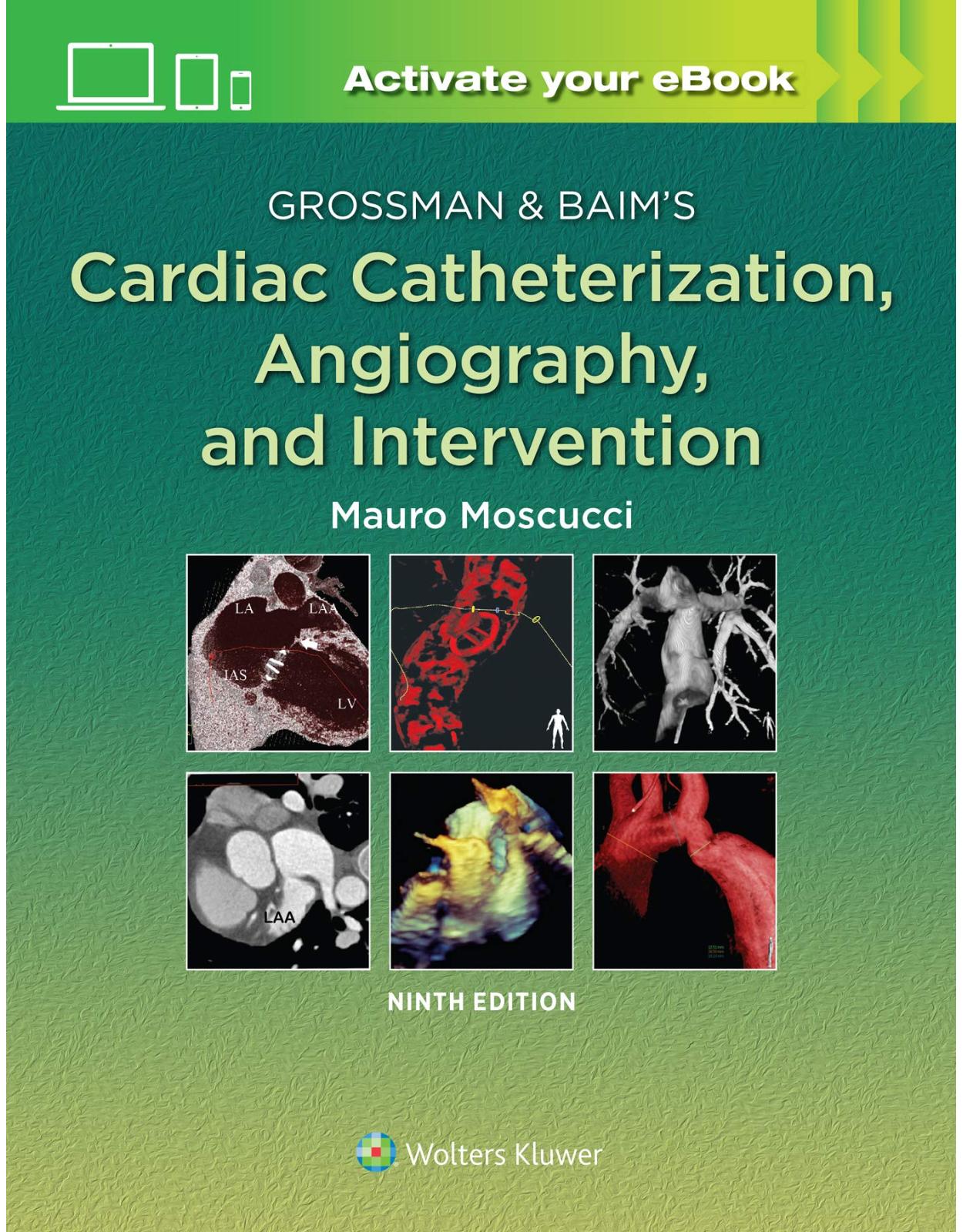
Grossman & Baim’s Cardiac Catheterization, Angiography, and Intervention
Livrare gratis la comenzi peste 500 RON. Pentru celelalte comenzi livrarea este 20 RON.
Disponibilitate: La comanda in aproximativ 4-6 saptamani
Autor: Mauro Moscucci
Editura: LWW
Limba: Engleza
Nr. pagini: 1216
Coperta: Hardcover
Dimensiuni: 21.59 x 2.54 x 25.4 cm
An aparitie: 9 Nov. 2020
Description:
The leading comprehensive reference on cardiac catheterization through eight outstanding editions, Grossman & Baim's Cardiac Catheterization, Angiography, and Intervention, Ninth Edition, continues to keep you up to date with every facet of this fast-changing field. Designed for quick access and easy reference, this text offers expert overviews of the theoretical and practical aspects of clinical issues, with emphasis given to hemodynamic data and tracings and interventional procedures. An impressive multimedia library with new videos and cases make this reference even more valuable for cardiologists and interventional cardiologists at all levels of experience.
Enrich Your Digital Reading Experience
- Read directly on your preferred device(s), such as computer, tablet, or smartphone.
- Easily convert to audiobook, powering your content with natural language text-to-speech.
Table of Contents:
Section I General Principles
1. Cardiac Catheterization History and Current Practice Standards
INTERVENTIONAL CARDIOLOGY
INDICATIONS FOR CARDIAC CATHETERIZATION
Research
Contraindications
Factors Influencing Choice of Approach
DESIGN OF THE CATHETERIZATION PROTOCOL
The Checklist
Preparation and Premedication of the Patient
Universal Protocol and Time-Out
THE CARDIAC CATHETERIZATION FACILITY
Location Within a Hospital Versus Freestanding
Outpatient Cardiac Catheterization
TRAINING STANDARDS
Physician and Laboratory Caseload
The Catheterization Laboratory Director and Quality Assurance
PERFORMING THE PROCEDURE
2. Cineangiographic Imaging, Radiation Safety, and Contrast Agents
BASIC X-RAY PHYSICS
CLINICAL MEASUREMENT OF PATIENT IRRADIATION
IMAGE FORMATION
Image Contrast
Image Noise
Image Sharpness
Scattered Radiation
OPTIMIZING PATIENT EXPOSURE AND IMAGE QUALITY
THE CINEFLUOROGRAPHIC SYSTEM
Radiation Production and Control
X-Ray Tubes
Spatial and Spectral Shaping of the X-Ray Beam
Imaging Modes
Automatic Dose Rate Control
CLINICAL PROGRAMS AND PROGRAMMING
IMAGE DETECTION, PROCESSING, AND RECORDING
Image Intensifiera
FLAT-PANEL X-RAY DETECTORS
IMAGE PROCESSING AND DISPLAY
DIGITAL IMAGING AND COMMUNICATION IN MEDICINE AND PICTURE ARCHIVING AND COMMUNICATION SYSTEM
THE ANGIOGRAPHIC ROOM
IMAGING EQUIPMENT QUALITY ASSURANCE
BIOLOGICAL EFFECTS OF RADIATION
Stochastic Effects
Radiogenic Cancer
Tissue Reactions
Patient Radiation Management
Clinical Dose Monitoring
Staged and Multiple Procedures
Patient Education, Consent, and Follow-Up
Staff Radiation Safety
Staff Tissue Reaction
Staff Cancer Risk
Basic Principles of Reducing Staff Radiation Exposure
Staff Radiation Monitoring
INTRAVASCULAR CONTRAST AGENTS
Iodinated Contrast Agents
Gadolinium
Carbon Dioxide
FUTURE DIRECTIONS
3. Integrated Imaging Modalities in the Cardiac Catheterization Lab
LIMITATIONS OF TRADITIONAL IMAGING SYSTEMS
EVOLUTION OF IMAGING NEEDS IN THE CARDIAC CATHETERIZATION SUITE
Value Assessment
NEW IMAGING MODALITIES
Echocardiography
Rotational Angiography
Intracardiac Echocardiography
Computed Tomography Angiography
Magnetic Resonance Imaging and Angiography
IMAGING COMPARATIVE ANATOMIC STRUCTURE AND FUNCTION
Fluoroscopy/Angiography Versus Echocardiography
Fluoroscopy/Angiography Volumetric Computed Tomography or Magnetic Resonance Imaging
IMAGE AND MODALITY COREGISTRATION
MODALITY SELECTION
VISUALIZATION: 2-DIMENSIONAL TO 3-DIMENSIONAL
3-Dimensional Fluoroscopy and Coregistration of Computed Tomography Imaging
Real-Time Echocardiographic 3-Dimensional
NEW FRONTIERS
4. Complications
OVERVIEW
DEATH
Death as a Complication of Diagnostic Catheterization
Left Main Disease
Left Ventricular Dysfunction
Valvular Heart Disease
Prior Coronary Artery Bypass Graft Surgery
Pediatric Patients
Death in the Course of an Interventional Procedure
MYOCARDIAL INFARCTION
Interventional Procedures
CEREBROVASCULAR COMPLICATIONS
LOCAL VASCULAR COMPLICATIONS
Femoral Artery Thrombosis
Femoral Vein Thrombosis
Hemorrhagic Complications
Retroperitoneal Bleeding
Femoral Neuropathy
Pseudoaneurysm and Arteriovenous Fistula
ARRHYTHMIAS OR CONDUCTION DISTURBANCE
Ventricular Fibrillation
Atrial Arrhythmias
Bradyarrhythmias
PERFORATION OF THE HEART OR GREAT VESSELS
INFECTIONS AND PYROGEN REACTIONS
ALLERGIC AND ANAPHYLACTOID REACTIONS
CONTRAST-INDUCED NEPHROPATHY/ACUTE KIDNEY INJURY
OTHER COMPLICATIONS
Hypotension
Volume Overload
Anxiety/Pain
Respiratory Insufficiency
Retained Equipment
CONCLUSION
5. Adjunctive Pharmacology for Cardiac Catheterization
ANTIPLATELET AGENTS
Aspirin
Adenosine Diphosphate Receptor Antagonists
Intravenous Glycoprotein IIb/IIIa Inhibitors
ANTITHROMBOTIC AGENTS
Unfractionated Heparin
Low-Molecular-Weight Heparin
Factor Xa Inhibitors
Direct Thrombin Inhibitors
Duration of Antiplatelet Therapy
Other Pharmacologic Agents
Section II Basic Techniques
6. Percutaneous Transfemoral, Transseptal, Transcaval, and Apical Approach
CATHETERIZATION VIA THE FEMORAL ARTERY AND VEIN
Patient Preparation
Selection of Puncture Site
Local Anesthesia
Femoral Vein Puncture
Catheterizing the Right Heart From the Femoral Vein
Femoral Artery Puncture
Catheterizing the Left Heart From the Femoral Artery
Control of the Puncture Site Following Sheath Removal
Contraindications to Femoral Approach to Left Heart Catheterization
ALTERNATIVE SITES FOR LEFT HEART CATHETERIZATION
Percutaneous Entry of the Axillary, Brachial, or Radial Arteries
Lumbar Aortic Puncture
Percutaneous Transcaval Access
Transseptal Puncture
Apical Left Ventricular Puncture
7. Radial Artery Approach
ANATOMICAL CONSIDERATIONS
TECHNICAL ASPECTS
Patient Preparation
Patient Positioning—Right Versus Left Radial Access
Sheath Selection
Radial Puncture Technique
Distal Transradial Approach
Learning Curve
Navigating the Upper Extremity Arterial System
CATHETER SELECTION
Diagnostic Angiography
Percutaneous Coronary Intervention
TRA for Peripheral Vascular Interventions
RADIAL HEMOSTASIS—PREVENTION OF RADIAL ARTERY OCCLUSION
Complications
Radial Artery Spasm
Hematoma and Bleeding
Radial Artery Perforation and Dissection
Radial Artery Pseudoaneurysm
TRANSRADIAL ACCESS AND RADIATION EXPOSURE
BRACHIAL VENOUS ACCESS FOR RIGHT HEART CATHETERIZATION
TRANSRADIAL ACCESS AND OUTCOMES
ECONOMICS—QUALITY OF LIFE—SAME-DAY DISCHARGE PCI
Economics
Quality of Life
Same-Day Discharge
CONCLUSION
8. Cutdown Approach: Brachial, Femoral, Axillary, Aortic, and Transapical
INDICATIONS
PREPROCEDURE EVALUATION
INCISION, ISOLATION OF VESSELS, AND CATHETER INSERTION
CATHETER SELECTION
Right Heart Catheters
Left Heart Catheters
ADVANCING THE RIGHT HEART CATHETER
ADVANCING THE LEFT HEART CATHETER
SPECIAL TECHNIQUES
Coronary Bypass Grafts
Internal Mammary Arteries
Anomalous Coronary Takeoff
Percutaneous Coronary Interventions
REPAIR OF VESSELS AND AFTERCARE
TROUBLESHOOTING
Loss of Radial Pulse
Hand Numbness
FEMORAL, AXILLARY, AORTIC, AND TRANSAPICAL ACCESS
Open Femoral Arterial Access
Axillary/Subclavian Artery Access
Direct Transthoracic Aortic Access
Left Ventricular Apical Access
9. Diagnostic Catheterization in Childhood and Adult Congenital Heart Disease
GENERAL PRINCIPLES IN THE CATHETERIZATION OF PATIENTS WITH CONGENITAL HEART DISEASE
Vascular Access/Vessel and Chamber Entry
Intracardiac Catheter Manipulation
Pressure Measurements and Oximetry
Angiography
SPECIAL CIRCUMSTANCES
Pregnancy
Down Syndrome
Pulmonary Ventricular Failure and Pulmonary Vascular Disease
Right Ventricular Outflow Failure
Cyanosis
Systemic Ventricular “Heart Failure”
Coronary Artery Disease
Vascular Anatomy Before Cardiac Surgery
Fontan
CONCLUSION
Section III Hemodynamic Principles
10. Pressure Measurement
THE INPUT SIGNAL: WHAT IS A PRESSURE WAVE?
PRESSURE MEASURING DEVICES
Sensitivity
Frequency Response
Natural Frequency and Damping
Linearity
WHAT FREQUENCY RESPONSE IS DESIRABLE?
EVALUATION OF FREQUENCY RESPONSE CHARACTERISTICS
TRANSFORMING PRESSURE WAVES INTO ELECTRICAL SIGNALS: THE ELECTRICAL STRAIN GAUGE
PRACTICAL PRESSURE TRANSDUCER SYSTEM FOR THE CATHETERIZATION LABORATORY
PHYSIOLOGIC CHARACTERISTICS OF PRESSURE WAVEFORMS
Reflected Waves
Wedge Pressures
NORMAL CONTOURS OF PRESSURE WAVEFORMS
Atrial Pressure
Pulmonary Wedge Pressure
Ventricular Pressure
Aortic Pressure
SOURCES OF ERROR AND ARTIFACT
Deterioration in Frequency Response
Catheter Whip Artifact
End Pressure Artifact
Catheter Impact Artifact
Systolic Pressure Amplification in the Periphery
Errors in Zero Level, Balancing, or Calibration
MICROMANOMETERS
PRESSURE TRACINGS IN VALVULAR AND NONVALVULAR HEART DISEASE
CONCLUSION
11. Blood Flow Measurement: Cardiac Output and Vascular Resistance
EXTRACTION RESERVE AND CARDIAC OUTPUT
Lower Limit of Cardiac Output
Upper Limit of Cardiac Output
Factors Influencing Cardiac Output in Normal Subjects
TECHNIQUES FOR DETERMINATION OF CARDIAC OUTPUT
Fick Oxygen Method
Indicator Dilution Methods
Thermodilution Method
Continuous Cardiac Output Monitoring
CLINICAL MEASUREMENT OF VASCULAR RESISTANCE
Poiseuille Law
Clinical Use of Vascular Resistance
Systemic Vascular Resistance
Total Pulmonary Resistance
Pulmonary Vascular Resistance
PULMONARY VASCULAR DISEASE IN PATIENTS WITH CONGENITAL CENTRAL SHUNTS
PULMONARY VASCULAR DISEASE IN PATIENTS WITH MITRAL STENOSIS
ASSESSMENT OF VASODILATOR DRUGS
12. Shunt Detection and Quantification
DETECTION OF LEFT-TO-RIGHT INTRACARDIAC SHUNTS
Measurement of Blood Oxygen Saturation and Content in the Right Heart (Oximetry Run)
Oximetry Run
Calculation of Pulmonary Blood Flow (Qp)
Calculation of Systemic Blood Flow (Qs)
Calculation of Left-to-Right Shunt
Examples of Left-to-Right Shunt Detection and Quantification
Flow Ratio
Calculation of Bidirectional Shunts
Limitations of the Oximetry Method
Other Indicators
Angiography
DETECTION OF RIGHT-TO-LEFT INTRACARDIAC SHUNTS
Angiography
Oximetry
Echocardiography
13. Calculation of Stenotic Valve Orifice Area
THE GORLIN FORMULA
MITRAL VALVE AREA
Example of Valve Area Calculation in Mitral Stenosis
Pitfalls
AORTIC VALVE AREA
Example
Pitfalls
AREA OF TRICUSPID AND PULMONIC VALVES
ALTERNATIVES TO THE GORLIN FORMULA
ASSESSMENT OF AORTIC STENOSIS IN PATIENTS WITH LOW CARDIAC OUTPUT
VALVE RESISTANCE
ACKNOWLEDGMENT
14. Pitfalls in the Evaluation of Hemodynamic Data
BASIC CONCEPTS
TRANSVALVULAR GRADIENT
EFFECTS OF CATHETER LOCATION
OTHER CONSIDERATIONS
CONCLUSION
Section IV Angiographic Techniques
15. Coronary Angiography
CURRENT INDICATIONS
GENERAL ISSUES
THE FEMORAL APPROACH
Insertion and Flushing of the Coronary Catheter
Damping and Ventricularization of the Pressure Waveform
Cannulation of the Left Coronary Ostium
Cannulation of the Right Coronary Ostium
Cannulation of Saphenous Vein and Arterial Grafts
Internal Mammary Artery Cannulation
Gastroepiploic Graft Cannulation
THE BRACHIAL OR RADIAL APPROACH
ADVERSE EFFECTS OF CORONARY ANGIOGRAPHY
INJECTION TECHNIQUE
ANATOMY, ANGIOGRAPHIC VIEWS, AND QUANTITATION OF STENOSIS
Coronary Anatomy
Angiographic Views
Lesion Quantification
Coronary Collaterals
BIPLANE AND ROTATIONAL CORONARY ANGIOGRAPHY
NONATHEROSCLEROTIC CORONARY ARTERY DISEASE
Coronary Vasospasm
Abnormal Coronary Vasodilator Reserve
MISTAKES IN INTERPRETATION
Inadequate Number of Projections
Inadequate Injection of Contrast Material
Superselective Injection
Catheter-Induced Coronary Spasm
Congenital Variants of Coronary Origin and Distribution
Myocardial Bridges
Total Occlusion
Complex Stenosis Classification and Risk Stratification
16. Coronary Artery Anomalies
DEFINITIONS
Anomalous Origin of a Coronary Artery From an Opposite Sinus of Valsalva, With an Intramural Course
Other Coronary Anomalies Frequently Encountered in the Adult Cath Lab: Coronary Fistulae and Myocardial Bridges
17. Cardiac Ventriculography
INJECTION CATHETERS
Pigtail Catheter
Straight Tip Left Ventriculographic Catheters
Balloon-Tip Ventriculographic Catheters
INJECTION SITE
INJECTION RATE AND VOLUME
FILMING PROJECTION AND TECHNIQUE
RIGHT VENTRICULOGRAPHY
ANALYSIS OF THE VENTRICULOGRAM
INTERVENTION VENTRICULOGRAPHY—HISTORICAL PERSPECTIVE
COMPLICATIONS AND HAZARDS
Arrhythmias
Intramyocardial Injection (Endocardial Staining)
Fascicular Block
Embolism
Complications of Contrast Media
ALTERNATIVES TO CONTRAST VENTRICULOGRAPHY
2D and Real-Time 3D Echocardiographic Visualization of the Left Ventricle
Radionuclide Imaging: First Pass, ERNA, and Gated SPECT
Magnetic Resonance Imaging and Computerized Tomographic Ventriculography
Electromechanical Mapping
Electrical Conductance Catheter
18. Pulmonary Angiography
Introduction
HISTORY
ANATOMY
TECHNICAL CONSIDERATIONS
Hemodynamic Monitoring
Percutaneous Venous Catheterization
Pulmonary Artery Catheterization
Catheter Exchange
Contrast Agents and Injection Rates
Imaging Modes
Complications and Contraindications
PULMONARY EMBOLISM
Diagnosis
OTHER INDICATIONS FOR PULMONARY ANGIOGRAPHY
Pulmonary Hypertension
Rare Indications
Meandering Pulmonary Vein
19. Angiography of the Aorta and Peripheral Arteries
PERIPHERAL IMAGING TECHNIQUES
CROSS-SECTIONAL ARTERIAL IMAGING
RADIOGRAPHIC IMAGING
VASCULAR ACCESS
RADIOLOGIC EQUIPMENT
CATHETERS AND GUIDEWIRES
CONTRAST AGENTS
THORACIC AORTA
Anatomy
Disorders of the Thoracic Aorta
Thoracic Aortography
ABDOMINAL AORTA
Anatomy
Abdominal Aortic Disease and the Role of Imaging
Abdominal Aortography
Selective Mesenteric Angiography
SUBCLAVIAN AND VERTEBRAL ARTERIES
Anatomy
Manifestations of Subclavian Disease
Subclavian and Vertebral Arteriography
CAROTID ARTERIES
Anatomy
RENAL ARTERIES
Anatomy
Atherosclerotic Renal Artery Stenosis
Renal Arteriography
PELVIC AND LOWER EXTREMITIES
Anatomy
Lower-Extremity Peripheral Artery Disease
Pelvic and Lower-Limb Angiography
CONCLUSION
Section V Evaluation of Cardiac Function
20. Stress Testing During Cardiac Catheterization: Exercise, Pacing, and Dobutamine Challenge
DYNAMIC EXERCISE
Oxygen Uptake and Cardiac Output
Exercise Index
Exercise Factor
Systemic and Pulmonary Arterial Pressure and Heart Rate
Upright Versus Supine Exercise
Left Ventricular Diastolic Function
Examples of the Use of Exercise to Evaluate Left Ventricular Failure in the Cardiac Catheterization Laboratory
Evaluation of Valvular Heart Disease
Performing a Dynamic Exercise Test
PACING TACHYCARDIA
Differences Between Pacing Tachycardia and Exercise Stress
Myocardial Metabolic Changes Induced by a Pacing Stress Test
Hemodynamic Changes During a Pacing Stress Test
DOBUTAMINE STRESS TESTING
21. Measurement of Ventricular Volumes, Ejection Fraction, Mass, Wall Stress, and Regional Wall Motion
VOLUMES
Technical Considerations
Biplane Formula
Single-Plane Formula
Magnification Correction: Single Plane
Magnification Correction: Biplane
Regression Equations
EJECTION FRACTION AND REGURGITANT FRACTION
OTHER TECHNIQUES FOR MEASURING VENTRICULAR VOLUME AND EJECTION FRACTION
LEFT VENTRICULAR MASS
NORMAL VALUES
WALL STRESS
PRESSURE-VOLUME CURVES
REGIONAL LEFT VENTRICULAR WALL MOTION
22. Evaluation of Systolic and Diastolic Function of the Ventricles and Myocardium
SYSTOLIC FUNCTION
Preload, Afterload, and Contractility
Isovolumic Indices
Pressure-Volume Analysis
Myocardial Deformation Analysis—Left Ventricular Strain
DIASTOLIC FUNCTION
Left Ventricular Diastolic Distensibility: Pressure-Volume Relationship
Clinical Conditions Influencing Diastolic Distensibility
Indices of Left Ventricular Diastolic Relaxation Rate
23. Evaluation of Tamponade, Constrictive, and Restrictive Physiology
NORMAL HEMODYNAMICS DURING THE RESPIRATORY CYCLE AND THE ROLE OF THE PERICARDIUM
TAMPONADE PHYSIOLOGY
Constrictive-Effusive Physiology
Low-Pressure Tamponade
Regional Cardiac Tamponade
CONSTRICTIVE PHYSIOLOGY
RESTRICTIVE PHYSIOLOGY
CONCLUSIONS
Section VI Special Catheter Techniques
24. Evaluation of Myocardial and Coronary Blood Flow and Metabolism
CONTROL OF MYOCARDIAL BLOOD FLOW: THE MYOCARDIAL OXYGEN SUPPLY AND DEMAND RELATIONSHIP
Determinants of Myocardial Oxygen Supply
MEASUREMENT OF MYOCARDIAL METABOLISM
Regulation of Coronary Blood Flow and Resistance
MEASUREMENTS OF INTRACORONARY PRESSURE AND FLOW VELOCITY USING SENSOR-TIPPED GUIDEWIRES
Technique of Angioplasty Sensor-Guidewire Use3
Coronary Hyperemia for Stenosis Assessment
Translesional Pressure–Derived Fractional Flow Reserve
Coronary Pulse Wave Analysis
NONHYPEREMIC PRESSURE RATIOS
Instantaneous Wave-Free Ratio
CLINICAL APPLICATIONS OF CORONARY BLOOD FLOW AND PRESSURE MEASUREMENTS
Validation and Threshold of Ischemia
Clinical Studies of Fractional Flow Reserve for Lesion Assessment
PHYSIOLOGIC LESION ASSESSMENT FOR CORONARY INTERVENTIONS
Left Main Stenosis
Fractional Flow Reserve and Ostial Branch Assessment
Fractional Flow Reserve and Saphenous Vein Graft Assessment
Assessment of Diffuse Atherosclerosis
Serial Epicardial Lesions
CLINICAL STUDIES OF INSTANTANEOUS WAVE-FREE RATIO
Instantaneous Wave-Free Ratio and Fractional Flow Reserve in Stable Ischemic Heart Disease
Instantaneous Wave-Free Ratio in Clinical Multivessel Disease
Instantaneous Wave-Free Ratio in Serial Lesions
Other Nonhyperemic Pressure Ratios
Post–Percutaneous Coronary Intervention Coronary Hemodynamic Measurements
Acute Coronary Syndromes
EVOLVING TECHNOLOGIES FOR IMAGING ASSESSMENT OF CORONARY STENOSIS HEMODYNAMICS
CORONARY PHYSIOLOGIC TOOLS LESS COMMONLY UTILIZED IN ROUTINE CLINICAL PRACTICE
Measuring Coronary and Myocardial Blood Flow in the Cardiac Cath Lab
MEASUREMENT OF CORONARY FLOW RESERVE
Coronary Doppler Flow Velocity
Guidewire Thermodilution Blood Flow Technique
Normal Coronary Flow and Flow Velocity Reserve
SIMULTANEOUS PRESSURE-FLOW VELOCITY INDICES
QUANTITATIVE ASSESSMENT OF COLLATERALS IN THE CATH LAB
CONCLUSIONS
25. Intravascular Imaging Techniques
INTRAVASCULAR ULTRASOUND
Imaging Systems
Image Acquisition Procedures
Image Interpretation
Quantitative Assessment
Qualitative Assessment
Interventional Applications
Safety and Limitations
Future Directions
OPTICAL COHERENCE TOMOGRAPHY
Imaging Systems
Image Acquisition Procedures
Image Interpretation
Quantitative Assessment
Qualitative Assessment
Interventional Applications
Safety and Limitations
Future Directions
ANGIOSCOPY
Imaging Systems and Procedures
Image Interpretation
Diagnostic Applications
Interventional Applications
Safety and Limitations
Future Directions
SPECTROSCOPY AND OTHER OPTICAL IMAGING
Imaging Systems and Procedures
Image Interpretation
Diagnostic Applications
Interventional Applications
Safety and Limitations
Future Directions
ACKNOWLEDGMENTS
26. Endomyocardial Biopsy
HISTORICAL PERSPECTIVE
MODERN BIOPTOMES
VASCULAR ACCESS FOR ENDOMYOCARDIAL BIOPSY
Internal Jugular Access
Right Subclavian Vein Access
Femoral Vein and Femoral Artery Access
BIOPSY METHODS
Right Internal Jugular Venous Approach—Preshaped Bioptome
Right Internal Jugular—Preformed Sheath
Left Internal Jugular Vein Approach—Flexible Sheath
Femoral Vein Approach—Preformed Sheath
Left Ventricular Biopsy—Femoral Artery Preformed Sheath
Left Ventricular Biopsy—Femoral Artery Guiding Catheter Approach
Left Ventricular Biopsy— Radial Artery Sheathless Approach
BIOPSY COMPLICATIONS
Perforation
Malignant Ventricular Arrhythmias
Supraventricular Arrhythmias
Heart Block
Pneumothorax
Puncture of the Carotid Artery or Subclavian Artery
Pulmonary Embolization
Nerve Paresis
Venous Hematoma
Arterial Venous Fistula
POSTPROCEDURE CARE
TISSUE PROCESSING
BIOPSY IN MYOCARDIAL DISEASE
Transplant Rejection
Adriamycin Cardiotoxicity
Dilated Cardiomyopathy
Myocarditis
Restrictive Versus Constrictive Disease
FUTURE DIRECTIONS
27. Percutaneous Mechanical Circulatory Support: Impella, Intra-aortic Balloon Counterpulsation, TandemHeart, Extracorporeal Bypass, and Right Ventricular Support Devices
HEMODYNAMIC PRINCIPLES OF CARDIOGENIC SHOCK
TRANSVALVULAR LEFT-VENTRICLE-TO-AORTIC PUMPS
Impella Hemodynamics
Insertion, Routine Care, and Weaning
Indications, Contraindications, and Complications
Clinical Results
Summary: Impella Devices
INTRA-AORTIC BALLOON PUMP
Intra-aortic Balloon Pump Hemodynamics
Insertion, Routine Care, and Weaning
Indications, Contraindications, and Complications
Clinical Results
Summary: Intra-aortic Balloon Pump
LEFT-ATRIAL-TO-AORTIC PUMPS
EXTRACORPOREAL MEMBRANE OXYGENATION/EXTRACORPOREAL CIRCULATORY LIFE SUPPORT
Extracorporeal Membrane Oxygenation Hemodynamics
Insertion, Monitoring, and Weaning
Indications, Contraindications, and Complications
Clinical Results
Summary: Veno-arterial Extracorporeal Membrane Oxygenation
RIGHT VENTRICULAR SUPPORT DEVICES
Impella RP
BiPella Support
Protek Duo
Summary: Percutaneous Right Ventricular Assist Devices
SUMMARY
Section VII Interventional Techniques
28. Percutaneous Balloon Angioplasty and General Coronary Intervention
GENERAL PRINCIPLES OF PCI
EQUIPMENT
Guiding Catheters
Guidewires
Dilatation Catheters
PROCEDURE
POSTPROCEDURE MANAGEMENT
MECHANISM OF PERCUTANEOUS TRANSLUMINAL CORONARY ANGIOPLASTY
ACUTE RESULTS OF ANGIOPLASTY
COMPLICATIONS
Periprocedural Myocardial Infarction
Coronary Artery Dissection
Abrupt Closure
Branch Vessel Occlusion
Coronary Perforation
Bleeding
Device Failures
THE HEALING RESPONSE TO CORONARY ANGIOPLASTY—RESTENOSIS
Brachytherapy
Drug-Eluting Stents
CURRENT INDICATIONS
Percutaneous Coronary Intervention to Improve Survival in Stable Disease
Percutaneous Coronary Intervention to Improve Symptoms in Stable Disease
Percutaneous Coronary Intervention in Acute Coronary Syndromes
Hybrid Coronary Revascularization
Complete Revascularization in Stable Disease
APPROPRIATENESS CRITERIA FOR USE OF PERCUTANEOUS CORONARY INTERVENTION IN CORONARY REVASCULARIZATION
QUALITY AND REGULATORY CONSIDERATIONS
29. Atherectomy, Thrombectomy, and Distal Protection Devices
ATHERECTOMY
Rotational Atherectomy and Orbital Atherectomy
Cutting Balloon Angioplasty
Scoring Balloon Angioplasty
ABLATIVE LASER TECHNIQUES
Laser Angioplasty
MECHANICAL THROMBECTOMY
Cut and Aspirate Devices
Venturi/Bernoulli Suction
Suction Thrombectomy
Ultrasonic Thrombectomy
EMBOLIC PROTECTION DEVICES
Distal Occlusion Systems
Distal Filters
Proximal Occlusion Systems
Embolic Protection During Acute Myocardial Infarction and Native Coronary Percutaneous Coronary Intervention
Embolic Protection Recommendations
30. Intervention for Acute Myocardial Infarction
HISTORICAL OVERVIEW
SYSTEMS OF CARE
Angioplasty With No Surgery Onsite
Regional Transfer Centers
Metropolitan Systems
FUNDAMENTAL CONCEPTS OF REPERFUSION
Time to Therapy
Clinical Risk Assessment
Optimal Preprocedural Therapy
Milieu for Reperfusion
Pre- and Postischemic Conditioning
PROCEDURAL ASPECTS
Angiography and Hemodynamic Assessment
Culprit Vessel Percutaneous Coronary Intervention
CONCLUSION
31. Coronary Stenting
BARE-METAL STENT OVERVIEW
Limitations of Balloon Angioplasty
Development of the Coronary Stent
Stent Design: Impact on Performance and Clinical Outcomes
Comparisons Between Bare-Metal Stents
INDICATIONS FOR CORONARY STENTING
DRUG-ELUTING STENT OVERVIEW
Limitations of Bare-Metal Stents
Components of Drug-Eluting Stents
GENERATIONS OF DRUG-ELUTING STENTS
First-Generation Drug-Eluting Stents
Second-Generation Drug-Eluting Stents
Everolimus-Eluting Stents (Xience V™/Promus™)
CONCERNS REGARDING SAFETY OF DRUG-ELUTING STENTS AND POOLED COMPARISONS OF DRUG-ELUTING STENTS AND BARE-METAL STENTS
BIODEGRADABLE POLYMER DRUG-ELUTING METAL STENTS
Biolimus A9-Eluting Stents (BioMatrix, Nobori Stent)
Everolimus-Eluting Platinum-Chromium (SYNERGY) Stent
Other Bioresorbable Polymer-Coated Metal Stents
POLYMER-FREE DRUG-ELUTING STENTS
BIOABSORBABLE DRUG-ELUTING STENTS
OTHER BIORESORBABLE SCAFFOLDS
DRUG-ELUTING STENT SUMMARY
STENT IMPLANTATION TECHNIQUE
Technical Aspects of Coronary Stent Implantation
COMPLICATIONS OF CORONARY STENTING
Stent Thrombosis
Restenosis
Other Complications of Coronary Stent Implantation
STENT USAGE IN SPECIFIC PATIENTS AND LESIONS
Acute ST-Segment Elevation Myocardial Infarctiona
Patients With Diabetes Mellitus
Multivessel and Left Main Disease
Chronic Total Occlusions
Bifurcation Lesions
Saphenous Vein Grafts
CONCLUSION: CURRENT PERSPECTIVES AND FUTURE DIRECTIONS
32. Peripheral Intervention
GENERAL CONSIDERATIONS
CAROTID ARTERIES
Concomitant Carotid and Coronary Artery Disease
Treatment Considerations and Technique
VERTEBRAL AND BASILAR ARTERIES
Treatment Considerations and Technique
VESSELS OF THE AORTIC ARCH
Subclavian, Common Carotid, and Innominate Arteries
Treatment Considerations and Technique
RENAL ARTERIES
Fibromuscular Dysplasia
Atherosclerotic Renal Artery Stenosis
Treatment Considerations and Technique
MESENTERIC ARTERIES
Indications for Treatment and Results
Mesenteric Artery Intervention
LOWER EXTREMITY
Clinical Presentation
Diagnosis
Indication for Intervention
AORTOILIAC OBSTRUCTIVE DISEASE
Stents for Aortoiliac Disease
Treatment Considerations and Technique
COMMON FEMORAL ARTERY
Treatment Considerations and Technique
PROFUNDA FEMORAL ARTERY
SUPERFICIAL FEMORAL AND POPLITEAL ARTERIES
Adjunct Therapies
Treatment Considerations and Technique
INFRAPOPLITEAL ARTERIES
Techniques
LOWER EXTREMITY BYPASS GRAFTS
Techniques
VENOUS DISEASE AND INTERVENTION
Techniques
TRAINING AND CREDENTIALING
33. General Overview of Interventions for Structural Heart Disease
CLASSIFICATION OF INTERVENTIONS FOR STRUCTURAL HEART DISEASE
Closure of Congenital and Acquired Cardiac Defects
Percutaneous Valve Interventions
Myocardial Interventions
Interventions for the Creation of Intracardiac Shunts
Pericardial Interventions
Miscellanea Intervention
TRAINING AND CREDENTIALING CRITERIA
INFORMED CONSENT AND THE USE OF APPROVED DEVICES FOR NONAPPROVED INDICATIONS
THE ROLE OF INTERVENTIONS FOR STRUCTURAL HEART DISEASE IN PATIENT MANAGEMENT: MULTIDISCIPLINARY PROGRAMS AND THE CARDIAC TEAM
CLINICAL REGISTRIES
ACADEMIC RESEARCH CONSORTIUM
CONCLUSION
34. Nonvalvular Interventions: Left Atrial Appendage Closure and Alcohol Septal Ablation
LEFT ATRIAL APPENDAGE CLOSURE
PLAATO Device
WATCHMAN Device
Amplatzer Amulet Device
LARIAT Device
ALCOHOL SEPTAL ABLATION
Patient Selection for Alcohol Septal Ablation
Procedure
Complications of ASA
Conclusion
35. Percutaneous Therapies for Aortic and Pulmonary Valvular Heart Disease
PERCUTANEOUS AORTIC VALVE THERAPIES
BALLOON AORTIC VALVULOPLASTY
Noncalcific Aortic Stenosis
Calcific Aortic Stenosis
Mechanism of Improved Aortic Orifice Area
Technique
Clinical Results and Complications
Long-Term Results
PERCUTANEOUS VALVE REPLACEMENT AND REPAIR
PERCUTANEOUS PULMONIC VALVE REPLACEMENT
PERCUTANEOUS AORTIC VALVE REPLACEMENT
Valve Construction
Patient Selection, Preparation, and Valve Delivery
36. Percutaneous Therapies for Mitral and Tricuspid Valve Disease
PERCUTANEOUS BALLOON MITRAL VALVULOPLASTY
Mechanisms
Patient Selection for Mitral Valvuloplasty
Contraindications
Anatomic Factors in Patient Selection for Mitral Valvuloplasty
Technique
Inoue Balloon Technique
Immediate Results
Long-Term Hemodynamic and Clinical Results
Comparison of Percutaneous Balloon Mitral Valvuloplasty and Surgery
Complications
PERCUTANEOUS MITRAL VALVE REPAIR
Percutaneous Annular Modification
Leaflet Repair or Plication
Anatomic Considerations
Contraindications
Mitraclip Procedure
Transcatheter Mitral Valve Replacement
PERCUTANEOUS APPROACHES TO TRICUSPID VALVE DISORDERS
Anatomy of the TV and Relevance for Transcatheter Procedures
Tricuspid Valve-in-Valve Implantation
Percutaneous Treatment of Functional Tricuspid Regurgitation
Annuloplasty Devices
Leaflet Approximation/Coaptation Devices
PASCAL TRANSCATHETER VALVE REPAIR SYSTEM
CONCLUSION
37. Intervention for Pediatric and Adult Congenital Heart Disease
THE CONGENITAL CATHETERIZATION LABORATORY
CONGENITAL OBSTRUCTIVE LESIONS
Obstructive Lesions of the Right Ventricular Outflow Tract
Branch Pulmonary Artery Stenosis
OBSTRUCTION OF THE LEFT VENTRICULAR OUTFLOW TRACT
Anatomy/Physiology
Transcatheter Therapy for Left-Sided Obstruction
COARCTATION OF THE AORTA
Pediatric Balloon Angioplasty Technique
Results
Coarctation of the Aorta in the Adult
Stent Angioplasty Procedure
Results
Congenital Mitral Stenosis
CONGENITAL LESIONS ASSOCIATED WITH SHUNTS
Atrial-Level Communications: Anatomy of the Atrial Septum
Pathophysiology of Atrial-Level Shunts
Transcatheter Closure of an Atrial Septal Defect
Transcatheter Closure of Patent Foramen Ovale
Special Techniques
Results—Atrial Septal Defect/Patent Foramen Ovale Closure
Ventricular Septal Defects
Transcatheter Closure of Ventricular Septal Defects
Technique of Muscular Ventricular Septal Defect Closure
Results—Ventricular Septal Defect Closure
POST–MYOCARDIAL INFARCTION VENTRICULAR SEPTAL RUPTURE
Transcatheter Embolization of Extracardiac Shunts
PATENT DUCTUS ARTERIOSUS
Transcatheter Closure of Patent Ductus Arteriosus
Technique of Patent Ductus Arteriosus Closure: AmplatzerTM Duct Occluder
Results—Patent Ductus Arteriosus Closure
TREATMENT OF OTHER EXTRACARDIAC SHUNTS
Systemic Arteriovenous Fistulas
Coronary Fistulas
Aortopulmonary (Bronchial) Collaterals
Old Surgical Shunts
Pulmonary Fistulas
Venovenous Collaterals
Techniques of Device Embolization
Results/Complications
CARDIAC CATHETERIZATION IN ADULT PATIENTS WITH FONTAN PHYSIOLOGY
Fontan Physiology
Hemodynamic Evaluation
SUMMARY
38. Cardiac Cell-Based Therapy: Methods of Application and Delivery Systems
STEM CELLS
DELIVERY APPROACHES AND SYSTEMS OF DELIVERY
Which Route of Cell Delivery Is the Best?
CELL TYPES USED FOR TRANSPLANTATION
IMAGING FOR CATHETER GUIDANCE
Magnetic Resonance Imaging
The NOGA System
DISEASE APPROACHES
Cell-Based Therapy in Acute Myocardial Infarction
Cell-Based Therapy in Angina Pectoris
Cell-Based Therapy in Ischemic Cardiomyopathy
Cell-Based Therapy in Nonischemic Cardiomyopathy
Cell-Based Therapy in Hibernating Myocardium
Cell-Based Therapy Associated With Coronary Artery Bypass Graft or Left Ventricular Assist Device Placement for Heart Failure
Cell-Based Therapy in Congenital Heart Disease
TRAINING IN TRANSENDOCARDIAL STEM CELL INJECTION
FUTURE DIRECTION
39. Endovascular Aortic Repair
INDICATIONS FOR REPAIR
ENDOGRAFT DESIGN
PREOPERATIVE EVALUATION
IMPLANTATION OF ENDOGRAFT
NOTABLE EARLY COMPLICATIONS
UNIQUE LATE COMPLICATIONS
CONCLUSION
40. Pericardial Interventions: Pericardiocentesis, Balloon Pericardiotomy, and Epicardial Approach to Cardiac Procedures
INTRODUCTION
PERICARDIOCENTESIS
Fluoroscopy-Guided Pericardiocentesis
Echocardiography-Guided Pericardiocentesis
Complications of Pericardiocentesis
PERCUTANEOUS BALLOON PERICARDIOTOMY
THERAPEUTIC INTRAPERICARDIAL INTERVENTION AND EPICARDIAL ACCESS
Anatomy of the Pericardial Space and Its Relation to Epicardial Access
Technical Aspects
Anterior and Posterior Approach
Fluoroscopic Navigation of the Epicardial Space
Epicardial Access in Patients With Prior Cardiac Surgery
Complications of Epicardial Access
Conclusion
41. Interventions for Cardiac Arrhythmias
CLASSIFICATION AND MECHANISMS OF ARRHYTHMIAS
GENERAL PRINCIPLES AND PERIPROCEDURAL CONSIDERATIONS
SUPRAVENTRICULAR TACHYCARDIA
Atrioventricular Nodal Reentrant Tachycardia
Atrioventricular Reentrant Tachycardia and Wolf-Parkinson-White
Focal Atrial Tachycardia
MACROREENTRANT ATRIAL TACHYCARDIAS
Nomenclature
Cavotricuspid Isthmus–Dependent Flutter
Atypical Atrial Flutter
ATRIAL FIBRILLATION
VENTRICULAR TACHYCARDIA
Indications for Ventricular Tachycardia Ablation
Preoperative Ventricular Tachycardia Localization
Periprocedural Imaging
Endocardial Left Ventricular Access and Anticoagulation
Ventricular Tachycardia Mapping Methods
Epicardial Access and Ablation
IDIOPATHIC VENTRICULAR TACHYCARDIA
Outflow Tract Ventricular Tachycardia
Nonoutflow Tract Focal Ventricular Tachycardia
Fascicular Ventricular Tachycardia
SCAR-RELATED VENTRICULAR TACHYCARDIA
Postinfarct Ventricular Tachycardia and Ischemic Cardiomyopathy
Nonischemic Idiopathic Dilated Cardiomyopathy
Other Nonischemic Cardiomyopathies
POLYMORPHIC VENTRICULAR TACHYCARDIA AND VENTRICULAR FIBRILLATION
CONCLUSION
Section VIII Clinical Profiles
42. Profiles in Valvular Heart Disease
MITRAL STENOSIS
The Second Stenosis
Catheterization Protocol
Interpretation
Interpretation
MITRAL REGURGITATION
Physiology
Hemodynamic Assessment
Angiographic Assessment
Catheterization Protocol
Interpretation
AORTIC STENOSIS
Hemodynamic Assessment
Angiographic Assessment
Catheterization Protocol
Interpretation
Interpretation
Interpretation
Paradoxical Low-Flow, Low-Gradient Aortic Stenosis
Aortic Regurgitation
Hemodynamic Assessment
Angiographic Assessment
Catheterization Protocol
TRICUSPID REGURGITATION
Hemodynamic Assessment
Angiographic Assessment
TRICUSPID STENOSIS
Hemodynamic Assessment
Angiographic Assessment
PULMONIC STENOSIS AND REGURGITATION
EVALUATION OF PROSTHETIC VALVES
Relative Stenosis of Prosthetic Valves
Catheter Passage Across Prosthetic Valves
Interpretation
43. Profiles in Coronary Artery Disease
STABLE CORONARY ARTERY DISEASE
ACUTE CORONARY SYNDROMES ST-SEGMENT ELEVATION MYOCARDIAL INFARCTION
Indications for Angiography and Percutaneous Coronary Intervention
Technical Considerations
NON–ST-SEGMENT ELEVATION ACUTE CORONARY SYNDROME
Indications for Angiography and Percutaneous Coronary Intervention
Management
UNPROTECTED LEFT MAIN CORONARY ARTERY DISEASE
Technical Considerations
CHRONIC TOTAL OCCLUSION
Indications for Coronary Arteriography and Percutaneous Revascularization
Technical Considerations
SAPHENOUS VEIN GRAFT DISEASE
Indications for Coronary Arteriography and Percutaneous Revascularization
Technical Considerations
44. Profiles in Pulmonary Hypertension and Pulmonary Embolism
PULMONARY HYPERTENSION
PATHOLOGY OF PULMONARY HYPERTENSION
MOLECULAR AND CELLULAR MECHANISM OF PULMONARY ARTERIAL HYPERTENSION
Prostanoids
Endothelin-1
Nitric Oxide Pathway
Serotonin
Novel Pathogenic Pathways
ETIOLOGIES
DIAGNOSIS
RIGHT HEART CATHETERIZATION IN PULMONARY ARTERIAL HYPERTENSION
Right Atrial Pressure
Pulmonary Artery and Pulmonary Capillary Wedge Pressures
Cardiac Output and Pulmonary Vascular Resistance
Vasodilator Testing
Other Considerations
TREATMENT
Risk Stratification
PULMONARY EMBOLISM
Diagnosis
Laboratory and Imaging Tests
Risk Stratification
Anticoagulation
Thrombolysis, Catheter Intervention, and Surgical Thromboendarterectomy
45. Profiles in Cardiomyopathy and Heart Failure
HEART FAILURE WITH REDUCED EJECTION FRACTION
HEART TRANSPLANTATION
46. Profiles in Pericardial Disease
PERICARDITIS, PERICARDIAL EFFUSION, AND TAMPONADE
DIAGNOSTIC AND THERAPEUTIC PERICARDIOCENTESIS
PERICARDIAL BIOPSY
Clinical Considerations
CONSTRICTIVE PERICARDITIS
Treatment
EFFUSIVE–CONSTRICTIVE PERICARDITIS
RESTRICTIVE CARDIOMYOPATHY
OTHER CONDITIONS ASSOCIATED WITH CONSTRICTIVE PHYSIOLOGY
ANOMALIES OF THE PERICARDIUM
47. Profiles in Congenital Heart Disease
ADULT WITH PULMONIC STENOSIS
Adult With Pulmonic Stenosis: Discussion
COARCTATION OF THE AORTA
Coarctation of the Aorta: Discussion
ATRIAL SEPTAL DEFECT
Atrial Septal Defect: Discussion
POST–MYOCARDIAL INFARCTION VENTRICULAR SEPTAL RUPTURE SHUNT REDUCTION
Post–Myocardial Infarction Ventricular Septal Rupture Shunt Reduction: Discussion
PATENT DUCTUS ARTERIOSUS
Patent Ductus Arteriosus: Discussion
CORONARY ARTERIOVENOUS FISTULA
Coronary Arteriovenous Fistula: Discussion
FAILING RIGHT VENTRICULAR OUTFLOW TRACT IN COMPLEX CONGENITAL HEART DISEASE
Failing Right Ventricular Outflow Tract in Complex Congenital Heart Disease: Discussion
FAILING RIGHT VENTRICULAR OUTFLOW TRACT IN COMPLEX CONGENITAL HEART DISEASE
Failing Right Ventricular Outflow Tract in Complex Congenital Heart Disease: Discussion
FAILING RIGHT VENTRICULAR OUTFLOW TRACT IN COMPLEX CONGENITAL HEART DISEASE
Failing Right Ventricular Outflow Tract in Complex Congenital Heart Disease: Discussion
ADVANCED PATHOLOGY OF AORTIC ARCH
Advanced Pathology of Aortic Arch: Discussion
48. Profiles in Peripheral Arterial Disease
STROKE INTERVENTION
AORTIC ARCH AND CERVICAL VESSELS
Extracranial Carotid Artery Intervention
Vertebral Artery Intervention
Subclavian and Innominate Artery Angioplasty
VISCERAL AND RENAL INTERVENTION
Chronic Mesenteric Ischemia
Renal Artery Intervention
LOWER EXTREMITY PERIPHERAL VASCULAR DISEASE
Aortoiliac Intervention
Femoral-Popliteal Intervention
Tibial-Peroneal Intervention
Index
| An aparitie | 9 Nov. 2020 |
| Autor | Mauro Moscucci |
| Dimensiuni | 21.59 x 2.54 x 25.4 cm |
| Editura | LWW |
| Format | Hardcover |
| ISBN | 9781496386373 |
| Limba | Engleza |
| Nr pag | 1216 |
| Versiune digitala | DA |







Clientii ebookshop.ro nu au adaugat inca opinii pentru acest produs. Fii primul care adauga o parere, folosind formularul de mai jos.