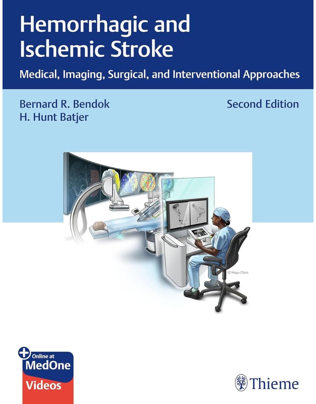
Hemorrhagic and Ischemic Stroke: Medical, Imaging, Surgical, and Interventional Approaches
Livrare gratis la comenzi peste 500 RON. Pentru celelalte comenzi livrarea este 20 RON.
Disponibilitate: La comanda in aproximativ 4-6 saptamani
Autor: Bernard Bendok, H. Batjer
Editura: Thieme
Limba: Engleza
Nr. pagini: 484
Coperta: Hardcover
Dimensiuni: 279 x 216 mm
An aparitie: 06.11.2024
A transdisciplinary approach to the medical, radiologic, interventional, and surgical treatment of stroke
In the last decade, advances in interventional, microsurgcial, radiosurgical techniques, medical approaches and imaging technology have dramatically changed the management of ischemic stroke. Perhaps the most important advance has been the increasing collaboration between multispecialty clinicians to deliver team-based stroke care. Hemorrhagic and Ischemic Stroke: Medical, Imaging, Surgical, and Interventional Approaches, Second Edition by renowned neurosurgeons Bernard R. Bendok and H. Hunt Batjer and acclaimed contributors provides a user-friendly, single resource for managing ischemic and hemorrhagic stroke. The second edition has been updated to reflect progress, most notably acute evaluation and triage, advanced imaging, critical care and medical support, and endovascular and surgical interventions. In addition to scientifically supported content, readers will benefit from pragmatic, real-world applications and concise, practical reviews.
Organized into four sections and 41 chapters, the text starts with the section Acute Triage, which includes early diagnosis and medical decision-making, advances in medical imaging, innovations in prehospital management of stroke, and the promise of telestroke networks. The second section, Critical Care, covers the importance of astute clinical examination, stabilization, and advanced neuromonitoring techniques. The third section, Open Surgical, Minimally Invasive, and Microsurgical Approaches, reviews recent surgical innovations that have the potential to be lifesaving and enhance quality of life in select patients. The fourth and final section, Neurointerventional Approaches, explores recent innovations in image-guided catheter techniques to deliver new therapies for stroke treatment and prevention.
Key Highlights:
The often difficult yet important adjudication of antithrombotic medication use
Insightful discussion of the importance of prehospital systems of care and stroke center certification
Insightful discussions of interventional and microsurgical approaches for hemorrhagic and ischemic stroke management
Medical illustrations and author-narrated videos provide a visual learning experience
Clinical pearls and case presentations enhance learning and retention of knowledge
The goal of this book is to increase collaborative stroke management among neuroradiologists, neurologists, neurosurgeons, neuroanesthesiolgists, neurologic critical care physicians, and allied health care professionals to achieve better patient outcomes.
Table of Contents:
Section I Acute Triage
1 Clinical Presentation and Acute Triage of Ischemic Stroke, Intracranial Hemorrhage, and Subarachnoid Hemorrhage
1.1 Clinical Case
1.2 Introduction
1.3 Acute Ischemic Stroke
1.3.1 Prehospital Setting
1.3.2 EMS Assessment and Management
1.3.3 Rapid Screening for Presence of Large Vessel Occlusion
1.3.4 Hospital Setting
1.3.5 Initial Stabilization
1.3.6 Clinical Examination
1.3.7 Determination of Thrombolysis Eligibility
1.3.8 Assessment of the Presence of LVO and Eligibility for Mechanical Thrombectomy
1.4 Intracranial Hemorrhage
1.4.1 Presentation
1.4.2 Prehospital Setting
1.4.3 Hospital Setting
1.5 Subarachnoid Hemorrhage
1.5.1 Presentation
1.6 Conclusions
1.7 Clinical Case Answers
1.8 References
2 Differential Diagnosis of Acute Ischemic and Hemorrhagic Stroke
2.1 Clinical Case
2.2 Introduction
2.3 Differential Diagnosis of Ischemic Strokes: Common Stroke Syndromes
2.4 Internal Carotid Artery Syndrome
2.4.1 Large Hemispheric Infarctions
2.4.2 Carotid Artery Dissection/Chronic ICA Occlusion/Hyperperfusion Syndromes
2.5 Anterior Cerebral Artery Syndrome
2.6 Middle Cerebral Artery Syndrome
2.7 Posterior Cerebral Artery Syndrome
2.8 Top of the Basilar Artery Syndrome and Brainstem Syndromes
2.9 Lacunar Strokes
2.10 Anterior Choroidal Artery Infarction
2.11 Behavioral Abnormalities Due to Acute Stroke
2.12 Intracerebral Hemorrhage
2.12.1 Hypertensive ICH
2.12.2 Cerebral Amyloid Angiopathy (CAA)
2.12.3 CNS Vascular Malformations
2.12.4 Medication-Induced Hemorrhage
2.13 Subarachnoid Hemorrhage
2.13.1 Aneurysmal SAH
2.13.2 Nonaneurysmal Perimesencephalic SAH
2.13.3 CNS Vascular Malformations
2.13.4 Cervicocephalic Arterial Dissection
2.13.5 Hematologic
2.14 Conclusion
2.15 Clinical Case Answers
2.16 References
2.17 Suggested Readings
3 Medical Imaging for Acute Ischemic and Hemorrhagic Stroke
3.1 Introduction
3.2 Imaging Modalities: Computed Tomography
3.2.1 Noncontrast Head CT
3.2.2 Computed Tomography Angiography
3.2.3 Computed Tomography Perfusion
3.3 Imaging Modalities: Magnetic Resonance Imaging
3.3.1 Diffusion-Weighted Imaging
3.3.2 Perfusion-Weighted Imaging
3.3.3 Magnetic Resonance Angiography
3.4 Summary
3.5 Clinical Case Answers
3.6 References
3.7 Suggested Readings
4 The Promise of Telestroke
4.1 Clinical Case
4.2 Introduction
4.3 Clinical Uses
4.3.1 Telestroke in the Emergency Department Setting
4.3.2 Telestroke in Settings Spanning the Continuum of Stroke Care
4.3.3 Telestroke in Clinical Research
4.3.4 Telestroke in Education
4.4 Technological Considerations
4.5 Telestroke Quality and Outcome Metrics
4.6 Telestroke Network Operational Considerations
4.6.1 Potential Obstacles
4.6.2 Health Economic Evaluations of Telestroke
4.7 Comprehensive Virtual Stroke Centers of the Future
4.8 Clinical Case Answers
4.9 References
4.10 Suggested Readings
5 Innovations in Prehospital Management of Stroke
5.1 Clinical Case
5.2 Emergency Medical System and Stroke Systems of Care
5.2.1 EMS Notification by the General Population
5.2.2 EMS Role in Stroke Triage
5.2.3 EMS Stroke Assessment: Prehospital Scales
5.2.4 Triage and Transport to the Target Stroke Center
5.2.5 Concept of the “Golden Hour”
5.3 The Mobile Stroke Unit
5.3.1 Mobile Stroke Ambulance: Ground and Air
5.3.2 Prehospital Brain Imaging
5.3.3 Prehospital Laboratory and Pharmacy
5.3.4 Cost-Effectiveness
5.4 Prehospital Stroke Management
5.4.1 Telemedicine in Prehospital Stroke Evaluation
5.4.2 Treatment of Acute Ischemic Stroke
5.4.3 Treatment of Hemorrhagic Stroke
5.4.4 Neuroprotection and Clinical Research in Prehospital Setting
5.5 Future Directions
5.6 Conclusion
5.7 Clinical Case Answers
5.8 Acknowledgements
5.9 References
5.10 Suggested Readings
6 Intravenous Fibrinolysis for Acute Ischemic Stroke
6.1 Clinical Case 1
6.2 Fibrinolysis
6.3 Fibrinolytic Drugs Studied for the Treatment of Acute Ischemic Stroke
6.4 History of Fibrinolysis Treatment for Acute Cerebral Ischemia
6.4.1 Clinical Case 2
6.4.2 Case Management Issues
6.5 Initial Restrictions for Administration of Intravenous Fibrinolytic Medication
6.5.1 Clinical Case 3
6.6 Increasing Utilization of Fibrinolysis for Treating Acute Ischemic Stroke Patients
6.6.1 Clinical Case 4
6.7 Revising Original Indications for the Use of Fibrinolytic Medication
6.7.1 Time from Stroke Onset, or Last Known to be at Usual Baseline
6.7.2 Age and Baseline Stroke Severity
6.7.3 Rapidly Improving Neurological Deficits
6.7.4 Use of Anticoagulation Medication
6.7.5 Mild, Nondisabling, Symptoms at Presentation
6.7.6 Patients without Capacity to Provide Consent to Treatment
6.7.7 Early Ischemic Changes on CT Imaging
6.7.8 Other Conditions Previously Considered by Some to be Too Risky for “Off-Label” Use of tPA
6.8 Clinical Situations where there is Considerable Uncertainty About Administering tPA to an Acute Ischemic Stroke Patient
6.9 Utilization of Telemedicine to Increase Use of Fibrinolytic Medication in the Treatment of Acute Ischemic Stroke
6.9.1 Clinical Case 5
6.10 Other Factors Impacting tPA Utilization Rates
6.10.1 Stroke Systems of Care
6.10.2 Teaching Hospitals
6.11 Future Strategies to Increase Utilization of Fibrinolysis for the Treatment of Acute Ischemic Stroke
6.12 Conclusion
6.13 Clinical Case 1 Answers
6.14 References
7 Identifying Patients for Stroke Thrombectomy
7.1 Clinical Case
7.2 Questions
7.3 Introduction
7.4 AHA/ASA 2019 Guidelines
7.5 Technical Details
7.6 Low ASPECTS Trials
7.7 Medium and Distal Occlusions
7.8 Intracranial Atherosclerotic Disease
7.9 Tandem Occlusion
7.10 Posterior Circulation Stroke
7.10.1 Basilar Artery Occlusion
7.10.2 Posterior Cerebral Artery Occlusion
7.11 Special Situations
7.11.1 Low National Institutes of Health Stroke Scale Score
7.11.2 Poor Baseline Modified Rankin Scale
7.11.3 Cancer
7.12 Future Directions
7.13 References
8 Stroke Center Certification
8.1 Clinical Case
8.2 Stroke Accreditation
8.2.1 Joint Commission Certification Process
8.2.2 Level 2 Acute Stroke Ready Hospital
8.2.3 Primary Stroke Center
8.2.4 Thrombectomy Capable
8.2.5 Comprehensive Stroke Center
8.3 Organization 2: Healthcare Facilities Accreditation Program
8.4 Organization 3: DNV GL
8.5 Clinical Case Answers
8.6 References
8.7 Suggested Readings
Section II Critical Care
9 Critical Care Management of Acute Ischemic Stroke
9.1 Clinical Case
9.2 Introduction
9.3 Critical Care after Acute Ischemic Stroke
9.3.1 Airway and Ventilation Management
9.3.2 Blood Pressure Monitoring and Goals
9.3.3 Antiplatelets, Systemic Anticoagulation, and Statin Therapy Initiation
9.3.4 Point-of-Care Ultrasound and Cardiac Telemetry
9.3.5 Neuromonitoring Modalities
9.4 Common Neurocritical Care Complications
9.4.1 Angioedema after IV tPA
9.4.2 Symptomatic Intracranial Hemorrhage
9.4.3 Malignant Cerebral Edema
9.4.4 Seizures
9.4.5 Other Complications
9.5 Conclusion
9.6 Clinical Case Answers
9.7 References
9.8 Suggested Reading
10 New Horizons for Hemorrhagic Stroke Critical Care Management
10.1 Clinical Case 1
10.2 Introduction
10.3 Etiology/Risk Factors
10.4 Medical Management
10.5 Clinical Severity Assessment
10.6 Emergency Department Management
10.7 The Importance of Intensive Medical Therapies
10.7.1 Fever
10.7.2 Hyperglycemia
10.7.3 Hypertension
10.7.4 Deep Veins Thrombosis
10.7.5 Steroids
10.7.6 Anticonvulsants
10.7.7 Cerebral Edema
10.7.8 Hydrocephalus and Intraventricular Hemorrhage
10.7.9 Anticoagulant-Related Intracranial Hemorrhage
10.8 Clinical Case 2
10.8.1 Heparin and Low-Molecular-Weight Heparins
10.8.2 Antiplatelet Agents
10.8.3 Factor Xa Inhibitors
10.8.4 Direct Thrombin Inhibitor
10.9 Resuming Anticoagulation Therapy after Intracerebral Hemorrhage
10.9.1 Outcome Predictions and Withdrawal of Life-Sustaining Treatments
10.10 Conclusion
10.11 Clinical Case Answers
10.12 References
10.13 Suggested Readings
11 Critical Care Management of Subarachnoid Hemorrhage and Vasospasm
11.1 Clinical Case
11.2 Introduction
11.3 Grading of Subarachnoid Hemorrhage
11.4 Diagnostic Challenges
11.5 Initial Stabilization
11.6 Common Complications
11.7 Seizures
11.8 Blood Pressure
11.9 Hydrocephalus
11.10 Rebleeding
11.11 Hyponatremia
11.12 Cardiac Arrhythmias and Cardiomyopathy
11.13 Glucose
11.14 Fever
11.15 Deep Vein Thrombosis
11.16 Vasospasm and Delayed Cerebral Ischemia
11.17 Prevention of Vasospasm
11.18 Detection of Vasospasm
11.19 Treatment of Vasospasm
11.20 Conclusion
11.21 References
11.22 Suggested Readings
12 Management of Anticoagulation and Antiplatelets in Hemorrhagic Stroke: Administration and Reversal
12.1 Clinical Case
12.1.1 Questions
12.2 Introduction
12.2.1 Pharmacology and Reversal of Antiplatelets
12.2.2 Pharmacology and Reversal of Anticoagulants
12.2.3 Challenges of Antiplatelets/Anticoagulants in SAH and ICH
12.3 Clinical Cases
12.3.1 Intracerebral Hemorrhage with a Mechanical Cardiac Valve
12.3.2 Acute Stenting with MCA Infarct
12.3.3 Atrial Fibrillation with Hemorrhagic Conversion of Infarct
12.4 Conclusion
12.5 Clinical Case Answers
12.6 References
13 Medical Management of Ischemic Stroke and Prevention
13.1 Clinical Case
13.2 General Medical Management
13.2.1 Initial Acute Management
13.2.2 Blood Pressure, Temperature, and Blood Glucose Management
13.2.3 Antiplatelet Treatment
13.2.4 Additional Screening and Prophylaxis
13.2.5 Stroke Mimics
13.2.6 Subsequent Workup and Monitoring
13.3 Etiology-Specific Management
13.3.1 Large Artery Atherosclerosis
13.3.2 Cardioembolic
13.3.3 Small Vessel Occlusion
13.3.4 Other Determined Causes of Ischemic Stroke
13.3.5 Cryptogenic
13.4 Secondary Prevention
13.5 Clinical Case Answers
13.6 References
13.7 Suggested Readings
14 Prognostication, Ethics, and Scales
14.1 Clinical Case
14.2 Prognostication in Stroke
14.3 Methods of Prognostication
14.4 Clinical Grading Scales
14.4.1 Ischemic Stroke
14.4.2 Intracerebral Hemorrhage
14.4.3 Subarachnoid Hemorrhage
14.5 Neuroimaging
14.6 Electrophysiology
14.7 Providing a Prognosis
14.8 Ethics
14.9 Clinical Case Answers
14.10 References
14.11 Links to Relevant Guidelines
14.12 Suggested Readings
15 Advanced Neuromonitoring in Acute Brain Injury
15.1 Clinical Case
15.2 Introduction
15.3 Placement of Brain Probes
15.4 Physiologic Principles
15.4.1 Intracranial Pressure and Monitoring
15.4.2 Brain Tissue Oxygen Monitoring
15.5 Neuroelectrophysiology
15.5.1 Electroencephalogram, Quantitative EEG, and Bispectral Index
15.5.2 Somatosensory Evoked Potentials
15.6 Case Presentation
15.7 Clinical Case Answers and Discussion
15.7.1 Highlights
15.8 References
15.9 Suggested Readings
16 Transcranial Doppler in the Intensive Care Unit
16.1 Clinical Case
16.2 Introduction
16.3 Physics
16.4 Traumatic Brain Injury
16.5 Subarachnoid Hemorrhage
16.6 Stroke
16.7 Brain Death Determination
16.8 Other Applications
16.9 Case Summary Discussion
16.10 References
16.11 Suggested Readings
17 Brain Death
17.1 Clinical Case 1
17.2 Brain Death: An Evolving Concept
17.3 Clinical Criteria
17.3.1 Prerequisites
17.3.2 Neurological Examination
17.4 Ancillary Testing
17.4.1 Electrophysiologic Tests
17.4.2 Neuroimaging
17.5 Clinical Case 2
17.6 Diagnosis of Brain Death in Pediatrics
17.7 Family Discussion
17.8 Physiologic Response to Brain Death and Organ Procurement
17.9 Clinical Case Answers
17.9.1 Case 1
17.9.2 Case 2
17.10 References
17.11 Suggested Further Readings
Section III Open Surgical, Minimally Invasive and Microsurgical Approaches
18 Anesthesia Considerations for Neurovascular Procedures
18.1 Clinical Case
18.2 Introduction
18.3 Preoperative Management
18.4 Intraoperative Management
18.4.1 Hemodynamic Management
18.4.2 Brain Bulk Management
18.4.3 Cerebral Protection
18.5 Anesthesia Considerations for Carotid Surgery
18.6 Anesthesia for Endovascular Treatment of Acute Ischemic Stroke
18.7 Postoperative Care
18.7.1 Systemic Hypertension on Emergence
18.7.2 Intracranial Hypertension on Emergence
18.8 Conclusion
18.9 Clinical Case Answers
18.10 References
18.11 Suggested Readings
19 Bypass for Acute and Chronic Ischemic States
19.1 Clinical Case
19.2 Introduction and Background
19.3 Patient Selection and Indications for Bypass Surgery
19.3.1 Diagnostic Workup
19.3.2 Indication
19.4 Treatment
19.4.1 Direct versus Indirect
19.5 Technique
19.5.1 Pre-, Peri-, and Postoperative Considerations
19.5.2 How We Do It: Combined Direct STA-MCA Anastomosis and EDAS
19.5.3 Vein Interposition
19.6 Clinical Case Answers
19.7 References
19.8 Suggested Readings
20 Moyamoya Disease
20.1 Clinical Case
20.2 Description
20.3 Epidemiology
20.4 Natural History
20.4.1 Adult Moyamoya Disease
20.4.2 Pediatric Moyamoya Disease
20.5 Radiographic Findings
20.6 Genetics
20.7 Treatment Options
20.7.1 Expectant
20.7.2 Medical
20.7.3 Surgical
20.8 Surgical Techniques
20.8.1 Direct Revascularization
20.8.2 Indirect Revascularization
20.9 Outcomes
20.10 Clinical Case Answers
20.11 References
20.12 Suggested Readings
21 Carotid Endarterectomy
21.1 Clinical Case
21.2 Natural History and Pathophysiology
21.3 History of Carotid Endarterectomy
21.4 Technique
21.4.1 Preparation
21.4.2 Positioning
21.4.3 Exposure
21.4.4 Vessel Occlusion
21.4.5 Plaque Removal
21.4.6 Shunt
21.4.7 Closure
21.4.8 Postoperative Care
21.5 Complications
21.5.1 Ischemia
21.5.2 Hemorrhage
21.5.3 Other
21.6 Patient Selection
21.6.1 Symptomatic Carotid Stenosis
21.6.2 Asymptomatic Carotid Stenosis
21.7 Clinical Case Answers
21.8 References
21.9 Suggested Readings
22 Microsurgical Revascularization of Extracranial Vertebral Artery Disease
22.1 Introduction
22.2 Anatomy
22.3 Clinical Manifestations
22.4 Indications for Treatment
22.5 Symptomatic Patients
22.6 Asymptomatic Patients
22.7 Microsurgical Revascularization of the Extracranial Artery
22.8 Surgical Technique for Proximal Vertebral Artery Revascularization
22.9 Microsurgical Revascularization versus Angioplasty and Stenting of the Proximal Vertebral Artery
22.10 Surgical Technique for Microsurgical Decompression of V2 Segment
22.11 Microsurgical Treatment of V3 Compression
22.12 Conclusion
22.13 References
23 Microsurgery for Unruptured Aneurysms
23.1 Clinical Case
23.2 Introduction
23.3 Diagnosis
23.3.1 Imaging Workup
23.3.2 Predictors of Rupture
23.3.3 Screening
23.3.4 Effect on Quality of Life and Other Considerations
23.4 Treatment Decision-Making
23.4.1 Risk of Treatment
23.4.2 International Study of Unruptured Intracranial Aneurysms
23.4.3 Unruptured Intracranial Aneurysm Treatment Score
23.4.4 Southwestern Aneurysm Severity Index
23.5 Treatment Options
23.6 Microsurgery Techniques
23.6.1 Mycotic Aneurysms
23.6.2 Mirror Aneurysms
23.7 Conclusion
23.8 Clinical Case Discussion
23.9 References
24 Microsurgery for Ruptured Aneurysms
24.1 Clinical Case
24.2 Introduction, Epidemiology, and Patient Selection
24.3 Pre- and Postoperative Management
24.4 Preoperative Imaging
24.5 Surgical Technique
24.5.1 Exposure
24.5.2 Temporary Clipping
24.5.3 Permanent Clip Selection
24.5.4 Anatomic Nuances: Anterior Circulation
24.5.5 Anatomic Nuances: Posterior Circulation Aneurysms
24.5.6 Other Anatomic Subtypes
24.6 Complication Avoidance and Management
24.6.1 Managing Intraoperative Rupture
24.6.2 Specific Anatomical Considerations
24.6.3 Conclusion
24.7 Clinical Case Answers
24.8 References
24.9 Suggested Readings
25 Microsurgery for Intracranial Arteriovenous Malformations
25.1 Clinical Case
25.2 Introduction
25.3 Epidemiology and Natural History
25.4 Evaluation of Arteriovenous Malformation
25.5 Grading of Arteriovenous Malformation
25.6 Decision-Making
25.7 Role of Preoperative Embolization
25.8 Microsurgery
25.9 Postoperative Management
25.10 Microsurgical Considerations at Specific Critical Locations
25.11 Surgical Complications and Management
25.12 Surgical Outcome
25.13 Future Directions
25.14 Clinical Case Answers
25.15 References
26 Microsurgery of Cavernous Malformations
26.1 Angioma Alliance 2017 Guidelines ▶ [615]
26.2 Clinical Case
26.3 Introduction
26.4 Radiographic Presentation
26.5 Surgical Indications
26.6 Microsurgical Techniques and Approaches
26.7 Microsurgical Technique
26.8 Surgical Approaches
26.8.1 Supracerebellar-Infratentorial Approach
26.8.2 Retrosigmoid Approach
26.8.3 Far-Lateral Approach
26.8.4 Interhemispheric Transcallosal Approach
26.9 Clinical Outcomes of Surgical Resection
26.10 Conclusion
26.11 Clinical Case Answers
26.12 References
26.13 Suggested Readings
27 Surgery for Intracranial Dural Arteriovenous Fistulas
27.1 Clinical Case
27.2 Introduction and Epidemiology
27.3 Proposed Pathogenesis
27.4 Presentation
27.5 Classification Schema
27.6 Imaging
27.7 Patient Selection and Treatment
27.7.1 Supratentorial
27.7.2 Infratentorial
27.8 Combined Surgical/Endovascular Treatment
27.9 Summary
27.10 Clinical Case Answers
27.11 References
27.12 Suggested Readings
28 Open Surgery and Craniectomy for Malignant Stroke and ICH
28.1 Clinical Case
28.2 Introduction
28.3 Craniectomy for Malignant Ischemic Stroke
28.3.1 Evidence on Craniectomy for Malignant Edema in Acute Ischemic Stroke
28.3.2 Guidelines
28.3.3 Surgical Technique
28.4 Open Surgery for Intracranial Hemorrhage
28.4.1 Intracranial Hemorrhage Clot Evacuation
28.4.2 Role of Craniectomy in Intracerebral Hemorrhage
28.4.3 Timing of Surgery
28.4.4 Guidelines
28.5 Cranioplasty
28.5.1 Stroke-Specific Studies
28.5.2 Large-Scale Studies
28.6 Conclusions
28.7 Clinical Case Answers
28.8 References
29 Minimally Invasive Approaches for Intracerebral Hemorrhage and Intraventricular Hemorrhage
29.1 Clinical Case
29.2 Introduction
29.3 Pathogenesis and Outcome Determinants
29.4 Acute Resuscitation and Medical Management
29.5 Blood Pressure Control
29.6 Reversal of Coagulopathy
29.7 Intracranial Pressure Management
29.8 Surgical Management
29.8.1 Open Craniotomy
29.8.2 Rationale for Minimally Invasive Procedures
29.9 Intracerebral Hemorrhage and MISTIE
29.9.1 Introduction
29.9.2 Clot Stability and Etiology Screening
29.9.3 Catheter Trajectories
29.9.4 Cannula Aspiration and Catheter Placement for Thrombolysis
29.9.5 Outcomes
29.9.6 Lesions Learned
29.10 Intraventricular Hemorrhage and CLEAR
29.10.1 Introduction
29.10.2 Rationale
29.10.3 CLEAR IVH and CLEAR III
29.10.4 Clot Stability and Etiology Screening
29.10.5 Catheter Placement
29.10.6 Lessons Learned
29.11 Other Trials
29.12 Conclusion
29.13 Clinical Case Answers
29.14 References
29.15 Suggested Readings
30 Radiosurgical Management of Arteriovenous Malformations
30.1 Clinical Case
30.2 Introduction
30.3 The Value of Stereotactic Radiosurgery
30.4 Limitations of Radiosurgery
30.5 How Does Radiosurgery Work?
30.6 Clinical Experience
30.7 How AVM Obliteration Affects Decision-Making
30.8 Timing
30.9 Radiosurgery Morbidity and Decision-Making
30.10 Options for Large AVMs: Staged Volume Radiosurgery and Embolization
30.11 Embolization and SRS
30.12 Radiosurgery First, then Embolization
30.13 Radiosurgery for Dural Arteriovenous Fistulas
30.14 Conclusion
30.15 Clinical Case Answers
30.16 References
Section IV Neurointerventional Approaches
31 Endovascular Thrombectomy: Revolution in the Care of Acute Ischemic Stroke
31.1 Clinical Case
31.2 Introduction
31.3 Intra-arterial Thrombolysis
31.4 PROACT I
31.5 PROACT II
31.6 Intra-arterial Thrombolysis in the Posterior Circulation
31.7 Mechanical Thrombectomy
31.8 Patient Selection
31.9 Devices, Techniques, and Clot Types
31.10 Anesthesia
31.11 Complications Management
31.12 Future Directions
31.13 Clinical Case Answers
31.14 References
31.15 Suggested Readings
32 Intracranial Atherosclerotic Disease
32.1 Clinical Case
32.2 Introduction
32.3 Epidemiology
32.4 Natural History
32.5 Clinical and Radiographic Evaluation
32.6 Treatment Options
32.6.1 Medical Management
32.6.2 Endovascular Management
32.6.3 Management of ICAD Underlying a Large-Vessel Occlusion
32.6.4 Outcomes
32.6.5 Surgical Management
32.7 Conclusion
32.8 Clinical Case Answers
32.9 References
32.10 Suggested Readings
33 Endovascular Stenting in Atherosclerotic Carotid Artery Stenosis
33.1 Clinical Case
33.2 Pathogenesis
33.3 Modes of Presentation
33.4 Diagnostic Tests
33.4.1 Calculating the Degree of Stenosis
33.4.2 Carotid Duplex Ultrasonography
33.4.3 Magnetic Resonance Angiography
33.4.4 Computed Tomography Angiography
33.4.5 Digital Subtraction Angiography
33.5 Natural History
33.6 Medical Management
33.7 Carotid Endarterectomy
33.8 Carotid Artery Stenting
33.9 Treatment Selection
33.10 Contraindications for Carotid Artery Stenting
33.11 Carotid Artery Stenting Technique
33.11.1 Stages of Carotid Artery Stenting
33.12 Stent Options
33.13 Embolic Protection Devices
33.14 Complications
33.15 Clinical and Radiographic Follow-up
33.16 Conclusion
33.17 Clinical Case Answers
33.18 References
33.19 Suggested Readings
34 Management of Vertebral Artery Origin Stenosis
34.1 Clinical Case 1
34.2 Introduction
34.3 Epidemiology
34.4 Risk Factors
34.5 Natural History
34.6 Presentation
34.7 Imaging
34.7.1 Noninvasive Vessel Imaging
34.7.2 Perfusion Imaging
34.7.3 Catheter Angiography
34.8 Medical Treatment of Vertebral Origin Stenosis
34.8.1 Blood Pressure Management
34.8.2 Statin Therapy
34.9 Revascularization Procedures
34.9.1 Surgical Revascularization
34.10 Endovascular Treatment of Vertebral Artery Origin Stenosis
34.10.1 General Patient Preparation
34.10.2 Anti-Aggregation
34.10.3 Anticoagulation
34.10.4 Anesthesia
34.10.5 Access
34.10.6 Measurements
34.10.7 Distal Embolic Protection
34.10.8 Stenting Strategies
34.11 Clinical Case 1 (Continued)
34.11.1 Technique
34.11.2 Self-expanding Intracranial Stents
34.12 Clinical Case 2
34.12.1 Clinical History
34.12.2 Technique
34.13 Stenting from the Vertebral Artery into the Subclavian Artery
34.14 Conclusion
34.15 Clinical Case Answers
34.16 References
34.17 Suggested Reading
35 Aneurysm Coiling
35.1 Clinical Case
35.2 Introduction
35.3 Aneurysm Coiling
35.4 Adjuncts to Coiling
35.4.1 Balloon-Assisted Coiling
35.4.2 Stent-Assisted Coiling
35.4.3 Adjunct Device-Assisted Coiling
35.5 Patient Selection
35.5.1 Unruptured Aneurysms
35.5.2 Ruptured Aneurysms
35.6 Coiling versus Clipping
35.7 Conclusion
35.8 Clinical Clinical Case Answers and Discussion
35.9 References
36 Vasospasm Management
36.1 Clinical Case
36.2 Introduction
36.3 Epidemiology
36.4 Pathophysiology
36.5 Clinical Presentation and Symptoms
36.6 Diagnosis
36.7 Vasospasm Prophylaxis
36.8 Vasospasm Medical Management
36.9 Vasospasm Endovascular Management
36.9.1 Timing of Endovascular Management
36.9.2 Types of Endovascular Intervention
36.9.3 Chemical Angioplasty (Intra-arterial Vasodilator Therapy)
36.9.4 Mechanical Angioplasty
36.9.5 Balloon Angioplasty
36.9.6 Stent Angioplasty
36.10 Role of Prophylactic Angioplasty
36.11 Experimental Therapies
36.12 Clinical Case Answers and Discussion
36.13 References
37 Flow Diversion
37.1 Clinical Case 1
37.1.1 Flow Diversion for a Communicating Segment Internal Carotid Artery Aneurysm Using Coils and Pipeline Flex
37.2 Introduction to Flow Diversion
37.3 Current Indications and Clinical Use
37.4 Safety and Efficacy
37.5 Devices: Design, Current Availability, and Technique
37.5.1 Pipeline Embolization Device
37.5.2 Silk Flow Diverter
37.5.3 SURPASS
37.5.4 Flow Re-direction Endoluminal Device
37.5.5 p64 Flow Modulation Device
37.5.6 Bravo
37.6 Special Considerations
37.6.1 Posterior Circulation Aneurysms
37.6.2 Distal Aneurysms
37.6.3 Ruptured Aneurysms
37.6.4 Blister Aneurysms
37.6.5 Giant Aneurysms
37.6.6 Carotid Cavernous Fistula
37.7 Antiplatelet Therapy
37.7.1 Platelet Function Testing and Alternative Regimens
37.8 Clinical Case 2
37.8.1 Flow Diversion for a Blister Type Dorsal Wall Internal Carotid Artery Aneurysm Using Multiple Pipeline Flex Devices
37.9 Clinical Case 1 Answers
37.10 Clinical Case 2 Answer
37.11 References
37.12 Suggested Readings
38 Approaches to Extracranial and Intracranial Dissection
38.1 Clinical Case 1
38.2 Pathophysiology and Natural History
38.3 Imaging of Cervical and Vertebral Artery Dissections
38.4 The Cervical Artery Dissection in Ischemic Stroke Patients Study Group: Medical Management of Dissections
38.5 Surgical Intervention and the Management of Dissections
38.6 Ischemic Presentation of Dissection and Surgical Intervention
38.7 Clinical Cases
38.8 Posterior Circulation Dissections and Surgical Treatment Options
38.9 Pseudoaneurysms and Subarachnoid Hemorrhage
38.10 Clinical Case
38.11 Technical Nuances of Endovascular Stenting, Stent-Assisted Coiling, and Flow Diversion
38.12 Conclusion
38.13 Clinical Case Answers and Discussion
38.14 References
39 Endovascular Management of Arteriovenous Malformations
39.1 Clinical Case
39.2 Historical Perspective
39.3 Pathophysiology
39.4 Epidemiology
39.5 Presenting Symptoms and Natural History
39.6 Anatomy and Classification
39.7 ARUBA Trial
39.8 Indications and Strategies for Endovascular Treatment
39.8.1 Preoperative Embolization
39.8.2 Pre- and Postradiosurgery Embolization
39.8.3 Stand-Alone Treatment
39.9 Technical Considerations and Complication Avoidance Embolic Agents
39.10 Microcatheters
39.11 Advanced Angiographic Techniques
39.11.1 Transvenous Approach
39.11.2 Double Arterial Catheterization
39.11.3 Balloon-Assisted Embolization
39.12 Complication Avoidance
39.13 Conclusion
39.14 Clinical Case Answers
39.15 References
40 Endovascular Approaches to Intracranial Dural Arteriovenous Fistulas
40.1 Clinical Case 1
40.2 Introduction
40.3 Pathogenesis
40.4 Presentation
40.5 Natural History
40.6 Anatomic Considerations
40.7 Intracranial Venous Anatomy
40.7.1 Deep Venous Drainage
40.7.2 Cortical Venous Drainage
40.7.3 Sinus Drainage
40.8 Arterial Supply of the Dura
40.8.1 Arterial Anastomoses
40.9 Imaging
40.9.1 Computed Tomography
40.9.2 Magnetic Resonance Imaging
40.9.3 Digital Subtraction Angiography
40.10 Grading
40.11 Patient Selection
40.12 Endovascular Embolization
40.12.1 Transarterial Embolization
40.13 Clinical Case 2
40.13.1 Transvenous Embolization
40.13.2 Transfemoral Venous Access
40.13.3 Head and Neck Direct Venous Access
40.14 Clinical Case 3
40.14.1 Outcome
40.15 Conclusion
40.16 Clinical Case 1 Answers
40.17 References
40.18 Suggested Readings
41 Venous Thrombectomy
41.1 Clinical Case
41.2 Introduction
41.3 Epidemiology and Pathogenesis
41.4 Pathophysiology
41.5 Clinical Presentation
41.6 Diagnosis
41.6.1 Laboratory Tests
41.6.2 Computed Tomography and Computed Tomography Venogram
41.6.3 Magnetic Resonance Imaging and Magnetic Resonance Venogram
41.6.4 Catheter Angiography
41.7 Treatment
41.7.1 Systemic Anticoagulation
41.7.2 Endovascular Intervention
41.7.3 Rheolytic Thrombectomy
41.7.4 Clot Aspiration System
41.7.5 Merci Clot Retriever with Microsnare
41.7.6 Balloon Extraction
41.7.7 Stent Retrievers
41.8 Outcome
41.9 Medical or Surgical Management of Secondary Complications
41.9.1 Seizure
41.9.2 Hydrocephalus
41.9.3 Intracranial Hypertension
41.9.4 Visual Loss
41.9.5 Arteriovenous Fistula
41.10 Conclusions
41.11 Clinical Case Answers
41.12 References
41.13 Suggested Readings
Contributors
Index
Imprint/Access Code
| An aparitie | 06.11.2024 |
| Autor | Bernard Bendok, H. Batjer |
| Dimensiuni | 279 x 216 mm |
| Editura | Thieme |
| Format | Hardcover |
| ISBN | 9781684200436 |
| Limba | Engleza |
| Nr pag | 484 |
| Versiune digitala | DA |





Clientii ebookshop.ro nu au adaugat inca opinii pentru acest produs. Fii primul care adauga o parere, folosind formularul de mai jos.