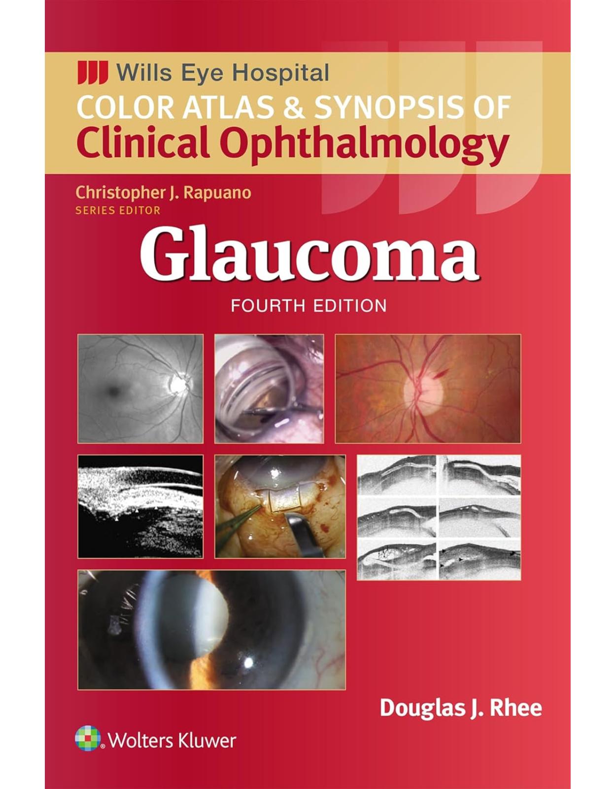
Glaucoma (Wills Eye Institute Atlas Series)
Livrare gratis la comenzi peste 500 RON. Pentru celelalte comenzi livrarea este 20 RON.
Disponibilitate: La comanda in aproximativ 4-6 saptamani
Autor: Douglas J. Rhee
Editura: LWW
Limba: Engleza
Nr. pagini: 550
Coperta: Paperback
Dimensiuni: 152 x 229 mm
An aparitie: 25 oct 2024
Developed at Philadelphia’s world-renowned Wills Eye Hospital, the Color Atlas and Synopsis of Clinical Ophthalmology series covers the most clinically relevant aspects of ophthalmology in a highly visual, easy-to-use format. Vibrant, full-color photos and a consistent outline structure present a succinct, high-yield approach to the seven topics covered by this popular series: Cornea, Retina, Glaucoma, Oculoplastics, Neuro-Ophthalmology, Pediatrics, and Uveitis. This in-depth, focused approach makes each volume an excellent companion to the larger Wills Eye Manual as well as a practical stand-alone reference for students, residents, and practitioners in every area of ophthalmology.
Table of Contents:
Chapter 1 Basic Science of Aqueous Humor Outflow
Aqueous Flow
Optic Nerve
Chapter 2 Tonometry
Introduction
Goldmann Applanation Tonometer
Schiötz Tonometer
Perkins Tonometer
Noncontact Tonometers
Tono-Pen
Pneumotonometer
Dynamic Contour Tonometry
iCare Tonometer
Promising Developments
Chapter 3 Gonioscopy
Introduction
Direct Gonioscopy
Indirect Gonioscopy
Estimating the Anterior Chamber Depth
Anterior Segment Imaging
Gonioscopy Technique
Elements of the Angle Anatomy
Identification of Angle Structures
Classification of the Angle
Pigment Deposition and Gonioscopy
Iridocorneoendothelial Syndrome (Ice Syndrome)
Use of Gonioscopy in Trauma
Traumatic Intraocular Foreign Bodies
Postoperative Evaluation
Chapter 4 Anterior Segment Imaging (UBM and Ant Seg OCT)
Introduction
Angle-Closure Glaucoma
Open-Angle Glaucoma
Other Conditions
Surgery and Glaucoma
Aqueous Outflow Pathway
Chapter 5 Optic Nerve Imaging
Introduction
Stereophotography
Confocal Scanning Laser Ophthalmoscopy
Scanning Laser Polarimetry
Optical Coherence Tomography (Structural)
Optical Coherence Tomography (Angiography)
Conclusion
Chapter 6 Retinal Nerve Fiber Layer Imaging: Peripapillary, and Macular Regions
Introduction
Structural Tests
Photography
Optical Coherence Tomography
Acknowledgments
Chapter 7 Psychophysical Testing
Introduction
Purpose of Test
Description
Glaucoma Progression Analysis
Common Optic Nerve Localizing Visual Fields Found in Patients With Glaucoma
Newer Psychophysical Testing: Frequency-Doubling Perimetry and Short-Wave Automated Perimetry
Electrophysiology Tests
Chapter 8 Blood Flow in Glaucoma
Introduction
Optical Coherence Tomography Angiography
Retinal Oximetry
Doppler Optical Coherence Tomography
Laser Doppler Flowmetry
Other Hemodynamic Imaging Techniques
Laser Speckle Flowgraphy
Mitochondrial Activity
Fluorescein and Indocyanine Green Scanning Laser Ophthalmoscope Angiography
Color Doppler Imaging
Pulsatile Assessments of Ocular Blood Flow
Acknowledgments
Disclosures
Chapter 9 Chidlhood Glaucoma
Introduction
Primary Congenital Glaucoma
Glaucoma Associated With Congenital Anomalies
Aniridia
Axenfeld Anomaly
Rieger Anomaly
Rieger Syndrome
Peters Anomaly
Marfan Syndrome
Microspherophakia
Sturge–Weber Syndrome (Encephalotrigeminal Angiomatosis)
Neurofibromatosis (Von Recklinghausen Disease and Bilateral Acoustic Neurofibromatosis)
Congenital Anterior Staphyloma and Keratectasia
Sclerocornea
Ectopia Lentis and Ectopia Lentis Et Pupillae
Chapter 10 Primary Open-Angle Glaucoma and Low Tension Glaucoma
Introduction
Definition
Epidemiology
Pathophysiology
History
Clinical Examination
Special Tests
Treatment
A Conceptual Approach to the Treatment of Glaucoma
Reversibility of Glaucoma
Chapter 11 Secondary Open-Angle Glaucoma (Steroid-induced, PXF, PDS)
Pigment Dispersion Syndrome
Pseudoexfoliation Syndrome
Steroid-Responsive Glaucoma
Chapter 12 Uveitic Glaucomas
Introduction
Epidemiology
Etiology
Open-Angle Mechanisms
Closed-Angle Mechanisms
Diagnosis
Management
Management of Angle-Closure Glaucoma
Specific Entities
Fuchs Heterochromic Iridocyclitis
Glaucomatocyclitic Crisis (Posner–Schlossman Syndrome)
Herpetic Keratouveitis
Diagnostic Evaluation
Syphilitic Interstitial Keratitis
Juvenile Idiopathic Arthritis
Lens-Induced Uveitis and Glaucoma
Sarcoidosis
Chapter 13 Lens-Associated Glaucomas
Introduction
Lens Protein or Phacolytic Glaucoma
Lens Particle Glaucoma
Lens-Associated Uveitis (Phacoanaphylaxis)
Phacomorphic Glaucoma
Chapter 14 Traumatic Glaucoma
Introduction
Traumatic Hyphema
Angle Recession
Cyclodialysis Cleft
Chapter 15 Primary Acute Angle Closure and Primary Angle Closure Glaucoma
Background
Primary Acute Angle Closure
Chapter 16 Secondary Angle–Closure Glaucoma
Neovascular Glaucoma
Iridocorneal Syndromes
Aqueous Misdirection Syndrome (Malignant Glaucoma)
Uveitis Glaucoma Hyphema Syndrome
Chapter 17 Glaucoma Secondary to Elevated Episcleral Venous Pressure
Introduction
Carotid–Cavernous Fistula
Sturge-Weber Syndrome
Idiopathic Elevated Episcleral Venous Pressure (Radius-Maumenee Syndrome)
Other Causes of Elevated Episcleral Venous Pressure
Chapter 18 Medical Management
Introduction
Description and Physiology
Alpha-Agonists
Beta-Blockers
Carbonic Anhydrase Inhibitors
Hyperosmolar Agents
Miotics
Prostaglandins
Rho Kinase Inhibitors
Sympathomimetic Agents
Combination Agents
Extended-Release Prostaglandin Analogue Depot
Technique of Drop Instillation
Chapter 19 Laser Trabeculoplasty
Introduction
Laser Overview
Mechanism of Action
Efficacy
Chapter 20 Microinvasive Glaucoma Surgery: Anterior Chamber–Schlemm Canal Stents (iStent, iStent inject, Hydrus)
Introduction
Indications
iStent
iStent Inject/Infinite
Hydrus
CHAPTER 21 Schlemm Canal Surgery—Dilation and Ablation
Introduction
Indications
Canaloplasty
Trabecular Meshwork Ablating Procedures: Direct Blade Approach and Catheter Approach
Chapter 22 Deep Sclerectomy Surgery for Glaucoma
Introduction
Studies
Surgical Technique
Postoperative Care
Conclusion
Chapter 23 Trabeculectomy, the Ex-Press Mini Glaucoma Shunt, the Xen, and the Preserflo Microshunts
Trabeculectomy
Surgical Technique
Intraoperative Application of Antimetabolites
Postoperative Care
Antivascular Endothelial Growth Factor Therapy for Bleb Vascularization
Bleb Needling
Complications
Ex-Press Mini Glaucoma Shunt
Surgical Technique
Xen Glaucoma Implant
Preserflo Glaucoma Implant
Acknowledgment
Chapter 24 Glaucoma Drainage Devices
Description
Surgical Technique
Chapter 25 Cyclodestructive Procedures for Glaucoma
Introduction
Techniques
Postprocedure Care
Complications
Chapter 26 Late Complications of Incisional Glaucoma Surgery
Introduction
Hypotony
Bleb Leaks
Giant Blebs
The Failing Bleb: Encapsulation
Circumferential Bleb
Tube Shunt Erosions
Index
| An aparitie | 25 oct 2024 |
| Autor | Douglas J. Rhee |
| Dimensiuni | 152 x 229 mm |
| Editura | LWW |
| Format | Paperback |
| ISBN | 9781975214814 |
| Limba | Engleza |
| Nr pag | 550 |
-
-

-
 47600 lei 42300 lei
 -
-


Clientii ebookshop.ro nu au adaugat inca opinii pentru acest produs. Fii primul care adauga o parere, folosind formularul de mai jos.