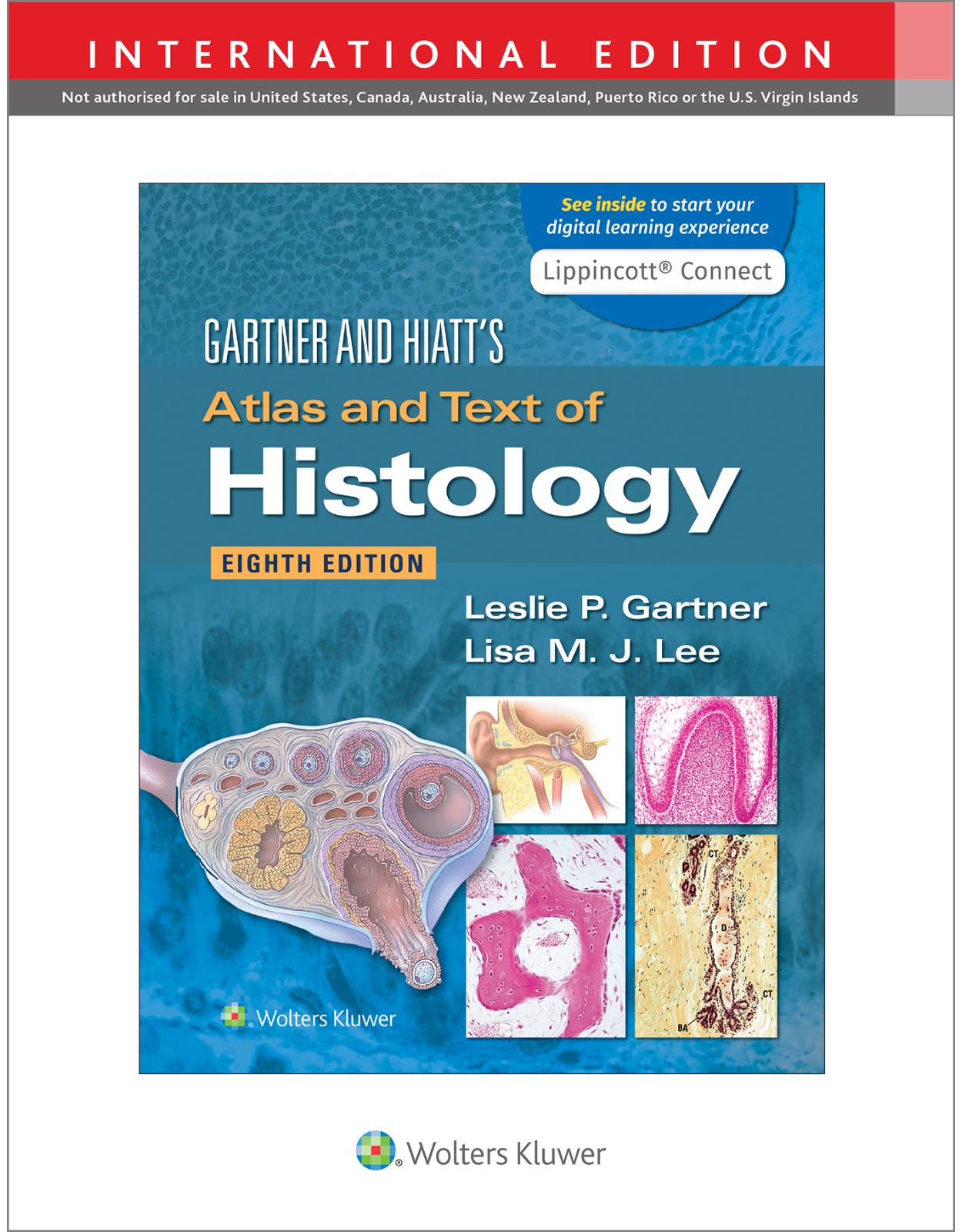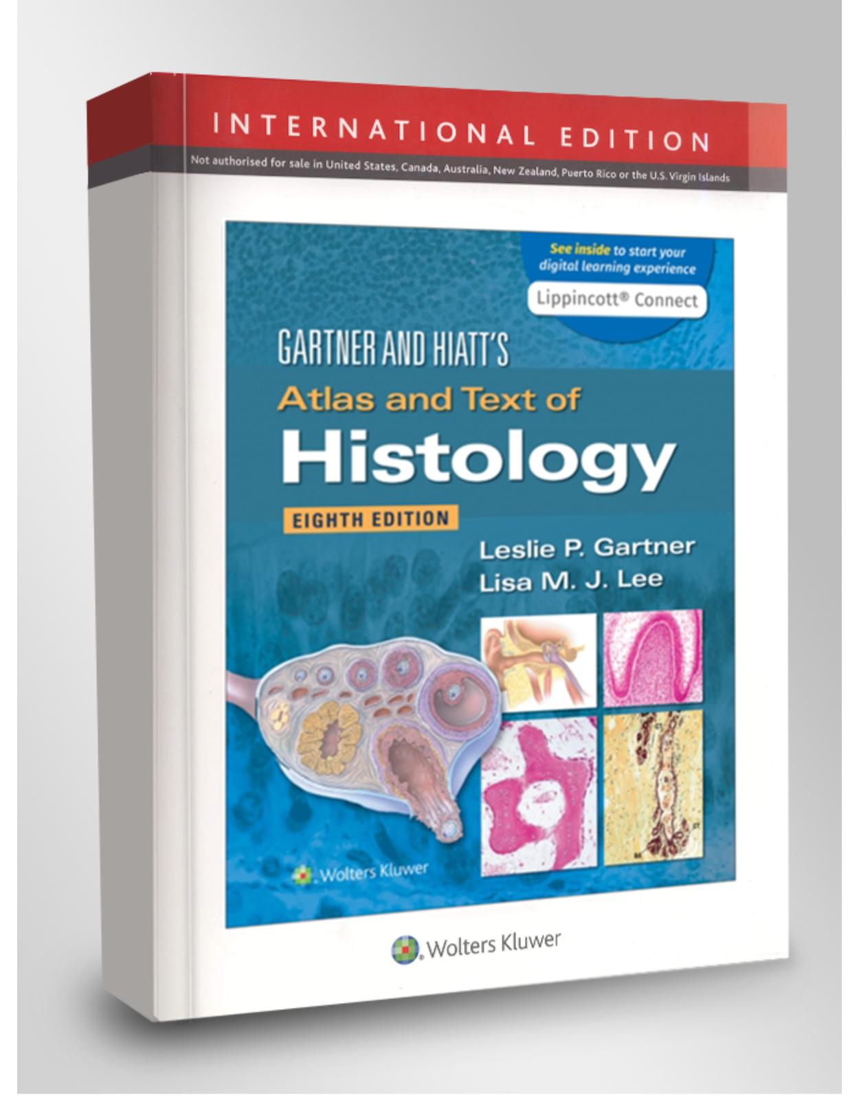
Gartner & Hiatt’s Atlas and Text of Histology
Livrare gratis la comenzi peste 500 RON. Pentru celelalte comenzi livrarea este 20 RON.
Disponibilitate: La comanda in aproximativ 4-6 saptamani
Editura: LWW
Limba: Engleza
Nr. pagini: 608
Coperta: Paperback
Dimensiuni: 213 x 276 mm
An aparitie: 25/04/2022
Description:
The go-to tool for mastering histology, Gartner & Hiatt's Atlas and Text of Histology, 8th Edition, equips medical, dental, allied health, and biology students with a concise review of all of the major tissue classes and body systems in an engaging approach optimized for superior classroom and clinical success. A consistent presentation combines relevant text and detailed photomicrographs to facilitate understanding and provide valuable review for in-class and licensing examinations. Helpful explanatory text in each chapter details Histophysiology, Clinical Considerations, Summaries of Histologic Organization, and more, accompanied by more than 700 vivid, full-color, high-quality images.
Lippincott® Connect features:
- Full access to the digital version of the book with the ability to highlight and take notes on key passages for a more personal, efficient study experience.
- Carefully curated resources, such as interactive diagrams, audio and video tutorials, and self-assessment, all designed to facilitate further comprehension.
Table of Contents:
CHAPTER 1 INTRODUCTION TO HISTOLOGIC TECHNIQUES
Light Microscopy
Terminology of Staining for Light Microscopy
Common Stains Used in Histology
Transmission Electron Microscopy
Scanning Electron Microscopy
Interpreting Microscopic Sections
CHAPTER 2 CELL BIOLOGY
Plasmalemma
Cytoplasm
Mitochondria
Ribosomes
Endoplasmic Reticulum
Golgi Apparatus
CLINICAL CONSIDERATIONS 2-1
CLINICAL CONSIDERATIONS 2-2
CLINICAL CONSIDERATIONS 2-3
CLINICAL CONSIDERATIONS 2-4
Endosomes
Lysosomes
Peroxisomes
CLINICAL CONSIDERATIONS 2-5
Proteasomes
Cytoskeleton
CLINICAL CONSIDERATIONS 2-6
Inclusions
Nucleus
Nucleolus
Chromatin and Chromosomes
CLINICAL CONSIDERATIONS 2-7
CLINICAL CONSIDERATIONS 2-8
Cell Cycle
Mitosis
Meiosis
Necrosis and Apoptosis
PLATE
2-1A Typical Cell
2-1B Typical Cell
2-2A Cell Organelles and Inclusions
2-2B Cell Organelles and Inclusions
2-3A Cell Surface Modifications
2-3B Cell Surface Modifications
2-4A Mitosis, Light and Electron Microscopy
2-4B Mitosis, Light and Electron Microscopy
2-5 Typical Cell, Electron Microscopy
Chapter Review Questions
CHAPTER 3 EPITHELIUM AND GLANDS
Epithelial Tissue
Epithelial Tissue Characteristics
Epithelial Tissue Classification
Epithelial Cell Membrane Specializations
Glands
CLINICAL CONSIDERATIONS 3-1
CLINICAL CONSIDERATIONS 3-2
CLINICAL CONSIDERATIONS 3-3
CLINICAL CONSIDERATIONS 3-4
PLATE
3-1A Simple Epithelia and Pseudostratified Epithelium
3-1B Simple Epithelia and Pseudostratified Epithelium
3-2 Stratified Epithelium and Transitional Epithelium
3-3A Epithelial Junctions, Electron Microscopy
3-3B Epithelial Junctions, Electron Microscopy
3-4A Glands
3-4B Glands
3-4C Glands
3-4D Glands
Selected Review of Histologic Images
Review Plate 3-1A
Review Plate 3-1B
Review Plate 3-1C
Summary of Histologic Organization
I. Epithelium
A. Types
B. General Characteristics
II. Glands
A. Exocrine Glands
B. Endocrine Glands
Chapter Review Questions
CHAPTER 4 CONNECTIVE TISSUE
Extracellular Matrix
Fibers
CLINICAL CONSIDERATIONS 4-1
CLINICAL CONSIDERATIONS 4-2
CLINICAL CONSIDERATIONS 4-3
Ground Substance
Cells
CLINICAL CONSIDERATIONS 4-4
CLINICAL CONSIDERATIONS 4-5
Connective Tissue Types
Mesenchymal and Mucous Connective Tissues
Loose (Areolar) Connective Tissue
Dense Irregular Connective Tissue
Dense Regular Connective Tissue
Reticular Connective Tissue
Elastic Connective Tissue
Adipose Tissue
CLINICAL CONSIDERATIONS 4-6
CLINICAL CONSIDERATIONS 4-7
PLATE
4-1 Fibroblasts and Collagen, Transmission Electron Microscopy
4-2 Mast Cell, Transmission Electron Microscopy
4-3 Mast Cell Degranulation, Electron Microscopy
4-4 Developing Fat Cell, Transmission Electron Microscopy
4-5 Connective Tissues
4-6 Connective Tissues
4-7 Connective Tissues and Cells
Selected Review of Histologic Images
Review Plate 4-1A
Review Plate 4-1B
Summary of Histologic Organization
I. Embryonic Connective Tissue
A. Mesenchymal Connective Tissue (Mesenchyme)
B. Mucous Connective Tissue
II. Connective Tissue Types
A. Loose (Areolar) Connective Tissue
B. Reticular Connective Tissue
C. Adipose Tissue
III. Extracellular Matrix
A. Dense Irregular Connective Tissue
B. Dense Regular Connective Tissue
C. Elastic Connective Tissue
Chapter Review Questions
CHAPTER 5 CARTILAGE AND BONE
Cartilage
Cartilage Extracellular Matrix
Cartilage Cellular Component
Perichondrium
Cartilage Growth
Cartilage Types
Bone
CLINICAL CONSIDERATIONS 5-1
CLINICAL CONSIDERATIONS 5-2
Bone Extracellular Matrix
Periosteum and Endosteum
Bone Cells
Osteogenesis
CLINICAL CONSIDERATIONS 5-3
CLINICAL CONSIDERATIONS 5-4
CLINICAL CONSIDERATIONS 5-5
Hormonal Influences on Bone
PLATE
5-1A Cartilage
5-1B Cartilage
5-2A Bone
5-2B Bone
5-2C Bone
5-3B Cells of the Bone
5-3B Cells of the Bone
5-3B Cells of the Bone
Selected Review of Histologic Images
Review Plate 5-1A
Review Plate 5-1B
Review Plate 5-2
Summary of Histologic Organization
I. Cartilage
A. Embryonic Cartilage
B. Hyaline Cartilage
C. Elastic Cartilage
D. Fibrocartilage
II. Bone
A. Compact Bone
B. Cancellous (Spongy, Medullary) Bone
C. Osteogenesis
Chapter Review Questions
CHAPTER 6 BLOOD AND HEMOPOIESIS
Blood
Formed Elements of Blood
CLINICAL CONSIDERATIONS 6-1
CLINICAL CONSIDERATIONS 6-2
CLINICAL CONSIDERATIONS 6-3
CLINICAL CONSIDERATIONS 6-4
CLINICAL CONSIDERATIONS 6-5
CLINICAL CONSIDERATIONS 6-6
CLINICAL CONSIDERATIONS 6-7
Plasma and Serum
Hemopoiesis
Erythrocytic Series
Granulocytic Series
Lymphoid Series
Hemopoietic Growth Factors
CLINICAL CONSIDERATIONS 6-8
PLATE
6-1A Circulating Blood
6-1B Circulating Blood
6-2 Circulating Blood (Drawing)
6-3 Blood and Hemopoiesis
6-4A Bone Marrow
6-4B Blood Smear and Bone Marrow Smear
6-5 Erythropoiesis
6-6 Granulocytopoiesis
Selected Review of Histologic Images
Review Plate 6-1A
Review Plate 6-1B
Review Plate 6-2A
Review Plate 6-2B
Summary of Histologic Organization
I. Circulating Blood
A. Erythrocytes (RBC)
B. Agranulocytes
C. Granulocytes
D. Platelets
E. Erythrocytic Series
F. Granulocytic Series
Chapter Review Questions
CHAPTER 7 MUSCLE
Skeletal Muscle
Myofilaments
CLINICAL CONSIDERATIONS 7-1
Sliding Filament Model of Skeletal Muscle Contraction
CLINICAL CONSIDERATIONS 7-2
CLINICAL CONSIDERATIONS 7-3
Skeletal Muscle Motor Innervation
Cardiac Muscle
CLINICAL CONSIDERATIONS 7-4
Morphologic Components of Cardiac Muscle Cells
CLINICAL CONSIDERATIONS 7-5
Smooth Muscle
Smooth Muscle Contraction
PLATE
7-1A Skeletal Muscle
7-1B Skeletal Muscle
7-2 Skeletal Muscle, Electron Microscopy
7-3A Myoneural Junction, Light Microscopy
7-3B Myoneural Junction, Electron Microscopy
7-4 Muscle Spindle, Light and Electron Microscopy
7-5A Smooth Muscle
7-5B Smooth Muscle
7-6 Smooth Muscle, Electron Microscopy
7-7A Cardiac Muscle
7-7B Cardiac Muscle
7-8 Cardiac Muscle, Electron Microscopy
Selected Review of Histologic Images
Review Plate 7-1A
Review Plate 7-1B
Review Plate 7-2A
Review Plate 7-2B
Review Plate 7-3A
Review Plate 7-3B
Summary of Histologic Organization
I. Skeletal Muscle
A. Longitudinal Section
B. Transverse Section
II. Cardiac Muscle
A. Longitudinal Section
B. Transverse Section
III. Smooth Muscle
A. Longitudinal Section
B. Transverse Section
Chapter Review Questions
CHAPTER 8 NERVOUS TISSUE
Nervous System
Neurons
CLINICAL CONSIDERATIONS 8-1
CLINICAL CONSIDERATIONS 8-2
CLINICAL CONSIDERATIONS 8-3
CLINICAL CONSIDERATIONS 8-4
Histologic Basis for Neuroanatomy
CLINICAL CONSIDERATIONS 8-5
Membrane Resting Potential
CLINICAL CONSIDERATIONS 8-6
CLINICAL CONSIDERATIONS 8-7
Action Potential
Neuromuscular (Myoneural) Junction
Neurotransmitters and Neuromodulators
Blood-Brain Barrier
CLINICAL CONSIDERATIONS 8-8
PLATE
8-1 Central Nervous System
8-2 Cerebrum and Neuroglial Cells
8-3 Sympathetic Ganglia and Sensory Ganglia
8-4 Peripheral Nerve
8-5 Peripheral Nerve, Electron Microscopy
8-6 Neuron Cell Body, Electron Microscopy
Selected Review of Histologic Images
Review Plate 8-1
Review Plate 8-2A
Review Plate 8-2B
Summary of Histologic Organization
I. Neurons
A. Cell Body/Soma/Perikaryon
B. Dendrite
C. Axon
II. Neuroglia
A. CNS
B. PNS
III. Spinal Cord
A. White Matter (Outer Layer)
B. Gray Matter (Core)
IV. Brain
A. Gray Matter (Outer Layer)
B. White Matter (Core)
C. Ventricles
D. Meninges
V. Dorsal Root Ganglia
VI. Peripheral Nerve
A. Longitudinal Section
B. Transverse Section
Chapter Review Questions
CHAPTER 9 CIRCULATORY SYSTEM
Cardiovascular System
Histologic Layers of Blood Vessels
Heart
CLINICAL CONSIDERATIONS 9-1
Arteries
Elastic Arteries
Muscular Arteries
CLINICAL CONSIDERATIONS 9-2
CLINICAL CONSIDERATIONS 9-3
CLINICAL CONSIDERATIONS 9-4
Arterioles
CLINICAL CONSIDERATIONS 9-5
Capillaries
CLINICAL CONSIDERATIONS 9-6
Veins
Lymph Vascular System
PLATE
9-1A Elastic Artery
9-1B Elastic Artery
9-2A Muscular Artery, Vein
9-2B Muscular Artery and Large Vein
9-3A Arterioles and Venules
9-3B Capillaries and Lymph Vessels
9-4A Heart: Endocardium and Myocardium
9-4B Heart Valve
9-5 Capillary, Transmission Electron Microscopy
9-6 Freeze Etch, Fenestrated Capillary, Electron Microscopy
Selected Review of Histologic Images
Review Plate 9-1A
Review Plate 9-1B
Review Plate 9-2
Summary of Histologic Organization
I. Elastic Artery (Conducting Artery)
A. Tunica Intima
B. Tunica Media
C. Tunica Adventitia
II. Muscular Artery (Distributing Artery)
A. Tunica Intima
B. Tunica Media
C. Tunica Adventitia
III. Arterioles
A. Tunica Intima
B. Tunica Media
C. Tunica Adventitia
IV. Capillaries
V. Venules
A. Tunica Intima
B. Tunica Media
C. Tunica Adventitia
VI. Medium-Sized Veins
A. Tunica Intima
B. Tunica Media
C. Tunica Adventitia
VII. Large Veins
A. Tunica Intima
B. Tunica Media
C. Tunica Adventitia
VIII. Heart
IX. Lymphatic Vessels
Chapter Review Questions
CHAPTER 10 LYMPHOID (IMMUNE) SYSTEM
General Plan of the Immune System
CLINICAL CONSIDERATIONS 10-1
Antibodies
CLINICAL CONSIDERATIONS 10-2
Immune Response
Cells of the Adaptive and Innate Immune Systems
CLINICAL CONSIDERATIONS 10-3
CLINICAL CONSIDERATIONS 10-4
CLINICAL CONSIDERATIONS 10-5
Diffuse Lymphoid Tissue
Lymphoid Follicles (Lymphoid Nodules)
Lymphoid Organs
CLINICAL CONSIDERATIONS 10-6
CLINICAL CONSIDERATIONS 10-7
CLINICAL CONSIDERATIONS 10-8
CLINICAL CONSIDERATIONS 10-9
CLINICAL CONSIDERATIONS 10-10
CLINICAL CONSIDERATIONS 10-11
PLATE
10-1A Lymphatic Infiltration
10-1B Lymphoid Nodules
10-2A Lymph Node
10-2B Lymph Node
10-3A Lymph Node
10-3B Tonsils
10-4 Lymph Node, Electron Microscopy
10-5A Thymus
10-5B Thymus
10-6A Spleen
10-6B Spleen
Selected Review of Histologic Images
Review Plate 10-1A
Review Plate 10-1B
Review Plate 10-2A
Review Plate 10-2B
Summary of Histologic Organization
I. Lymph Node
A. Capsule
B. Cortex
C. Paracortex
D. Medulla
E. Reticular Fibers
II. Tonsils
A. Palatine Tonsils
B. Pharyngeal Tonsils
C. Lingual Tonsils
III. Spleen
A. Capsule
B. White Pulp
C. Marginal Zone
D. Red Pulp
E. Reticular Fibers
IV. Thymus
A. Capsule
B. Cortex
C. Medulla
D. Involution
E. Reticular Fibers and Sinusoids
Chapter Review Questions
CHAPTER 11 ENDOCRINE SYSTEM
Pituitary Gland
Vascular Supply of the Pituitary Gland
Pars Anterior (Pars Distalis)
Pars Intermedia
Pars Tuberalis
Pars Nervosa and Infundibular Stalk
Thyroid Gland
Thyroid Hormone
Synthesis of Thyroid Hormone
CLINICAL CONSIDERATIONS 11-1
Release of Thyroid Hormone
Parathyroid Glands
CLINICAL CONSIDERATIONS 11-2
CLINICAL CONSIDERATIONS 11-3
Suprarenal Glands
Cortex
Medulla
Pineal Gland
CLINICAL CONSIDERATIONS 11-4
Mechanism of Hormonal Action
Nonsteroid-Based Hormones and Amino Acid Derivatives
Steroid-Based Hormones
Selected Review of Histologic Images
Review Plate 11-1A
Review Plate 11-1B
Review Plate 11-2A
Review Plate 11-2B
Review Plate 11-3A
Review Plate 11-3B
Review Plate 11-4A
Review Plate 11-4B
Review Plate 11-5A
Review Plate 11-5B
Summary of Histologic Organization
I. Pituitary Gland
A. Adenohypophysis (Anterior Pituitary)
B. Neurohypophysis (Posterior Pituitary)
II. Thyroid Gland
A. Capsule
B. Parenchymal Cells
C. Connective Tissue
III. Parathyroid Gland
A. Capsule
B. Parenchymal Cells
C. Connective Tissue
IV. Suprarenal (Adrenal) Gland
A. Cortex
B. Medulla
V. Pineal Gland
A. Capsule
B. Parenchymal Cells
C. Brain Sand (Corpora Arenacea)
Chapter Review Questions
CHAPTER 12 INTEGUMENT
Skin
Epidermis
CLINICAL CONSIDERATIONS 12-1
CLINICAL CONSIDERATIONS 12-2
Dermis
CLINICAL CONSIDERATIONS 12-3
Derivatives of Skin
Selected Review of Histologic Images
Review Plate 12-1A
Review Plate 12-1B
Review Plate 12-1C
Review Plate 12-1C
Review Plate 12-1D
Summary of Histologic Organization
I. Skin
A. Epidermis
B. Dermis
II. Appendages
A. Hair
B. Sebaceous Glands
C. Arrector Pili Muscle
D. Sweat Glands
E. Nail
Chapter Review Questions
CHAPTER 13 RESPIRATORY SYSTEM
Conducting Portion of the Respiratory System
Respiratory Epithelium
Extrapulmonary Region
Intrapulmonary Region
Respiratory Portion of the Respiratory System
Mechanism of Gaseous Exchange
CLINICAL CONSIDERATIONS 13-1
CLINICAL CONSIDERATIONS 13-2
CLINICAL CONSIDERATIONS 13-3
CLINICAL CONSIDERATIONS 13-4
Selected Review of Histologic Images
Review Plate 13-1A
Review Plate 13-1B
Review Plate 13-1C
Summary of Histologic Organization
I. Conducting Portion
A. Nasal Cavity
B. Larynx
C. Trachea
D. Extrapulmonary Bronchi
E. Intrapulmonary Bronchi
F. Bronchioles
G. Terminal Bronchioles
II. Respiratory Portion
A. Respiratory Bronchiole
B. Alveolar Ducts
C. Alveolar Sacs
D. Alveolus
Chapter Review Questions
CHAPTER 14 DIGESTIVE SYSTEM I
Oral Region: Oral Cavity
Oral Mucosa
Lips
CLINICAL CONSIDERATIONS 14-1
Salivary Glands
Palate
Teeth
Structure of Teeth
CLINICAL CONSIDERATIONS 14-2
Supporting Apparatus of the Tooth
CLINICAL CONSIDERATIONS 14-3
CLINICAL CONSIDERATIONS 14-4
Odontogenesis
Morphology of Odontogenesis
CLINICAL CONSIDERATIONS 14-5
CLINICAL CONSIDERATIONS 14-6
Tongue
CLINICAL CONSIDERATIONS 14-7
Lingual Papillae
Taste Buds and Taste Perception
PLATE
14-1A Lip
14-1B Lip
14-2A Tooth
14-2B Pulp
14-3A Periodontal Ligament
14-3B Gingiva
14-4A Tooth Development
14-4B Tooth Development
14-5A Tongue
14-5B Tongue
14-6A Tongue
14-6B Palate
14-7A Teeth
14-7B Nasal Aspect of the Hard Palate
14-8 Scanning Electron Micrograph of Tooth Enamel
14-9 Scanning Electron Micrograph of Tooth Dentin
Selected Review of Histologic Images
Review Plate 14-1A
Review Plate 14-1B
Review Plate 14-2
Summary of Histologic Organization
I. Lips
A. External Surface
B. Transitional Zone
C. Mucous Membrane
D. Core of the Lip
II. Teeth
A. Enamel
B. Dentin
C. Cementum
D. Pulp
III. Gingiva
IV. Tongue
A. Oral Region (Anterior Two-Thirds)
B. Pharyngeal Region (Posterior One-Third)
V. Palate
VI. Tooth Development
Chapter Review Questions
CHAPTER 15 DIGESTIVE SYSTEM II
Layers of the Wall of the Alimentary Canal
Mucosa
Submucosa
Muscularis Externa
Serosa or Adventitia
Regions of the Alimentary Canal
Esophagus
CLINICAL CONSIDERATIONS 15-1
Stomach
CLINICAL CONSIDERATIONS 15-2
Small Intestine
Large Intestine
Rectum, Anal Canal, and Appendix
Progress of Food Through the Alimentary Canal
Microbiota of the Large Intestine
Digestion and Absorption
Carbohydrates
Proteins
Lipids
Water and Ions
Composition of Feces
CLINICAL CONSIDERATIONS 15-3
PLATE
15-1A Esophagus
15-1B Esophagus and Esophagogastric Junction
15-2A Stomach
15-2B Stomach
15-3A Stomach
15-3B Stomach
15-4A Duodenum
15-4B Duodenum
15-5A Jejunum
15-5B Ileum
15-6A Colon
15-6B Appendix
15-7 Colon, Electron Microscopy
15-8 Colon, Scanning Electron Microscopy
Selected Review of Histologic Images
Review Plate 15-1A
Review Plate 15-1B
Review Plate 15-2A
Review Plate 15-2B
Summary of Histologic Organization
I. Esophagus
A. Mucosa
B. Submucosa
C. Muscularis Externa
D. Adventitia
II. Stomach
A. Mucosa
B. Submucosa
C. Muscularis Externa
D. Serosa
III. Small Intestine
A. Mucosa
B. Submucosa
C. Muscularis Externa
D. Serosa
IV. Large Intestine
A. Colon
B. Appendix
C. Anal Canal
Chapter Review Questions
CHAPTER 16 DIGESTIVE SYSTEM III
Major Salivary Glands
Parotid Gland
Submandibular Gland
Sublingual Gland
Tubarial Gland
Pancreas
CLINICAL CONSIDERATIONS 16-1
Vascular Supply of the Pancreas
CLINICAL CONSIDERATIONS 16-2
Exocrine Pancreas
Endocrine Pancreas
Liver
CLINICAL CONSIDERATIONS 16-3
CLINICAL CONSIDERATIONS 16-4
CLINICAL CONSIDERATIONS 16-5
Kupffer Cells and Perisinusoidal Stellate Cells
Perisinusoidal Space (Space of Disse)
Hepatocytes
Functions of the Liver
Gallbladder
CLINICAL CONSIDERATIONS 16-6
CLINICAL CONSIDERATIONS 16-7
CLINICAL CONSIDERATIONS 16-8
CLINICAL CONSIDERATIONS 16-9
PLATE
16-1A Salivary Glands
16-1B Salivary Glands
16-2A Pancreas
16-2B Pancreas
16-3A Liver
16-3B Liver
16-4A Liver
16-4B Gallbladder
16-5 Salivary Gland, Electron Microscopy
16-6 Liver, Electron Microscopy
16-7 Pancreatic Islet of Langerhans, Electron Microscopy
Selected Review of Histologic Images
Review Plate 16-1A
Review Plate 16-1B
Review Plate 16-2A
Review Plate 16-2B
Summary of Histologic Organization
I. Major Salivary Glands
A. Parotid Gland
B. Submandibular Gland
C. Sublingual Gland
II. Pancreas
III. Liver
A. Capsule
B. Lobules
IV. Gallbladder
A. Epithelium
B. Lamina Propria
C. Muscularis Externa
D. Serosa
Chapter Review Questions
CHAPTER 17 URINARY SYSTEM
Kidney
Uriniferous Tubule
CLINICAL CONSIDERATIONS 17-1
CLINICAL CONSIDERATIONS 17-2
Concentration of Urine in the Nephron (Countercurrent Multiplier System)
Function of the Vasa Recta (Countercurrent Exchange System)
Collecting Tubules
CLINICAL CONSIDERATIONS 17-3
CLINICAL CONSIDERATIONS 17-4
Extrarenal Excretory Passages
CLINICAL CONSIDERATIONS 17-5
CLINICAL CONSIDERATIONS 17-6
PLATE
17-1A Kidney, Survey and General Morphology
17-1B Kidney Cortex and Its Vascular Supply
17-2A Renal Cortex
17-2B Renal Cortex
17-3 Glomerulus, Scanning Electron Microscopy
17-4 Renal Corpuscle, Electron Microscopy
17-5A Renal Medulla
17-5B Renal Medulla
17-6A Ureter
17-6B Urinary Bladder
Selected Review of Histologic Images
Review Plate 17-1A
Review Plate 17-1B
Review Plate 17-2
Summary of Histologic Organization
I. Kidney
A. Capsule
B. Cortex
C. Medulla
D. Renal Pelvis
II. Extrarenal Passages
A. Ureter
B. Bladder
Chapter Review Questions
CHAPTER 18 FEMALE REPRODUCTIVE SYSTEM
Ovary
Ovarian Follicles
Corpus Luteum and Corpus Albicans
Genital Ducts
Oviduct
Uterus
Fertilization and Implantation
Placenta
Fetal Component
Maternal Component
Vagina
External Genitalia
CLINICAL CONSIDERATIONS 18-1
CLINICAL CONSIDERATIONS 18-2
Mammary Gland
CLINICAL CONSIDERATIONS 18-3
CLINICAL CONSIDERATIONS 18-4
CLINICAL CONSIDERATIONS 18-5
PLATE
18-1 Ovary and Corpus Luteum
18-2 Ovary and Oviduct
18-3 Oviduct, Electron Microscopy
18-4 Uterus
18-5 Uterus
18-6 Mammary Gland
Selected Review of Histologic Images
Review Plate 18-1A
Review Plate 18-1B
Review Plate 18-2A
Review Plate 18-2B
Summary of Histologic Organization
I. Ovary
A. Cortex
B. Medulla
C. Corpus Luteum
D. Corpus Albicans
II. Genital Ducts
A. Oviduct
B. Uterus
C. Placenta
D. Vagina
E. Mammary Glands
Chapter Review Questions
CHAPTER 19 MALE REPRODUCTIVE SYSTEM
Testes
Seminiferous Tubules
CLINICAL CONSIDERATIONS 19-1
CLINICAL CONSIDERATIONS 19-2
CLINICAL CONSIDERATIONS 19-3
Genital Ducts
CLINICAL CONSIDERATIONS 19-4
CLINICAL CONSIDERATIONS 19-5
Accessory Genital Glands
Seminal Vesicle
Prostate Gland
Bulbourethral Glands (Cowper’Glands)
Urethra
CLINICAL CONSIDERATIONS 19-6
CLINICAL CONSIDERATIONS 19-7
CLINICAL CONSIDERATIONS 19-8
Penis
Erection
Ejaculation
PLATE
19-1A Testis
19-1B Testis
19-2A Testis
19-2B Epididymis
19-3A Epididymis and Ductus Deferens
19-3B Seminal Vesicle
19-4A Prostate
19-4B Penis and Urethra
19-5 Epididymis, Electron Microscopy
Selected Review of Histologic Images
Review Plate 19-1A
Review Plate 19-1B
Review Plate 19-2A
Review Plate 19-2B
Summary of Histologic Organization
I. Testes
A. Capsule
B. Seminiferous Tubules
C. Stroma
II. Genital Ducts
A. Tubuli Recti
B. Rete Testis
C. Epididymis
D. Ductus (Vas) Deferens
III. Accessory Glands
A. Seminal Vesicles
B. Prostate Gland
C. Bulbourethral Glands
IV. Penis
V. Urethra
A. Epithelium
B. Lamina Propria
Chapter Review Questions
CHAPTER 20 SPECIAL SENSES
Eye
Fibrous Tunic
CLINICAL CONSIDERATIONS 20-1
Vascular Tunic
CLINICAL CONSIDERATIONS 20-2
Retinal Tunic
CLINICAL CONSIDERATIONS 20-3
CLINICAL CONSIDERATIONS 20-4
CLINICAL CONSIDERATIONS 20-5
Accessory Structures of the Eye
Ear
External and Middle Ear
Inner Ear
CLINICAL CONSIDERATIONS 20-6
Selected Review of Histologic Images
Review Plate 20-1A
Review Plate 20-1B
Review Plate 20-2A
Review Plate 20-2B
Summary of Histologic Organization
I. Eye
A. Fibrous Tunic
B. Vascular Tunic
C. Retinal Tunic
D. Lens
E. Lacrimal Gland
F. Eyelid
II. Ear
A. External Ear
B. Middle Ear
C. Inner Ear
Chapter Review Questions
Appendix A
Appendix B
Index
| An aparitie | 25/04/2022 |
| Autor | Leslie P. Gartner PhD, Lisa M.J. Lee PhD |
| Dimensiuni | 213 x 276 mm |
| Editura | LWW |
| Format | Paperback |
| ISBN | 9781975192037 |
| Limba | Engleza |
| Nr pag | 608 |
| Versiune digitala | DA |

