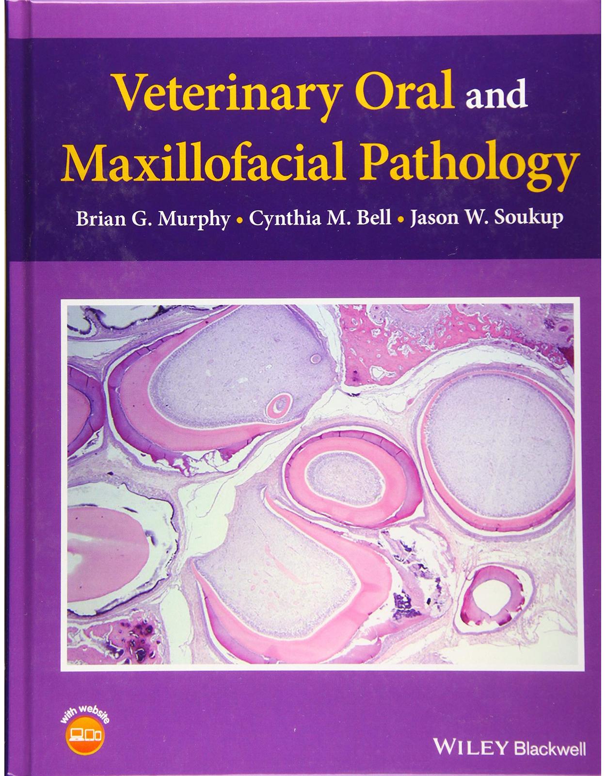
Veterinary Oral and Maxillofacial Pathology
Livrare gratis la comenzi peste 500 RON. Pentru celelalte comenzi livrarea este 20 RON.
Disponibilitate: La comanda in aproximativ 4 saptamani
Editura: Wiley
Limba: Engleza
Nr. pagini: 272
Coperta: Hardcover
Dimensiuni: 21.59 x 1.52 x 27.94 cm
An aparitie: 21 Nov. 2019
DESCRIPTION:
Veterinary Oral and Maxillofacial Pathology focuses on methods for establishing a diagnosis and set of differential diagnoses.
Provides the only text dedicated solely to veterinary oral and maxillofacial pathology
Guides the pathologist through the thought process of diagnosing oral and maxillofacial lesions
Focuses on mammalian companion animals, including dogs, cats and horses, with some coverage of ruminants, camelids, and laboratory animal species
Features access to video clips narrating the process of histological diagnosis on a companion website
Table of Contents:
1 A Philosophical Approach to Establishing a Diagnosis 1
2 Histological Features of Normal Oral Tissues 3
2.1 Oral Mucosa 3
2.2 Gingiva 3
2.3 Periodontal Apparatus 6
2.4 Enamel 7
2.5 Dentin 9
2.6 Cementum 9
2.7 Odontoblasts and Pulp Stroma 9
2.8 Maxillary and Mandibular Bone 10
3 Tooth Development (Odontogenesis) 13
3.1 Species Differences 18
4 Conditions and Diseases of Teeth 21
4.1 Odontogenic Developmental Anomalies and Attrition 21
4.1.1 Primary Enamel Disorders 21
4.1.2 Primary Dentin Disorders 23
4.1.3 Abnormalities in Tooth Number 24
4.1.4 Abnormalities in Tooth Shape 26
4.1.5 Tooth Discoloration 28
4.1.6 Dental Attrition, Abrasion, and Erosion 29
4.2 Degenerative and Inflammatory Disorders of Teeth 31
4.2.1 Pulpitis 31
4.2.2 Pulp Degeneration 32
4.2.3 Periapical Periodontitis 33
4.2.4 Caries 34
4.2.5 Plaque and Calculus 34
4.2.6 Tooth Resorption 35
4.2.6.1 Tooth Resorption in Cats 36
4.2.6.2 Tooth Resorption in Dogs 38
4.2.7 Odontogenic Dysplasia 39
4.3 Equine Dental Diseases 42
4.3.1 Equine Odontoclastic Tooth Resorption and Hypercementosis 42
4.3.2 Periodontitis and Pulpitis of Cheek Teeth 43
4.3.3 Nodular Hypercementosis (Cementoma) 44
4.3.4 Tooth Fractures 45
4.3.5 Caries 45
5 Inflammatory Lesions of the Oral Mucosa and Jaws 49
5.1 Inflammation of the Oral Mucosa 49
5.1.1 Gingivitis and Periodontitis 49
5.1.2 Feline Chronic Gingivostomatitis 52
5.1.2.1 Clinical and Gross Presentation of FCGS 52
5.1.2.2 Pathogenesis of FCGS 53
5.1.2.3 Histologic Features of FCGS 54
5.1.2.4 Clinical Management of FCGS 56
5.1.3 Virus‐Associated Stomatitis in Cats 56
5.1.4 Canine Stomatitis 57
5.1.5 Immune‐Mediated Dermatoses with Oral Involvement 60
5.1.6 Mucosal Drug Reactions 64
5.1.7 Mucocutaneous Pyoderma 64
5.1.8 Eosinophilic Stomatitis 65
5.1.9 Granulomatous Stomatitis 65
5.1.10 Oral Candidiasis 67
5.1.11 Uremia‐Associated Stomatitis 68
5.1.12 Oral inflammation Due to Chronic or Systemic Disease 69
5.2 Inflammation of the Jaw 72
5.2.1 Periodontal Osteomyelitis 72
5.2.2 Lumpy Jaw (Actinomycosis) 75
5.2.3 Mandibulofacial/Maxillofacial Abscesses of Mice 76
5.2.4 Periostitis Ossificans 77
6 Trauma and Physical Injury 79
6.1 Soft Tissue Injury 79
6.1.1 Abrasions and Lacerations 79
6.1.2 Traumatic “Granuloma” 79
6.1.2.1 Clinical Features 82
6.1.3 Thermal and Chemical Burns 83
6.2 Traumatic Lesions of the Teeth and Jaws 85
6.2.1 Disrupted Tooth Development 85
6.2.2 Aneurysmal Bone Cyst (Pseudocyst) 86
6.2.3 Dentoalveolar Trauma 87
6.2.4 Fractures of the Jaw 88
7 Odontogenic Tumors 91
7.1 Approach to Odontogenic Neoplasms 91
7.1.1 Odontogenic Epithelium 91
7.1.2 Mineralized Dental Matrices 93
7.1.3 Dental Papilla 94
7.1.4 Dental Follicle 94
7.1.5 Induction 94
7.1.6 Diagnosing Odontogenic Neoplasms – the Process 95
7.2 Tumors Composed of Odontogenic Epithelium and Fibrous Stroma 98
7.2.1 Conventional Ameloblastoma (CA) 98
7.2.1.1 Clinical Features 100
7.2.1.2 Ameloblastic Carcinoma and Malignant Ameloblastoma 100
7.2.2 Canine Acanthomatous Ameloblastoma (CAA) 102
7.2.2.1 Clinical Features 104
7.2.3 Amyloid‐Producing Ameloblastoma (CEOT/APOT) 105
7.2.3.1 Clinical Features 108
7.3 Tumors Composed of Odontogenic Epithelium, Ectomesenchyme of the Dental Papilla and Follicle 109
7.3.1 Ameloblastic Fibroma 109
7.3.1.1 Clinical Features 110
7.3.2 Feline Inductive Odontogenic Tumor 111
7.3.2.1 Clinical Features 112
7.4 Odontogenic Tumors Composed of Odontogenic Epithelium, Ectomesenchyme of the Dental Papilla, and Mineralized Dental Matrices 113
7.4.1 Odontoma 113
7.4.1.1 Compound Odontoma 114
7.4.1.2 Complex Odontoma 115
7.4.2 Ameloblastic Fibro‐Odontoma 119
7.4.3 Odontoameloblastoma 122
7.5 Cementoblastoma 124
7.6 Odontogenic Myxoma 126
7.6.1 Clinical Features 127
8 Tumors Arising from the Soft Tissues 129
8.1 Melanocytic Tumors 129
8.1.1 Oral Melanocytoma 129
8.1.2 Oral Melanoma 130
8.1.2.1 Clinical Features 133
8.2 Oral Fibroma/Fibrosarcoma 134
8.2.1 Oral Fibroma 134
8.2.2 Oral Fibrosarcoma in Dogs 134
8.2.3 Canine Biologically High‐Grade/Histologically Low‐Grade Fibrosarcoma 137
8.2.3.1 Clinical Features 137
8.2.4 Oral Fibrosarcoma in other Species 137
8.3 Oral Squamous Cell Carcinoma 139
8.3.1 Oral Squamous Cell Carcinoma in Dogs 139
8.3.1.1 Clinical Features 143
8.3.2 Oral Squamous Cell Carcinoma in Domestic Cats 143
8.3.2.1 Clinical Features 145
8.3.3 Oral Squamous Cell Carcinoma in Horses, Cattle, and Other Species 145
8.4 Oral Papilloma 149
8.4.1 Oral Papillomas in Dogs 149
8.4.1.1 Clinical Features 151
8.4.2 Oral Papillomas in Felids 152
8.4.3 Oral Papillomas in Cattle and Other Species 152
8.5 Oral Lymphoma 154
8.5.1 Canine Oral Lymphoma 155
8.5.2 Feline Oral Lymphoma 156
8.5.3 Oral Lymphoma of Ruminants 157
8.6 Neuroendocrine Carcinoma (Carcinoid) 158
8.7 Granular Cell Tumor 159
8.8 Mast Cell Tumor 161
8.9 Plasmacytoma 163
8.10 Vascular Tumors 164
8.10.1 Hemangioma and Vascular Malformations 164
8.10.2 Hemangiosarcoma 164
9 Tumors of the Jaw 167
9.1 Maxillofacial Osteosarcoma 167
9.1.1 Tumor‐Associated Osteoidal Matrix 167
9.1.2 Central Osteosarcoma 168
9.1.3 Osteosarcoma Subtypes 169
9.1.4 Central Low‐Grade Osteosarcoma 171
9.1.5 Peripheral Osteosarcoma 172
9.1.6 Parosteal Osteosarcoma 172
9.1.7 Periosteal Osteosarcoma 172
9.2 Osteoma174
9.3 Multilobular Sarcoma of Bone 178
9.4 Chondrosarcoma 182
10 Tumor‐Like Proliferative Lesions of the Oral Mucosa and Jaws 185
10.1 Tumor‐Like Proliferative Lesions of the Oral Mucosa 185
10.1.1 Calcinosis Circumscripta 185
10.1.2 Ectopic Sebaceous Tissue 186
10.1.3 Follicular Lymphoid Hyperplasia 187
10.1.4 Tonsillar Polyp 188
10.1.5 Sublingual Nodules and Polyps 188
10.1.6 Histiocytic Foam Cell Nodules 189
10.1.7 Nodular Chondroid Hyperplasia/ Degeneration of the Larynx and Epiglottis 192
10.2 Tumor‐Like Proliferative Lesions of the Tooth‐Bearing Regions of the Jaw 194
10.2.1 Gingival Hyperplasia and Focal Fibrous Hyperplasia 194
10.2.2 Craniomandibular Osteopathy 195
10.2.3 Peripheral Giant Cell Granuloma 196
10.2.4 Fibromatous Epulis of Periodontal Ligament Origin (FEPLO)/ Peripheral Odontogenic Fibroma (POF) 197
10.2.4.1 Clinical Presentation and Gross Pathology 198
10.2.4.2 Histological Features 199
10.2.4.3 Clinical Features 201
10.2.5 Proliferative Fibro‐Osseous Lesions of the Oral Cavity and Jaws 201
10.2.5.1 Ossifying Fibroma 201
10.2.5.2 Fibrous Dysplasia 203
11 Odontogenic Cysts 207
11.1 Radiologic Features of Odontogenic Cysts 207
11.2 Histological Features of Odontogenic Cysts 207
11.3 Types of Odontogenic Cysts 209
11.3.1 Dentigerous Cyst 209
11.3.2 Lateral Periodontal Cyst and Gingival Cyst 209
11.3.3 Periapical (Radicular) Cyst 212
11.3.4 Keratin‐Filled Cysts 213
11.4 Treatment and Considerations for Biopsy of Oral Cystic Lesions 213
11.5 Pathogenesis of Odontogenic Cysts 214
11.6 Do Odontogenic Tumors Arise from Odontogenic Cysts? 215
12 Lesions of the Salivary Gland 217
12.1 Microanatomy and Physiology 217
12.2 Salivary Duct Obstruction, Cysts, and Pseudocysts 218
12.3 Salivary Gland Infarction (Necrotizing Sialometaplasia) 220
12.4 Salivary Gland Inflammation (Sialoadenitis) 221
12.5 Salivary Gland Neoplasia 224
12.5.1 Benign Salivary Gland Tumors 225
12.5.2 Pleomorphic Adenoma (Salivary Gland Mixed Tumor) 225
12.5.3 Malignant Salivary Gland Tumors 225
12.5.4 Mucoepidermoid Carcinoma 226
12.5.5 Adenocarcinoma 227
12.5.6 Acinic Cell Carcinoma 227
12.5.7 Basal Cell Adenocarcinoma 228
Appendix 231
A.1 Processing Teeth Submitted as Biopsy Specimens 231
A.2 Face Decalcification of Paraffin Blocks 232
A.3 Recommended Best Practices for “Grossing” Mandibulectomy and Maxilectomy Surgical Specimens 232
A.4 Specimen Immobilization 234
A.5 Cutting Instrumentation 234
A.6 Orienting the Specimen 236
A.7 Decalcification 236
Index 239
| An aparitie | 21 Nov. 2019 |
| Autor | Brian G. Murphy, Cynthia M. Bell, Jason W. Soukup |
| Dimensiuni | 21.59 x 1.52 x 27.94 cm |
| Editura | Wiley |
| Format | Hardcover |
| ISBN | 9781119221258 |
| Limba | Engleza |
| Nr pag | 272 |







Clientii ebookshop.ro nu au adaugat inca opinii pentru acest produs. Fii primul care adauga o parere, folosind formularul de mai jos.