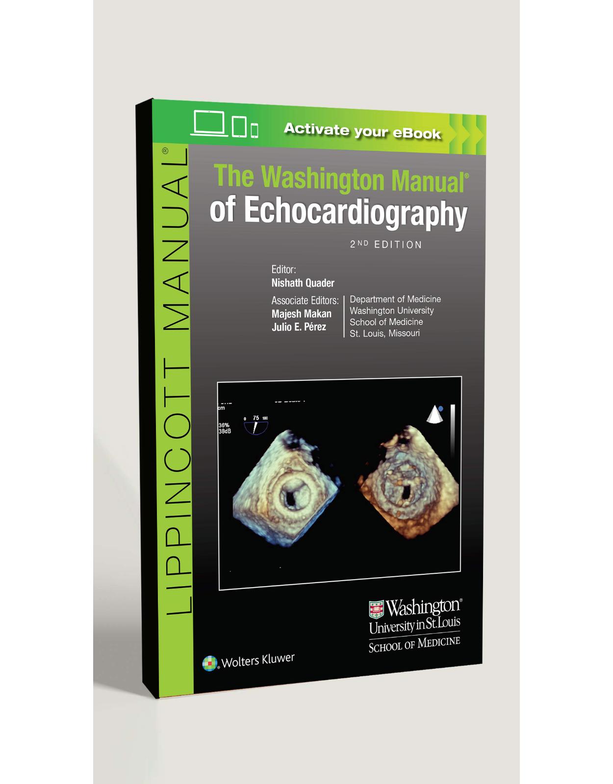
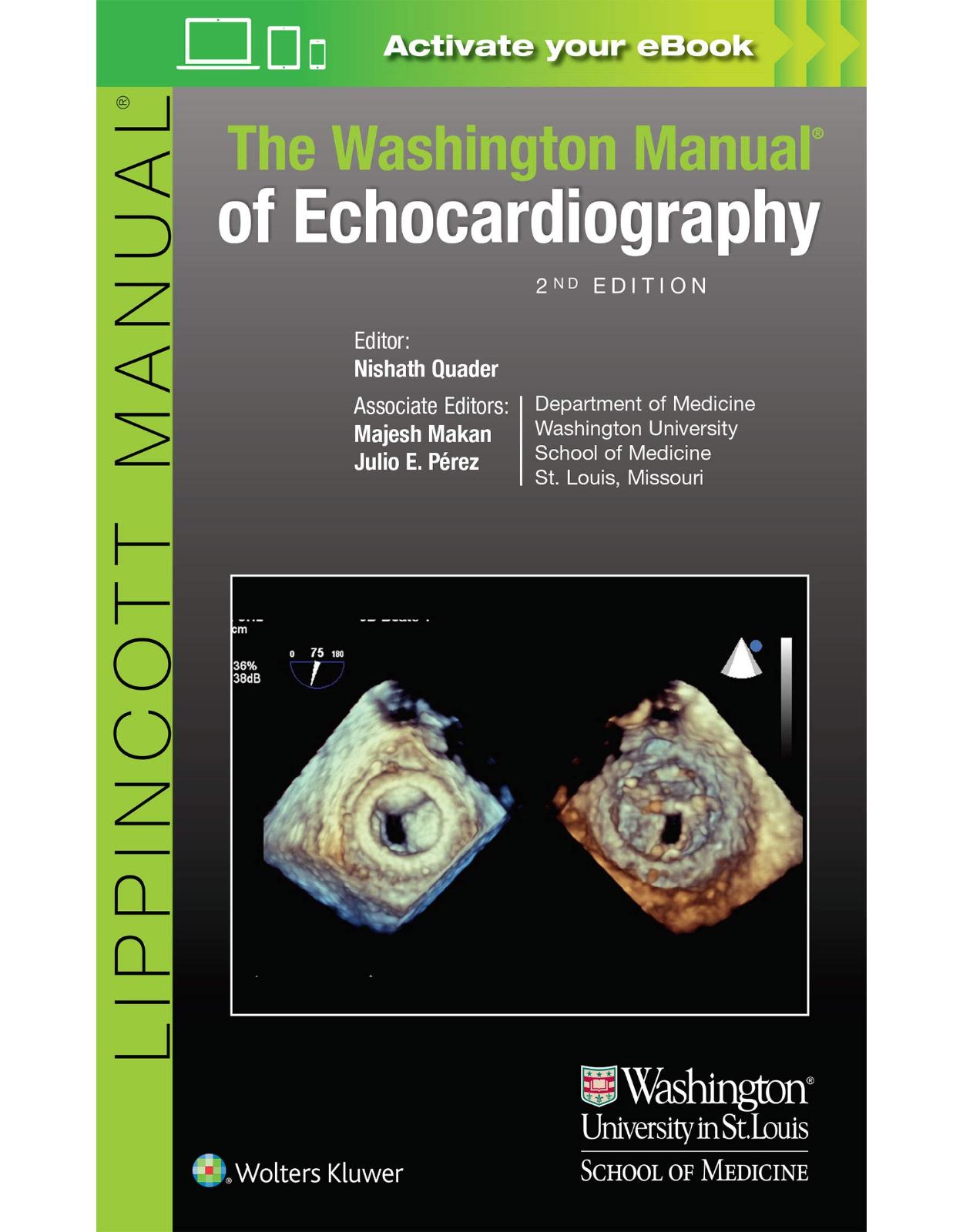
The Washington Manual of Echocardiography
Livrare gratis la comenzi peste 500 RON. Pentru celelalte comenzi livrarea este 20 RON.
Disponibilitate: La comanda in aproximativ 4-6 saptamani
Editura: LWW
Limba: Engleza
Nr. pagini: 368
Coperta: Paperback
Dimensiuni: 12.7 x 1.52 x 20.32 cm
An aparitie: 1 Aug. 2016
Description:
Concise, portable, and user-friendly, The Washington Manual® of Echocardiography, Second Edition focuses on the essential information you need to know to successfully perform and read echocardiograms, as well as to identify valvular heart disease, cardiac myopathies, and congenital anomalies. Supervised and edited by faculty from the Washington University School of Medicine, this highly regarded reference has been completely updated throughout, with a new, streamlined layout as well more echo clips, and an update on the current guidelines. You'll find expert guidance, practical tips, and up-to-date information on all aspects of echocardiography – all in one convenient and easily accessible source. •New chapters on hypertrophic cardiomyopathy and cardiac devices include the latest ACC/AHA and ASE guidelines.•Extensively revised sections on LV systolic and diastolic function, valve assessment, and TEE.•Hundreds of updated, high-quality images enhance visual learning and improve diagnostic accuracy.•Reorganized content emphasizes clarity and consistency, so you can find what you need more quickly.•Tips and pearls from cardiologists and sonographers offer guidance on proper set-up, correct transduction technique, and image optimization.•A handy, portable resource for cardiology fellows, as well as anesthesia fellows, cardiac sonographers, residents, attendings, and intensive care and emergency department physicians who utilize handheld ultrasound. Now with the print edition, enjoy the bundled interactive eBook edition, which can be downloaded to your tablet and smartphone or accessed online and includes features like:•Complete content with enhanced navigation•Powerful search tools and smart navigation cross-links that pull results from content in the book, your notes, and even the web•Cross-linked pages, references, and more for easy navigation•Highlighting tool for easier reference of key content throughout the text•Ability to take and share notes with friends and colleagues•Quick reference tabbing to save your favorite content for future use
Table of Contents:
Title Page
Copyright Information
Dedication
Contributors
Foreword
Preface
Videos
Chapter 2
Movie 2.1
Movie 2.2
Movie 2.3
Movie 2.4
Movie 2.5
Movie 2.6
Movie 2.7
Movie 2.8
Figure 2.9
Movie 2.10
Movie 2.11
Movie 2.12
Movie 2.13
Figure 2.14
Movie 2.15
Figure 2.16
Chapter 3
Figure 3.1a
Figure 3.1b
Movie 3.2a
Movie 3.2b
Movie 3.3a
Movie 3.3b
Movie 3.4a
Movie 3.4b
Movie 3.5a
Movie 3.5b
Movie 3.5c
Movie 3.5d
Movie 3.6a
Movie 3.6b
Movie 3.6c
Movie 3.7a
Movie 3.7b
Movie 3.8
Movie 3.9
Movie 3.10a
Movie 3.10b
Movie 3.11
Movie 3.12
Movie 3.13
Movie 3.14
Movie 3.15
Movie 3.16a
Movie 3.16b
Movie 3.17a
Movie 3.17b
Figure 3.18
Chapter 4
Movie 4.1a
Figure 4.1b
Movie 4.2a
Movie 4.2b
Movie 4.3a
Movie 4.3b
Movie 4.4a
Figure 4.4b
Figure 4.4c
Movie 4.5a
Figure 4.5b
Figure 4.5c
Movie 4.6a
Movie 4.6b
Movie 4.6c
Movie 4.7
Movie 4.8a
Movie 4.8b
Figure 4.8c
Movie 4.8d
Figure 4.8e
Movie 4.8f
Chapter 5
Movie 5.1
Movie 5.2a
Movie 5.2b
Movie 5.2c
Figure 5.2d
Movie 5.3a
Movie 5.3b
Movie 5.3c
Movie 5.3d
Movie 5.3e
Movie 5.3f
Figure 5.3g
Figure 5.3h
Figure 5.3i
Movie 5.4a
Movie 5.4b
Movie 5.4c
Movie 5.4d
Movie 5.5
Movie 5.6
Movie 5.7a
Movie 5.7b
Movie 5.7c
Movie 5.7d
Chapter 6
Movie 6.1
Movie 6.2a
Movie 6.2b
Figure 6.2c
Movie 6.3a
Movie 6.3b
Movie 6.4a
Movie 6.4b
Movie 6.5a
Movie 6.5b
Movie 6.5c
Movie 6.5d
Movie 6.6a
Movie 6.6b
Movie 6.7a
Movie 6.7b
Movie 6.8
Movie 6.9a
Movie 6.9b
Movie 6.9c
Chapter 7
Movie 7.1
Movie 7.2a
Movie 7.2b
Movie 7.3a
Movie 7.3b
Movie 7.3c
Movie 7.3d
Figure 7.3e
Movie 7.4
Movie 7.5a
Movie 7.5b
Movie 7.5c
Movie 7.5d
Movie 7.6a
Movie 7.6b
Movie 7.7
Chapter 8
Movie 8.1a
Movie 8.1b
Movie 8.2
Movie 8.3a
Movie 8.3b
Movie 8.4a
Movie 8.4b
Movie 8.4c
Movie 8.5
Movie 8.6
Figure 8.7
Movie 8.8a
Movie 8.8b
Movie 8.8c
Movie 8.9a
Movie 8.9b
Movie 8.9c
Movie 8.10
Movie 8.11
Movie 8.12a
Movie 8.12b
Chapter 9
Movie 9.1
Movie 9.2
Movie 9.3
Movie 9.4
Chapter 10
Movie 10.1a
Movie 10.1b
Movie 10.2a
Movie 10.2b
Movie 10.2c
Movie 10.3a
Movie 10.3b
Movie 10.4a
Movie 10.4b
Movie 10.5
Movie 10.6
Movie 10.7a
Movie 10.7b
Movie 10.7c
Movie 10.7d
Movie 10.8
Movie 10.9a
Movie 10.9b
Movie 10.10a
Movie 10.10b
Movie 10.10c
Figure 10.10d
Figure 10.10e
Figure 10.10f
Movie 10.10g
Movie 10.10h
Movie 10.11a
Movie 10.11b
Figure 10.11c
Figure 10.11d
Figure 10.11e
Figure 10.11f
Figure 10.12
Movie 10.13a
Movie 10.13b
Movie 10.13c
Figure 10.13d
Figure 10.13e
Movie 10.14a
Figure 10.14b
Movie 10.15a
Movie 10.15b
Movie 10.15c
Movie 10.15d
Movie 10.15e
Figure 10.16
Movie 10.17
Chapter 11
Movie 11.1a
Movie 11.1b
Movie 11.1c
Movie 11.1d
Movie 11.1e
Movie 11.2a
Movie 11.2b
Movie 11.3a
Movie 11.3b
Movie 11.3c
Figure 11.3d
Movie 11.4
Movie 11.5a
Movie 11.5b
Movie 11.5c
Movie 11.6a
Movie 11.6b
Movie 11.7a
Movie 11.7b
Movie 11.7c
Movie 11.7d
Movie 11.8a
Movie 11.8b
Movie 11.8c
Movie 11.8d
Figure 11.8e
Figure 11.8f
Movie 11.9
Movie 11.10a
Figure 11.10b
Movie 11.10c
Movie 11.11a
Movie 11.11b
Movie 11.11c
Movie 11.12a
Movie 11.12b
Movie 11.12c
Movie 11.13
Movie 11.14a
Movie 11.14b
Movie 11.14c
Chapter 12
Movie 12.1a
Figure 12.1b
Movie 12.2a
Movie 12.2b
Figure 12.2c
Movie 12.2d
Movie 12.2e
Movie 12.3a
Movie 12.3b
Figure 12.3c
Figure 12.3d
Movie 12.4a
Movie 12.4b
Figure 12.4d
Figure 12.4e
Movie 12.4f
Movie 12.4g
Figure 12.4h
Movie 12.4i
Chapter 13
Movie 13.1b
Figure 13.1c
Movie 13.1d
Figure 13.1e
Movie 13.2a
Movie 13.2b
Movie 13.3a
Movie 13.3b
Movie 13.3c
Figure 13.3d
Movie 13.3e
Movie 13.3f
Movie 13.4a
Movie 13.4b
Movie 13.4c
Movie 13.5a
Movie 13.5b
Chapter 14
Movie 14.1a
Movie 14.1b
Movie 14.1c
Figure 14.1d
Movie 14.1e
Movie 14.1f
Movie 14.2a
Figure 14.2b
Movie 14.3a
Movie 14.3b
Movie 14.4a
Movie 14.4b
Movie 14.5a
Movie 14.5b
Movie 14.5c
Movie 14.5d
Movie 14.6a
Movie 14.6b
Movie 14.6c
Movie 14.7a
Movie 14.7b
Movie 14.8a
Movie 14.8b
Movie 14.8c
Movie 14.8d
Movie 14.9a
Movie 14.9b
Movie 14.9c
Movie 14.9d
Figure 14.9e
Movie 14.9f
Movie 14.10a
Movie 14.10b
Figure 14.10c
Movie 14.10d
Movie 14.10e
Figure 14.10f
Movie 14.10g
Movie 14.11a
Movie 14.11b
Figure 14.11c
Movie 14.12a
Movie 14.12b
Movie 14.12c
Movie 14.12d
Movie 14.12e
Figure 14.12f
Movie 14.13
Movie 14.14a
Movie 14.14b
Movie 14.14c
Movie 14.14d
Movie 14.15a
Movie 14.15b
Movie 14.15c
Chapter 15
Movie 15.1a
Movie 15.1b
Movie 15.1c
Movie 15.1d
Movie 15.1e
Movie 15.1f
Movie 15.1g
Movie 15.1h
Movie 15.2a
Movie 15.2b
Figure 15.2c
Movie 15.2d
Movie 15.2e
Movie 15.3a
Movie 15.3b
Movie 15.3c
Movie 15.3d
Movie 15.3e
Movie 15.3f
Movie 15.4a
Movie 15.4b
Movie 15.5a
Movie 15.5b
Movie 15.5c
Movie 15.5d
Movie 15.5e
Chapter 16
Movie 16.1
Movie 16.2a
Movie 16.2b
Movie 16.2c
Movie 16.2d
Movie 16.2e
Movie 16.2f
Movie 16.3a
Movie 16.3b
Movie 16.4a
Movie 16.4b
Movie 16.5
Movie 16.6a
Movie 16.6b
Movie 16.7
Movie 16.8a
Movie 16.8b
Movie 16.8c
Movie 16.8d
Figure 16.8e
Movie 16.9a
Movie 16.9b
Movie 16.9c
Movie 16.10a
Figure 16.10b
Movie 16.10c
Movie 16.10d
Figure 16.10e
Figure 16.10f
Figure 16.10g
Figure 16.10h
Figure 16.10i
Movie 16.10j
Figure 16.10k
Figure 16.11
Movie 16.12a
Figure 16.12b
Movie 16.12c
Movie 16.12d
Movie 16.13a
Movie 16.13b
Movie 16.13c
Movie 16.13d
Movie 16.13e
Movie 16.13f
Figure 16.13g
Figure 16.13h
Figure 16.13i
Figure 16.13j
Movie 16.13k
Movie 16.13l
Movie 16.13m
Movie 16.13n
Movie 16.14
Chapter 17
Movie 17.1
Movie 17.2
Movie 17.3
Movie 17.4
Movie 17.5a
Movie 17.5b
Movie 17.5c
Figure 17.5d
Movie 17.5e
Movie 17.5f
Movie 17.5g
Figure 17.5h
Movie 17.5i
Figure 17.5j
Movie 17.6a
Movie 17.6b
Movie 17.6c
Movie 17.7a
Movie 17.7b
Movie 17.8
Movie 17.9a
Movie 17.9b
Movie 17.10
Movie 17.11a
Figure 17.11b
Movie 17.12a
Figure 17.12b
Movie 17.13a
Figure 17.13b
Movie 17.13c
Figure 17.13d
Movie 17.14a
Figure 17.14b
Figure 17.14c
Figure 17.14d
Movie 17.15
Movie 17.16a
Movie 17.16b
Movie 17.16c
Movie 17.17
Movie 17.18a
Movie 17.18b
Movie 17.19
Movie 17.20a
Movie 17.20b
Movie 17.21
Chapter 18
Movie 18.1a
Movie 18.1b
Movie 18.2a
Movie 18.2b
Movie 18.2c
Figure 18.2d
Figure 18.2e
Movie 18.3a
Movie 18.3b
Movie 18.4
Movie 18.5
Movie 18.6
Movie 18.7a
Movie 18.7b
Movie 18.7c
Movie 18.7d
Movie 18.7e
Movie 18.7f
Figure 18.7g
Movie 18.8
Movie 18.9
Movie 18.10a
Movie 18.10b
Movie 18.11a
Movie 18.11b
Movie 18.11c
Movie 18.12a
Movie 18.12b
Movie 18.13a
Movie 18.13b
Movie 18.13c
Movie 18.13d
Figure 18.13e
Figure 18.13f
Movie 18.14a
Movie 18.14b
Movie 18.14c
Figure 18.14d
Movie 18.14e
Movie 18.15a
Movie 18.15b
Movie 18.15c
Movie 18.15d
Movie 18.15e
Movie 18.15f
Movie 18.16
Movie 18.17a
Movie 18.17b
Movie 18.17c
Movie 18.17d
Chapter 19
Movie 19.1
Movie 19.2
Movie 19.3a
Movie 19.3b
Movie 19.3c
Movie 19.3d
Movie 19.4a
Movie 19.4b
Movie 19.4c
Movie 19.4d
Movie 19.5a
Movie 19.5b
Movie 19.5c
Movie 19.6
Movie 19.7
Movie 19.8a
Movie 19.8b
Movie 19.9a
Movie 19.9b
Movie 19.10a
Movie 19.10b
Movie 19.10c
Movie 19.11a
Movie 19.11b
Movie 19.11c
Movie 19.12
Movie 19.13
Movie 19.14
Movie 19.15
Movie 19.16a
Movie 19.16b
Movie 19.17a
Movie 19.17b
Movie 19.17c
Movie 19.18
Movie 19.19a
Movie 19.19b
Movie 19.20
Movie 19.21
Movie 19.22a
Movie 19.22b
Movie 19.22c
Movie 19.22d
Figure 19.22e
Movie 19.23a
Movie 19.23b
Movie 19.23c
Movie 19.23d
Movie 19.24a
Movie 19.24b
Movie 19.24c
Figure 19.24d
Movie 19.25a
Movie 19.25b
Movie 19.25c
Movie 19.26
Movie 19.27
Chapter 20
Movie 20.1a
Movie 20.1b
Movie 20.2a
Movie 20.2b
Movie 20.3a
Movie 20.3b
Movie 20.3c
Movie 22.4a
Movie 22.4b
Movie 22.5
Movie 22.6a
Movie 20.6
Movie 20.7a
Movie 20.7b
Chapter 21
Movie 21.1a
Movie 21.1b
Movie 21.1c
Movie 21.1d
Movie 21.1e
Movie 21.1f
Movie 21.1g
Movie 21.1h
Movie 21.1i
Movie 21.1j
Movie 21.1k
Movie 21.1l
Movie 21.2a
Movie 21.2b
Movie 21.2c
Movie 21.2d
Movie 21.3a
Movie 21.3b
Movie 21.3c
Movie 21.4a
Movie 21.4b
Movie 21.5a
Movie 21.5b
Movie 21.6
Movie 21.7
Movie 21.8a
Movie 21.8b
Movie 21.8c
Movie 21.8d
Movie 21.9a
Movie 21.9b
Movie 21.9c
Movie 21.9d
Movie 21.9e
Figure 21.9f
Figure 21.9g
Movie 21.10a
Movie 21.10b
Movie 21.10c
Movie 21.10d
Movie 21.11a
Movie 21.11b
Movie 21.12a
Movie 21.12b
Movie 21.12c
Movie 21.12d
Movie 21.12e
Movie 21.12f
Figure 21.12g
Movie 21.13a
Movie 21.13b
Movie 21.13c
Movie 21.14a
Movie 21.14b
Movie 21.15a
Movie 21.15b
Movie 21.16a
Movie 21.16b
Movie 21.16c
Figure 21.16d
Movie 21.17a
Movie 21.17b
Movie 21.18a
Movie 21.18b
Movie 21.18c
Movie 21.19a
Movie 21.19b
Movie 21.19c
Movie 21.20a
Movie 21.20b
Movie 21.21a
Movie 21.21b
Movie 21.21c
Movie 21.21d
Movie 21.22a
Movie 21.22b
Movie 21.22c
Movie 21.22d
Movie 21.22e
Movie 21.22f
Movie 21.22g
Movie 21.22h
Movie 21.23a
Movie 21.23b
Movie 21.23c
Movie 21.24a
Figure 21.24b
Movie 21.25
Movie 21.26a
Movie 21.26b
Figure 21.26c
Movie 21.26d
Movie 21.26e
Movie 21.26f
Figure 21.26g
Movie 21.27
Movie 21.28
Movie 21.29a
Movie 21.29b
Movie 21.29c
Movie 21.30
Movie 21.31
Movie 21.32
Movie 21.33
Movie 21.34
Movie 21.35a
Movie 21.35b
Movie 21.35c
Movie 21.35d
Movie 21.35e
Movie 21.35f
Movie 21.35g
Movie 21.35h
Movie 21.35i
Movie 21.35j
Movie 21.35k
Movie 21.35l
Movie 21.35m
Movie 21.35n
Movie 21.35o
Chapter 22
Movie 22.1
Movie 22.2
Movie 22.3
Movie 22.4
Movie 22.5
Movie 22.6
Chapter 1: Introduction to Echocardiographic Principles
Chapter 1 Introduction
General Principles
Figure 1-1
Imaging Modalities
M-Mode Echocardiography
Two-dimensional Echocardiography
Three-dimensional Echocardiography
Figure 1-2
Doppler Principles and Applications
Doppler Effect
Pulsed-wave Doppler
Continuous-wave Doppler
High-pulse Repetition Frequency (HPRF) Pulsed Doppler
Color Doppler
Tissue Doppler Imaging (TDI)
Figure 1-3
Figure 1-4
Figure 1-5
Table 1-1: Characteristics of the Different Doppler Modes
Figure 1-6
Useful Hemodynamic Principles and Applications
Stroke Volume (SV) and Other Flow Volumes
Bernoulli Principle and Estimation of Pressure in Cardiac Chambers
Continuity Principle
Figure 1-7
Figure 1-8: dfsdfdfsf
Table 1-2: Assessing Cardiac Hemodynamic Indices by Echocardiography
Chapter 2: The Comprehensive Transthoracic Echocardiographic Examination
Getting Started
Figure 2-1
Exhibit 752
Adjusting the Image
Two-dimensional Gain
Figure 2-2
Image Depth
Color Doppler
Color Gain
Frequency
Focus
Pulsed-wave Doppler Sample Volume Sizes
Spectral Gain
Sweep Speed
Figure 2-3
Figure 2-4
Figure 2-5
Figure 2-6
Figure 2-7
Figure 2-8
The Views: Parasternal Long-Axis View (PLAX)
Figure 2-9
Exhibit 764
Right Ventricular Inflow Tract
Figure 2-10
Exhibit 767
Parasternal Short Axis (PSAX) View
Exhibit 769
Figure 2-11
Figure 2-12
Figure 2-13
Apical Four-Chamber (A4C) View
Figure 2-14
Exhibit 775
Apical Five-Chamber (A5C) View
Figure 2-15
Exhibit 778
Apical Two-Chamber View
Figure 2-16
Exhibit 781
Apical Parasternal Long-Axis (APLAX) View
Figure 2-17
Exhibit 784
Subcostal Coronal View
Figure 2-18
Exhibit 787
Subcostal Sagittal View
Figure 2-19
Exhibit 790
Suprasternal Notch (SSN) View
Figure 2-20
Exhibit 793
Chapter 3: The Role of Contrast in Echocardiography
Chapter 3 Introduction
General Principles
Figure 3-1
Figure 3-2
Figure 3-3
Figure 3-4
Preparing Definity (Lantheus Medical Imaging) Contrast
Preparing Optison (GE Healthcare) Contrast
Pitfalls
High Mechanical Index
Attenuation
LV Underfilled with Contrast
Figure 3-5
Figure 3-6
Agitated Bacteriostatic Saline Contrast
Reasons to Perform an Agitated Saline Contrast
Contraindications
Preparing Agitated Bacteriostatic Saline Contrast
Injection of Agitated Saline
Figure 3-7
Figure 3-8
Chapter 4: Quantification of Left Ventricular Systolic and Diastolic Function
Chapter 4 Introduction
Introduction
Tips for Optimizing Image Quality
Tips for Interpreting Studies
LV Dimensions
Plax
Apical Views—A4C and Apical Two Chamber (A2C)
Three-dimensional Echocardiography (3DE)
Figure 4-1
Table 4-1: Linear Dimensions
Figure 4-2
Figure 4-3
Table 4-2: Volumes and Mass
LV Systolic Function
Table 4-3: LV Functional Assessment
Figure 4-4
LV Mass
2D and 3D Methods Are Preferred over M-mode (Fig. 4-5A,B).
Linear Method: Uses LVIDd, SWT, PWT from PLAX view
Figure 4-5
Table 4-4: Relationships Between LV Mass and RWT (cm)
Diastolic Function Assessment
Mitral Inflow Pulsed-Wave (PW) Doppler: E and A Waves
Tissue Doppler of the Mitral Annulus
Pulmonary Vein (PV): S/D (Systolic to Diastolic Filling Ratio)
Diastolic Function Assessment in Special Populations
Table 4-5: Doppler Parameters and LV Diastolic Function
Figure 4-6
Chapter 5: Right Ventricular Function and Pulmonary Hemodynamics
Chapter 5 Introduction
Anatomy and Physiology of the Right Ventricle
Two-Dimensional Echocardiographic Assessment of Right Ventricular Size
Qualitative RV Size
Quantitative RV Size
Quantitative RVOT Size
Quantitative RV Thickness
Figure 5-1
Figure 5-2
Figure 5-3
Figure 5-4
Assessment of Right Ventricular Function
Two-dimensional Assessment
M-mode
Doppler Assessment
RV Diastolic Function
Figure 5-5
Figure 5-6
Figure 5-7
Right Ventricular Pathology
RV Volume Overload
RV Pressure Overload
RV Infarction
Arrhythmogenic Right Ventricular Dysplasia
Figure 5-8
Pulmonary Hemodynamics
Figure 5-9
Figure 5-10
Right Atrial Assessment
RA Size Assessment
Estimating Mean RA Pressure
Figure 5-11
Figure 5-12
Discrepancy between Doppler and Cardiac Catheterization
Chapter 6: Stress Testing for Ischemia and Viability
Chapter 6 Introduction
General Principles
Figure 6-1
Anatomy
Figure 6-2
Exercise Stress Echocardiography
Absolute Contraindications
Relative Contraindications
Absolute Indications to Terminate Exercise Testing
Relative Indications to Terminate Exercise Testing
Dobutamine Stress Echocardiography
Interpretation
Baseline Transthoracic Echocardiography
Peak Stress Transthoracic Echocardiography
False Results
Figure 6-3
Prognosis
Other Considerations
Stress Testing for Myocardial Viability
General Principles
Protocol
Interpretation
Other Considerations
Table 6-1: Characteristic Echocardiographic Responses to Dobutamine Infusion to Determine the Etiology of Regional Akinesis
Chapter 7: Ischemic Heart Disease and Complications of Myocardial Infarction
Chapter 7 Introduction
Evaluation of Chest Pain Syndrome in the Acute Setting
Mechanical Complications of Acute Myocardial Infarction
Left Ventricular Dysfunction and Cardiogenic Shock
Ventricular Septal Defect
Figure 7-1
Figure 7-2
Left Ventricular Free Wall Rupture
Figure 7-3
Figure 7-4
Papillary Muscle Rupture and Ischemic Mitral Regurgitation
Figure 7-5
Figure 7-6
Pericardial Effusion
Acute Dynamic Left Ventricular Outflow Tract Obstruction
Left Ventricular Pseudoaneurysm
Figure 7-7
Figure 7-8
Takotsubo Syndrome
Chronic Complications of Acute Myocardial Infarction
Left Ventricular Aneurysm and Thrombus
Right Ventricular Dysfunction
Chapter 8: Cardiomyopathies
Chapter 8 Introduction
Dilated Cardiomyopathy
Background
Echocardiographic Findings
Figure 8-1
Table 8-1: Chamber Dimensions
Restrictive Cardiomyopathy
Background
Echocardiographic Findings
Figure 8-2
Arrhythmogenic Right Ventricular Dysplasia
Background
Echocardiographic Findings
Figure 8-3
Takotsubo Cardiomyopathy
No Consensus Criteria, But Modified Mayo Clinic Criteria Often Used
Background
Echocardiographic Findings
Figure 8-4
Isolated Noncompaction of the Left Ventricle
Background
Echocardiographic Findings
Figure 8-5
Post Heart Transplant
Background
Echocardiographic Findings
Allograft Rejection
Cardiac Allograft Vasculopathy (CAV)
Chapter 9: Hypertrophic Cardiomyopathy
Chapter 9 Introduction
Background
Echocardiographic Findings
Table 9-1: Guidelines for Echocardiography in Patients with Hypertrophic Cardiomyopathy
Table 9-2: Screening Strategies with Echocardiography for Detection of Hypertrophic Cardiomyopathy with Left Ventricular Hypertrophy
Figure 9-1
Figure 9-2
Figure 9-3
Figure 9-4
Figure 9-5
Figure 9-6
Figure 9-7
Figure 9-8
Figure 9-8
Figure 9-8
Figure 9-8
Figure 9-9
Figure 9-10
Figure 9-11
Chapter 10: Aortic Valve Disease
Chapter 10 Introduction
Anatomy
Figure 10-1
Aortic Stenosis
Table 10-1: Severity of Aortic Stenosis
Figure 10-2
Figure 10-3
Figure 10-4
Figure 10-5
Figure 10-6
Figure 10-7
Aortic Regurgitation
Table 10-2: Severity of Aortic Regurgitation
Figure 10-8
Figure 10-9
Figure 10-10
Chapter 11: Mitral Valve Disease
Chapter 11 Introduction
Mitral Valve Apparatus
Figure 11-1
Mitral Regurgitation
Exhibit 932
Figure 11-2
Figure 11-3
Figure 11-4
Table 11-1: Criteria for Determining Severity of Mitral Valve Regurgitation
Figure 11-5
Figure 11-6
Figure 11-7
Figure 11-8
Figure 11-9
Figure 11-10
Mitral Stenosis
Table 11-2: Stages of Mitral Stenosis
Exhibit 945
Figure 11-11
Figure 11-12
Figure 11-13
Figure 11-14
Table 11-3: Wilkins Score Grading
Chapter 12: Pulmonic Valve
Chapter 12 Introduction
General Principles
Pulmonic Valve Stenosis
Etiology
2D Echocardiography
M-mode
Color Doppler
PW and CW Doppler
Figure 12-1
Exhibit 955
Figure 12-2
Figure 12-3
Table 12-1: Parameters for Determining Severity of Pulmonic Stenosis
Pulmonic Regurgitation
Etiology
2D Echocardiography
M-mode
CW and PW Doppler
Color Doppler
Table 12-2: Echocardiographic Assessment of Pulmonary Regurgitation
Exhibit 961
Figure 12-4
Figure 12-5
Figure 12-6
Chapter 13: Tricuspid Valve Disorders
Chapter 13 Introduction
General Principles
Figure 13-1
Two-Dimensional Assessment
TV Leaflets and Chordae
Chamber Dimensions and Ventricular Function
Interventricular Septum
IVC
Intracardiac Catheters or Pacemaker/Defibrillator Leads
Figure 13-2
Figure 13-3
Figure 13-4
Figure 13-5
Tricuspid Valve Regurgitation
Color Doppler
Pulse and CW Doppler
Table 13-1: Tricuspid Valve Regurgitation Evaluation
Table 13-2: Classification of Tricuspid Stenosis
Figure 13-6
Tricuspid Stenosis
M-mode
Doppler
Figure 13-7
Chapter 14: Evaluation of Prosthetic Valves
Chapter 14 Introduction
General Principles
Figure 14-1
Echocardiographic Evaluation
General Evaluation
Regurgitation
Color Doppler
High Gradients
Pressure Recovery
Figure 14-2
Figure 14-3
Figure 14-4
Figure 14-5
Table 14-1: Expected Values for Normally Functioning Aortic and Mitral Prosthetic Valves
Figure 14-6
Table 14-2: Expected Parameters for a Normal Mitral Valve Prosthesis and Parameters Suggesting Possible or Significant Stenosis
Figure 14-7
Figure 14-8
Chapter 15: Infective Endocarditis
Chapter 15 Introduction
Introduction
Transthoracic versus Transesophageal Echocardiography
Figure 15-1
Vegetations
Table 15-1: Echo Criteria for Defining Vegetation
Figure 15-2
Figure 15-3
Figure 15-4
Table 15-2: Complications of Endocarditis
Figure 15-5
Abscesses
Figure 15-6
Fistulas
Figure 15-7
Prosthetic Valves
Figure 15-8
Infected Intracardiac Devices
Embolization
Indications for Surgical Consultation
Monitoring of Infective Endocarditis
False Positives for Infective Endocarditis
Chapter 16: Pericardial Disease and Cardiac Tamponade
Chapter 16 Introduction
Definitions
Anatomy and Physiology of the Pericardium
Etiology of Pericardial Disease and Effusions
Differential Diagnosis of Echolucent Space Surrounding the Heart
Figure 16-1
Echocardiographic Assessment of Cardiac Tamponade
Figure 16-2
Figure 16-3
Figure 16-4
Figure 16-5
Figure 16-6
General Considerations
Evaluation of Constrictive Pericarditis
Figure 16-7
Figure 16-8
Differentiation of Constrictive Pericarditis and Restrictive Cardiomyopathy
Other Pericardial Pathology
Chapter 17: Diseases of the Great Vessels: Aorta and Pulmonary Artery
Chapter 17 Introduction
Aorta
Anatomy
Key TTE Views
Key TEE Views
Pathology
Aortic Aneurysm
Sinus of Valsalva Aneurysm
Acute Aortic Syndromes
Coarctation of the aorta
Aortic Atherosclerosis
Figure 17-1
Figure 17-2
Table 17-1: Normal Aortic Root Measurements for Men and Women Indexed to Body Surface Area
Figure 17-3
Figure 17-4
Figure 17-5
Figure 17-6
Figure 17-7
Figure 17-8
Figure 17-9
Figure 17-10
Figure 17-11
Figure 17-12
Figure 17-13
Figure 17-14
Figure 17-15
Pulmonary Artery
Anatomy
Key Views
Pathology
Pulmonary Artery Dilation
Pulmonary Embolism
2D and Doppler Findings of PE
Figure 17-16
Figure 17-17
Figure 17-18
Figure 17-19
Figure 17-20
Figure 17-21
Chapter 18: Congenital Heart Disease
Chapter 18 Introduction
Segmental Echocardiographic Exam
Figure 18-1
Shunts
Key Concepts
Atrial Septal Defect (ASD)
Types
Hemodynamics
Echocardiography
Ventricular Septal Defect (VSD)
Types
Hemodynamics
Echocardiography
Patent Ductus Arteriosus
Hemodynamics
Echocardiography
Endocardial Cushion Defect
Hemodynamics
Echocardiography
Figure 18-2
Figure 18-3
Figure 18-4
Figure 18-5
Figure 18-6
Figure 18-7
Figure 18-8
Figure 18-9
Figure 18-10
Figure 18-11
Obstructions
Bicuspid AoV
Key Concepts
Echocardiography
Cor Triatriatum and Congenital Mitral Stenosis (MS)
Aortic Coarctation
Hemodynamics
Echocardiography
Figure 18-12
Figure 18-13
Figure 18-14
Figure 18-15
Transposition of the Great Arteries (TGA)
D-TGA
Hemodynamics/Physiology
Echocardiography—Post Repair
L-TGA (congenitally corrected transposition)
Hemodynamics
Echocardiography
Exhibit 1077
Exhibit 1078
Exhibit 1079
Figure 18-16
Figure 18-17
Tetralogy of Fallot
Hemodynamics
Surgical Repair
Echocardiography
Figure 18-18
Truncus Arteriosus
Surgical Repair
Echocardiography
Ebstein Anomaly
Hemodynamics
Echocardiography
Fontan Procedure for Single Ventricle Physiology
Echocardiography
Figure 18-19
Chapter 19: Cardiac Masses
Chapter 19 Introduction
Artifacts
Figure 19-1
Normal Variants and Pathologic Variations in Normal Structures
Normal Variants
Pathologic Variations in Normal Structures
Figure 19-2
Figure 19-3
True Cardiac Masses
Thrombus
Vegetations
Tumors
Benign Tumors
Malignant Tumors
Establishing a Differential Diagnosis
Figure 19-4
Figure 19-5
Figure 19-6
Figure 19-7
Figure 19-8
Figure 19-9
Figure 19-10
Chapter 20: Cardiac Manifestations of Systemic Illness
Chapter 20 Introduction
Amyloidosis
Classifications
Primary (AL)
Secondary (AA)
Familial (ATTR)
Senile
Atrial (AANF)
Hemodialysis
Echocardiographic Findings (Fig. 20-1)
Figure 20-1
Figure 20-2
Carcinoid
Background
Echocardiographic Findings
Figure 20-3
Hypereosinophilic Syndrome
Background
Echocardiographic Findings
Figure 20-4
Marfan Syndrome
Background
Echocardiographic Findings
Figure 20-5
Sarcoidosis
Background
Echocardiographic Findings
Figure 20-6
Scleroderma
Background
Echocardiographic Findings
Systemic Lupus Erythematosus (SLE)
Background
Echocardiographic Findings
Radiation-Induced Cardiomyopathy
Background
Echocardiographic Findings
Chemotherapy-Induced Cardiotoxicity
Background
Echocardiographic Findings
Chapter 21: Transesophageal Echocardiography
Chapter 21 Introduction
Basic Principles
Figure 21-1
Patient Evaluation and Preparation
Tee Procedure
Machine Settings
Patient Settings and Positioning
Esophageal Intubation
The TEE Exam and Basic Views
The TEE Exam
Basic Views
Midesophageal 0-degree View
Midesophageal 30-degree View
Midesophageal 60-degree View (“bicommissural view”)
Midesophageal 90-degree View (“bicaval view”)
Midesophageal 120-degree View (“long-axis view”)
Transgastric 0-degree View
Transgastric 90-degree View
Deep Transgastric 0-degree View
Aortic Examination at 0 and 90 Degrees
Mitral Valve Scallops at Different Angles (Fig. 21-11)
Figure 21-2
Figure 21-3
Figure 21-4
Figure 21-5
Figure 21-6
Figure 21-7
Figure 21-8
Figure 21-9
Figure 21-10
Figure 21-11
Three-Dimensional TEE
Figure 21-12
Figure 21-13
Figure 21-14
Figure 21-15
Figure 21-16
Mini Atlas of TEE Images
Figure 21-17
Figure 21-18
Figure 21-19
Figure 21-20
Figure 21-21
Figure 21-22
Figure 21-23
Figure 21-24
Figure 21-25
Figure 21-26
Chapter 22: Cardiac Devices in Heart Failure
Chapter 22 Introduction
Durable Mechanical Circulatory Support
Echocardiography in Preimplantation Assessment
LV Assessment
RV Assessment
Valvular Assessment
Shunts
Intraoperative TEE in Guiding LVAD Placement
Routine Echocardiographic Assessment of the Normal Functioning LVAD
Echocardiographic Assessment of LVAD Complications
Figure 22-1
Figure 22-2
Figure 22-3
Figure 22-4
Figure 22-5
Figure 22-6
Table 22-1: Differential Diagnosis of Left Ventricular Assist Device Pump Alarms and Echo Correlations
Figure 22-7
Figure 22-8
Figure 22-9
Figure 22-10
Temporary Mechanical Cirulatory Support Devices
Abiomed Impella Devices
Intra-aortic balloon pump (IABP)
Extracorporeal membrane oxygenation (ECMO)
Figure 22-11
Figure 22-12
Appendix
Remarks
Glossary
Read Less
| An aparitie | 1 Aug. 2016 |
| Autor | Nishath Quader , Majesh Makan , Julio Perez |
| Dimensiuni | 12.7 x 1.52 x 20.32 cm |
| Editura | LWW |
| Format | Paperback |
| ISBN | 9781496321282 |
| Limba | Engleza |
| Nr pag | 368 |





Clientii ebookshop.ro nu au adaugat inca opinii pentru acest produs. Fii primul care adauga o parere, folosind formularul de mai jos.