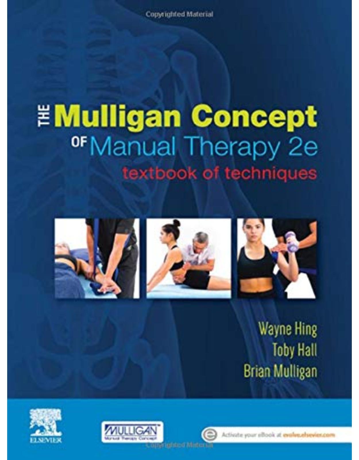
The Mulligan Concept of Manual Therapy
Livrare gratis la comenzi peste 500 RON. Pentru celelalte comenzi livrarea este 20 RON.
Disponibilitate: La comanda in aproximativ 4 saptamani
Editura: Elsevier
Limba: Engleza
Nr. pagini: 411
Coperta: Paperback
Dimensiuni: 19.05 x 1.91 x 26.04 cm
An aparitie: 22 Nov. 2019
|
Description: Endorsed by the Mulligan Concept Teachers Association (MCTA) The MCTA is the accredited body of Mulligan Concept teachers. A comprehensive and easy-to-follow resource for the manual therapist seeking to improve patients’ movement using pain-free hands-on techniques. The Mulligan Concept of manual therapy was developed by Brian Mulligan in 1983 and is now used by health practitioners globally to assist individuals in improving movement restrictions, pain with movement and functional restrictions. Designed as a companion to Mulligan Concept training courses, the text is divided by body regions, with techniques highlighting key information to assist with clinical reasoning and assessment, patient and practitioner positioning, guidelines for application and further adjustments. |
||
|
|
||
|
||
|
Table Of Contents:
|
||
|
|
||
|
|
||
|
Mulligan Concept annotations References Introduction Mobilisation With Movement Clinical Reasoning and the Mulligan Concept Patient-centred approach to healthcare Promotion of knowledge organisation Proposed Mechanisms by Which MWM Work MWMs and peripheral mechanisms Altered mechanoreceptive input during MWM MWMs and central mechanisms Does pain alleviation by MWM prove peripheral dysfunction? Extinguishing movement–pain associations Centrally mediated inhibitory mechanisms MWMs, placebo and reassurance MWMs and output (response) mechanisms Pain and the motor system Mechanoreceptors and the motor system The possibility of sustained benefit Conclusion Aims and Structure of the Book References 1 Cervicogenic headache Introduction Levels of evidence Level 2: four RCTs and one case report Flexion–Rotation Test C1 / 2 Self-SNAG Headache MWM Headache SNAG Reverse Headache SNAG Upper Cervical Traction References 2 Cervicogenic dizziness Introduction Levels of evidence Level 2: four RCTs C1 SNAG for Cervical Rotation Dizziness C2 SNAG for Cervical Extension Dizziness C2 SNAG for Nausea, Light-Headedness or Visual Disturbances (Rescue Manoeuvre) References 3 Cervical spine Introduction Levels of evidence Level 2: eight RCTs, one case report and two laboratory-based studies Cervical Snags C2–7 SNAGs for cervical motion restriction – flexion, extension, lateral flexion and rotation C5 / 6 or C6 / 7 transverse (positional) SNAG Fist Traction Natural Apophyseal Glides (NAGs) (Central and Unilateral) Reverse Nags (Central and Unilateral) Cervicothoracic Junction Mobilisation: Bridge Technique Cervical Traction: Upper Extremity Pain Spinal Mobilisation With Arm Movement (Smwam) Shoulder abduction Smwam: Horizontal Extension and Neurodynamic Dysfunction References 4 Temporomandibular joint Introduction Levels of evidence Level 4: one case series and one case report Temporomandibular Joint: MWM MWM for reduction of internal derangement limiting mandibular depression MWM for painful limitation of mandibular depression MWM scream stretch: movement limitation and pain on mandibular depression MWM for pain on jaw closure References 5 Shoulder complex Introduction Levels of evidence Level 2: five RCTs, one pilot RCT, one multi-case series, one case report MWM to Shoulder Girdle Scapular depression, retraction and downward rotation with clavicle and scapular approximation for s Scapular depression, retraction and downward rotation with clavicle and scapular approximation for s Acromioclavicular Joint MWM for Shoulder Flexion / Abduction / Scaption and / or Elevation Mid-range mobilisation in sitting: posterolateral glide Mid-range elevation mobilisation in sitting: posterolateral–inferior glide with a belt End-range elevation mobilisation in sitting: posteroinferior glide Movement Limitation: Hand Behind Back Inferior glide MWM to restore a loss of hand-behind-back (HBB) movement Movement Limitation: Internal or External Rotation Inferior glide MWM to restore a loss of internal rotation and HBB movement Sleeper stretch MWM to restore a loss of internal rotation Contraction combined with MWM to restore a loss of internal or external rotation References 6 Elbow region Introduction Levels of evidence Level 2: six RCTs, two case series Tennis Elbow: Lateral Elbow Pain Lateral elbow pain: manual lateral glide MWM with gripping Proximal radioulnar joint posteroanterior MWM Golfer’s Elbow: Medial Elbow Pain Olecranon medial and lateral tilt (lateral and medial rotation) Elbow Movement Dysfunction Elbow extension manual lateral and medial glide Elbow flexion manual lateral and medial glide Elbow flexion and extension manual olecranon lateral tilt / medial rotation Elbow flexion and extension manual olecranon medial tilt / lateral rotation Forearm: Treated Proximally Proximal radioulnar joint posteroanterior MWM to improve supination and pronation References 7 Wrist and hand Introduction Levels of evidence Level 4: three case series, three case studies Distal Forearm / Wrist Inferior radioulnar joint: pain or limitation of movement during pronation or supination Carpal lateral glide for non-weight-bearing wrist flexion and extension Carpal medial glide for non-weight-bearing wrist flexion and extension Carpal lateral glide for weight-bearing wrist extension Carpal rotation for wrist flexion and extension Scaphoid PA or AP glide non-weight-bearing Hand Metacarpal PA and AP glide with fist clenching Finger – Proximal Interphalangeal (PIP) Joint Pain and / or Restriction Finger PIP joint pain / restriction with flexion manual lateral / medial glide References 8 Thoracic spine and rib cage Introduction Levels of evidence Level 4: one case series and one case report Thoracic Spine Thoracic traction with a belt Thoracic SNAG central (and unilateral) for flexion, extension, lateral flexion or rotation Thoracic SNAG – central for flexion Thorax – Rib and Spine Upper and lower rib MWM Costovertebral MWM for first or second rib References 9 Sacroiliac joint Introduction Levels of evidence Level 2: three RCTs, one case series and two case studies Posterior Glide MWM Innominate in Relation to Sacrum With Trunk Extension in Prone Lying Lateral Glide MWM Innominate in Relation to Sacrum With Trunk Extension in Prone Lying Posterior Glide and / or Posterior Rotation MWM Innominate in Relation to Sacrum During Walking Taping: Posterior Glide and / or Posterior Rotation Innominate in Relation to Sacrum Taping: Anterior Glide and / or Anterior Rotation Innominate in Relation to Sacrum Home Exercise: Posterior Rotation Innominate MWM in Step Standing Posterior Rotation Innominate / Anterior Rotation Sacrum With Appropriate Glides for Trunk Extension Anterior Rotation Innominate for Trunk Movement in Standing Posterior Rotation Innominate / Anterior Rotation Sacrum With Appropriate Glides for Hip Flexion in Posterior Rotation Innominate / Anterior Rotation Sacrum With Appropriate Glides for Hip Extension i References 10 Lumbar spine Introduction Levels of evidence Level 2: five RCTs and two pilot RCTs Lumbar Snags L1–5 SNAGs for lumbar motion pain and / or restriction for extension (or flexion and lateral flexi SNAG in four-point kneeling (‘lion position’) Lumbar Spine Pain With Leg Symptoms SLR-induced symptoms proximal to the knee Gate (two-leg rotation) technique Bent leg raise (BLR) Traction straight leg raise (TrSLR) Spinal Mobilisation With Leg Movement (SMWLM) for SLR SLR-induced distal leg symptoms SLR SMWLM in side lying Femoral nerve test-induced anterior leg symptoms Femoral SMWLM in side lying References 11 Hip region Introduction Levels of evidence Level 2: four RCTs and one case report Lateral Glide MWM for Hip Flexion and Internal / External Rotation in Supine Lying Lateral glide MWM for hip flexion in supine lying Lateral Glide MWM for Hip Extension in Weight-Bearing Lateral Glide MWM for Hip Flexion in Weight-Bearing Lateral Glide for Hip Internal and External Rotation in Weight-Bearing Hip MWM in Supine Lying for Abduction and Adduction Hip Extension and Rectus Femoris / Hip Flexor MWM in Prone, Supine and Side Lying Hip extension and rectus femoris MWM in prone lying Hip extension and rectus femoris / hip flexor stretch MWM in side lying References 12 Knee Introduction Levels of evidence Level 3: six RCTs, two case series, two case reports Lateral and Medial Glide (Non-Weight-Bearing / Weight-Bearing) – Flexion and Extension (Supine) Lateral glide MWM for flexion / extension Medial glide MWM for flexion / extension Knee Anteroposterior MWM for Flexion and Posteroanterior MWM for Extension Knee anteroposterior MWM for flexion Knee posteroanterior MWM for extension Tibial Rotation – Non-Weight-Bearing / Weight-Bearing Internal rotation MWM for flexion Internal / external rotation MWM for extension Internal rotation MWM for extension Knee Squeeze Technique for Meniscal Pain Proximal Fibular MWM – Ventral or Posterior Glide During Knee Flexion and Extension References 13 Ankle and foot Introduction Levels of evidence Level 2: six RCTs, three case series, two case reports Talus posterior glide with dorsiflexion Posterocephalad fibula glide Talocrural Joint Anteroposterior glide for ankle dorsiflexion in non-weight-bearing Ankle dorsiflexion MWM in weight-bearing Plantarflexion MWM in non-weight-bearing Inferior Tibiofibular Joint – Ankle Sprain Fibula posterior glide MWM for dorsiflexion / plantarflexion–inversion in non-weight-bearing Mid-Tarsal Medial – cuneiform on navicular MWM dorsal / plantar glide medial cuneiform on navicular Lateral – fifth metatarsal on cuboid MWM dorsal / ventral glide fifth metatarsal on cuboid First Metatarsophalangeal Joint Lateral glide for flexion and extension References 14 Pain release phenomenon Introduction Levels of evidence Trapezium – First Metacarpal Joint PRP Tennis Elbow PRP With a Muscle Contraction (Lateral Epicondylalgia) Chronic Painful Shoulder PRP Hip Pain (Faber Position) PRP Hip Pain (Posterior Shear) PRP References Index A B C D E F G H I J K L M N O P R S T U V W Z
|
||
| An aparitie | 22 Nov. 2019 |
| Autor | Wayne Hing PhD MSc(Hons) ADP(OMT) DipMT Dip Phys FNZCP, Toby Hall PT PHD MSc FACP |
| Dimensiuni | 19.05 x 1.91 x 26.04 cm |
| Editura | Elsevier |
| Format | Paperback |
| ISBN | 9780729542821 |
| Limba | Engleza |
| Nr pag | 411 |
| Versiune digitala | DA |
-
 34700 lei 32000 lei
 -
-

Clientii ebookshop.ro nu au adaugat inca opinii pentru acest produs. Fii primul care adauga o parere, folosind formularul de mai jos.